Acinar Cell Cystadenoma (Acinar Cystic Transformation) of the Pancreas: the Radiologic-Pathologic Features
- Affiliations
-
- 1Department of Radiology, Ataturk Training and Research Hospital, Ankara, Turkey. droktayalgin@gmail.com
- 2Department of Pathology, Selcuk University, Selcuklu Medical Faculty, Konya, Turkey.
- 3Department of General Surgery, Ataturk Training and Research Hospital, Ankara, Turkey.
- KMID: 991691
- DOI: http://doi.org/10.3348/kjr.2011.12.1.129
Abstract
- Acinar cystic transformation of the pancreas is also known as acinar cell cystadenoma (ACC), and this is an extremely rare benign lesion that was first described in April 2002. We report here on a case of a previously asymptomatic patient with pancreatic ACC and this was diagnosed by computed tomography (CT) and magnetic resonance imaging (MRI). To the best of our knowledge, there is no previous report concerning the CT or MRI features of ACC in the medical literature. We present here the CT, MRI and pathological findings of pancreatic ACC.
Keyword
MeSH Terms
Figure
-
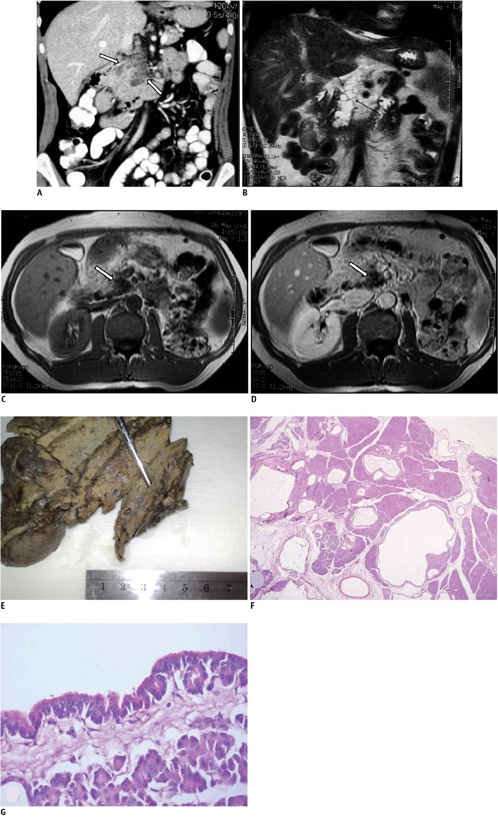
Fig. 1 Pancreas acinar cell cystadenoma in 52-year-old man. A. Contrast-material enhanced, coronal reformatted multidetector CT image. Multiple hypodense cystic lesions are clearly seen in pancreas (arrows). B-D. Coronal half-Fourier acquisition single-shot turbo spin-echo (HASTE) (B), axial precontrast (C) and postcontrast T1 weighted (D) MR images of pancreas. Nonenhancing cystic lesions are seen in pancreas (arrows). E. Image of macroscopic specimen. Numerous cysts with thin translucent wall are seen in body and head of pancreas. F, G. Numerous cystic spaces are diffusely distributed in pancreas tissue (F). Note small clusters of acinar cells forming acini that open into cyst lumens (G) (Hematoxylin & Eosin stain, original magnification, × 20 [F], 600 [G]).
Cited by 1 articles
-
췌장의 장액낭샘종으로 오인된 췌장의 샘꽈리세포낭종 1예
Jong Hyun Lee, Soo Jin Jung, Yo Han Park, Sung Jae Park, Jung Sik Choi
Korean J Gastroenterol. 2021;78(2):138-143. doi: 10.4166/kjg.2021.044.
Reference
-
1. Hamilton SR, Aaltonen LA. WHO classification of tumours. Pathology and genetics of tumours of digestive system. 2000. Lyon, France: IARC Press.2. Huang Y, Cao YF, Lin JL, Gao F, Li F. Acinar cell cystadenocarcinoma of the pancreas in a 4-year-old child. Pancreas. 2006. 33:311–312.3. Solcia E, Capella C, Kloppel G. Tumors of the pancreas. Atlas of tumor pathology. 1997. Washington, DC: AFIP;103–114. 3rd Series, Fascicle 20.4. Adsay NV. Cystic lesions of the pancreas. Mod Pathol. 2007. 20:S71–S93.5. Cantrell BB, Cubilla AL, Erlandson RA, Fortner J, Fitzgerald PJ. Acinar cell cystadenocarcinoma of human pancreas. Cancer. 1981. 47:410–416.6. Albores-Saavedra J. Acinar cystadenoma of the pancreas: a previously undescribed tumor. Ann Diagn Pathol. 2002. 6:113–115.7. Zamboni G, Terris B, Scarpa A, Kosmahl M, Capelli P, Klimstra DS, et al. Acinar cell cystadenoma of the pancreas: a new entity? Am J Surg Pathol. 2002. 26:698–704.8. Chatelain D, Paye F, Mourra N, Scoazec JY, Baudrimont M, Parc R, et al. Unilocular acinar cell cystadenoma of the pancreas an unusual acinar cell tumor. Am J Clin Pathol. 2002. 118:211–214.9. Couvelard A, Terris B, Hammel P, Palazzo L, Belghiti J, Lévy P, et al. Acinar cystic transformation of the pancreas (or acinar cell cystadenoma), a rare and recently described entity. Ann Pathol. 2002. 22:397–400. [French].10. Tatli S, Mortele KJ, Levy AD, Glickman JN, Ros PR, Banks PA, et al. CT and MRI features of pure acinar cell carcinoma of the pancreas in adults. AJR Am J Roentgenol. 2005. 184:511–519.11. Brugge WR. Cystic neoplasms of the pancreas. Endoscopic oncology. 2006. Totowa, New Jersey: Humana Press Inc;289–294. Chapter 24.12. Sahani DV, Kadavigere R, Saokar A, Fernandez-del Castillo C, Brugge WR, Hahn PF. Cystic pancreatic lesions: a simple imaging-based classification system for guiding management. Radiographics. 2005. 25:1471–1484.13. Acar M, Tatli S. Cystic tumors of the pancreas: a radiological perspective. Diagn Interv Radiol. 2010. [Epub ahead of print]. DOI: 10.4261/1305-3825.DIR.3254-09.1.14. Brugge WR, Lauwers GY, Sahani D, Fernandez-del Castillo C, Warshaw AL. Cystic neoplasms of the pancreas. N Engl J Med. 2004. 351:1218–1226.15. Kazumori H, Sizuku T, Ueki T, Uchida Y, Yamamoto S. Lymphoepithelial cyst of the pancreas. J Gastroenterol. 1997. 32:700–703.16. Chiou YY, Chiang JH, Hwang JI, Yen CH, Tsay SH, Chang CY. Acinar cell carcinoma of the pancreas: clinical and computed tomography manifestations. J Comput Assist Tomogr. 2004. 28:180–186.17. Chung WJ, Byun JH, Lee SS, Lee MG. Imaging findings in a case of mixed acinar-endocrine carcinoma of the pancreas. Korean J Radiol. 2010. 11:378–381.
- Full Text Links
-
- Actions
-
Cited
- CITED
-
- Close
- Share
-
- Similar articles
-
- Acinar Cell Cystadenoma of the Pancreas: Report of a Case with Metaplastic Ossification
- Pancreatic Acinar Cell Cystadenoma Mimicking Pancreatic Serous Cystadenoma
- Mixed acinar-endocrine carcinoma of the pancreas: a case report
- Acinar Cell Carcinoma of the Pancreas: A case report
- Cystic Neoplasm of the Pancreas: Its Diagnosis and Treatment
