Confident Diagnosis of Bronchobiliary Fistula Using Contrast-Enhanced Magnetic Resonance Cholangiography
- Affiliations
-
- 1Department of Radiology, Pamukkale University Medical Center, Denizli, Turkey. nkarabulut@yahoo.com
- KMID: 984907
- DOI: http://doi.org/10.3348/kjr.2010.11.4.493
Abstract
- We report the utility of contrast-enhanced magnetic resonance cholangiography (MRC) using gadoxetic acid (Gd-EOB-DTPA) in the diagnosis of bronchobiliary fistula associated with liver hydatid cyst. Contrast-enhanced MRC clearly delineated the leakage of contrast agent from the biliary duct and its communication with the bronchial tree. Providing functional information about physiologic or pathologic biliary flow in addition to the display of biliary anatomy, contrast-enhanced MRC stands as a robust technique in confidently detecting bronchobiliary fistula and bile leaks.
MeSH Terms
Figure
-
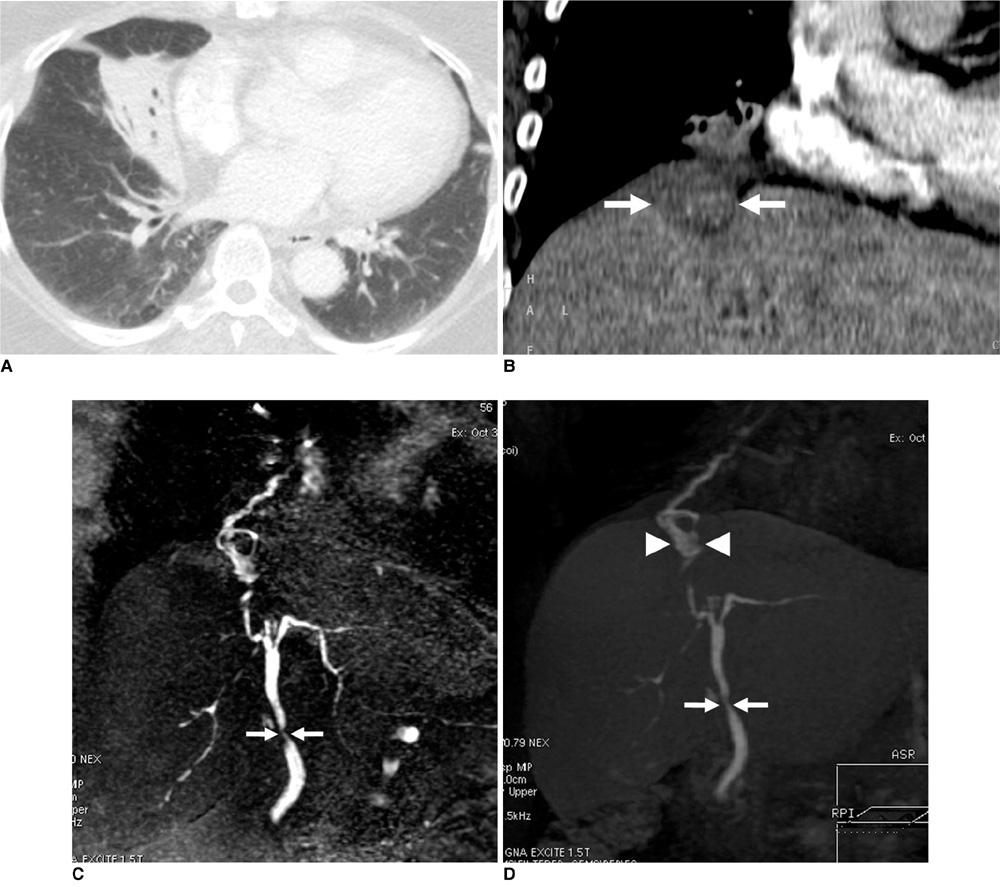
Fig. 1 Bronchobiliary fistula in 56-year-old woman. A. Chest CT shows distal subsegmental atelectasis and air bronchograms in right middle lobe. B. Targeted coronal reconstruction of upper abdominal CT shows obscure subdiaphragmatic hypodense lesion (arrows) adjacent to inferior surface of right middle lobe. C. Coronal 3D-maximum intensity projection reconstruction of conventional MR cholangiography demonstrates stenosis of common bile duct (arrows) as well as fistulous bronchobiliary communication. D. Coronal 3D-maximum intensity projection reconstruction of contrast-enhanced MR cholangiography reveals contrast agent leaking from ventrocranial branch of right hepatic duct and further into subphrenic liver cyst (arrowheads), which transdiaphragmatically communicates with bronchial tree. Stricture of common bile duct is also demonstrated (arrows).
Cited by 1 articles
-
Two cases of bronchobiliary fistula: Case report
Jae Ryong Shim, Sung-Sik Han, Hyung Min Park, Eung Chang Lee, Sang-Jae Park, Joong-Won Park
Ann Hepatobiliary Pancreat Surg. 2018;22(2):169-172. doi: 10.14701/ahbps.2018.22.2.169.
Reference
-
1. Ragozzino A, De Rosa R, Galdiero R, Maio A, Manes G. Bronchobiliary fistula evaluated with magnetic resonance imaging. Acta Radiol. 2005. 46:452–454.2. Oettl C, Schima W, Metz-Schimmerl S, Függer R, Mayrhofer T, Herold CJ. Bronchobiliary fistula after hemihepatectomy: cholangiopancreaticography, computed tomography and magnetic resonance cholangiography findings. Eur J Radiol. 1999. 32:211–215.3. Eryigit H, Oztas S, Urek S, Olgac G, Kurutepe M, Kutlu CA. Management of acquired bronchobiliary fistula: 3 case reports and a literature review. J Cardiothorac Surg. 2007. 2:52.4. Senturk H, Mert A, Ersavasti G, Tabak F, Akdogan M, Ulualp K. Bronchobiliary fistula due to alveolar hydatid disease: report of three cases. Am J Gastroenterol. 1998. 93:2248–2253.5. George TK, Carignan JR. Bronchobiliary fistula after hepatic resection for metastatic colon cancer. J Surg Oncol. 1984. 25:198–200.6. Moreira VF, Arocena C, Cruz F, Alvarez M, San Roman AL. Bronchobiliary fistula secondary to biliary lithiasis. Treatment by endoscopic sphincterotomy. Dig Dis Sci. 1994. 39:1994–1999.7. Ergen FB, Akata D, Sarikaya B, Kerimoglu U, Hayran M, Akhan O, et al. Visualization of the biliary tract using gadobenate dimeglumine: preliminary findings. J Comput Assist Tomogr. 2008. 32:54–60.8. Dahlström N, Persson A, Albiin N, Smedby O, Brismar TB. Contrast-enhanced magnetic resonance cholangiography with Gd-BOPTA and Gd-EOB-DTPA in healthy subjects. Acta Radiol. 2007. 48:362–368.9. Aduna M, Larena JA, Martin D, Martinez-Guereñu B, Aguirre I, Astigarraga E. Bile duct leaks after laparoscopic cholecystectomy: value of contrast-enhanced MRCP. Abdom Imaging. 2005. 30:480–487.10. Hoeffel C, Azizi L, Lewin M, Laurent V, Aubé C, Arrivé L, et al. Normal and pathologic features of the postoperative biliary tract at 3D MR cholangiopancreatography and MR imaging. Radiographics. 2006. 26:1603–1620.11. Karabulut N, Elmas N. Contrast agents used in MR imaging of the liver. Diagn Interv Radiol. 2006. 12:22–30.
- Full Text Links
-
- Actions
-
Cited
- CITED
-
- Close
- Share
-
- Similar articles
-
- A case of bronchobiliary fistula caused by choledocholithiasis
- Why is my phlegm green? A rare case of bronchobiliary fistula
- Surgical Treatment of Bronchobiliary Fistula with Pulmonary Resection and Omentopexy
- Congenital Bronchobiliary Fistula: A case report
- Successful percutaneous management of bronchobiliary fistula after radiofrequency ablation of metastatic cholangiocarcinoma in a patient who has a postoperative stricture of hepaticojejunostomy site
