Imaging Features of Pediatric Pentastomiasis Infection: a Case Report
- Affiliations
-
- 1Department of Radiology, Children's Hospital, Zhejiang University School of Medicine and the Zhejiang Key Laboratory for the Diagnosis and Therapy of Neonatal Diseases, Zhejiang 310003, China. laican1@126.com
- 2Department of Pathology, Children's Hospital, Zhejiang University School of Medicine and the Zhejiang Key Laboratory for the Diagnosis and Therapy of Neonatal Diseases, Zhejiang 310003, China.
- KMID: 984904
- DOI: http://doi.org/10.3348/kjr.2010.11.4.480
Abstract
- We report here a case of pentastomiasis infection in a 3-year-old girl who had high fever, abdominal pain, abdominal tension and anemia. Ultrasound scanning of the abdomen revealed disseminated hyperechoic nodules in the liver and a small amount of ascites. Abdominal MRI showed marked hepatomegaly with disseminated miliary nodules of high signal intensity throughout the hepatic parenchyma on T2-weighted images; retroperitoneal lymphadenopathy and disseminated miliary nodules on the peritoneum were also noted. Chest CT showed scattered small hyperdense nodules on both sides of the lungs. The laparoscopy demonstrated diffuse white nodules on the liver surface and the peritoneum. After the small intestinal wall and peritoneal biopsy, histological examination revealed parenchymal tubercles containing several larvae of pentastomids and a large amount of inflammatory cell infiltration around them. The pathological diagnosis was parasitic granuloma from pentastomiasis infection.
MeSH Terms
Figure
-
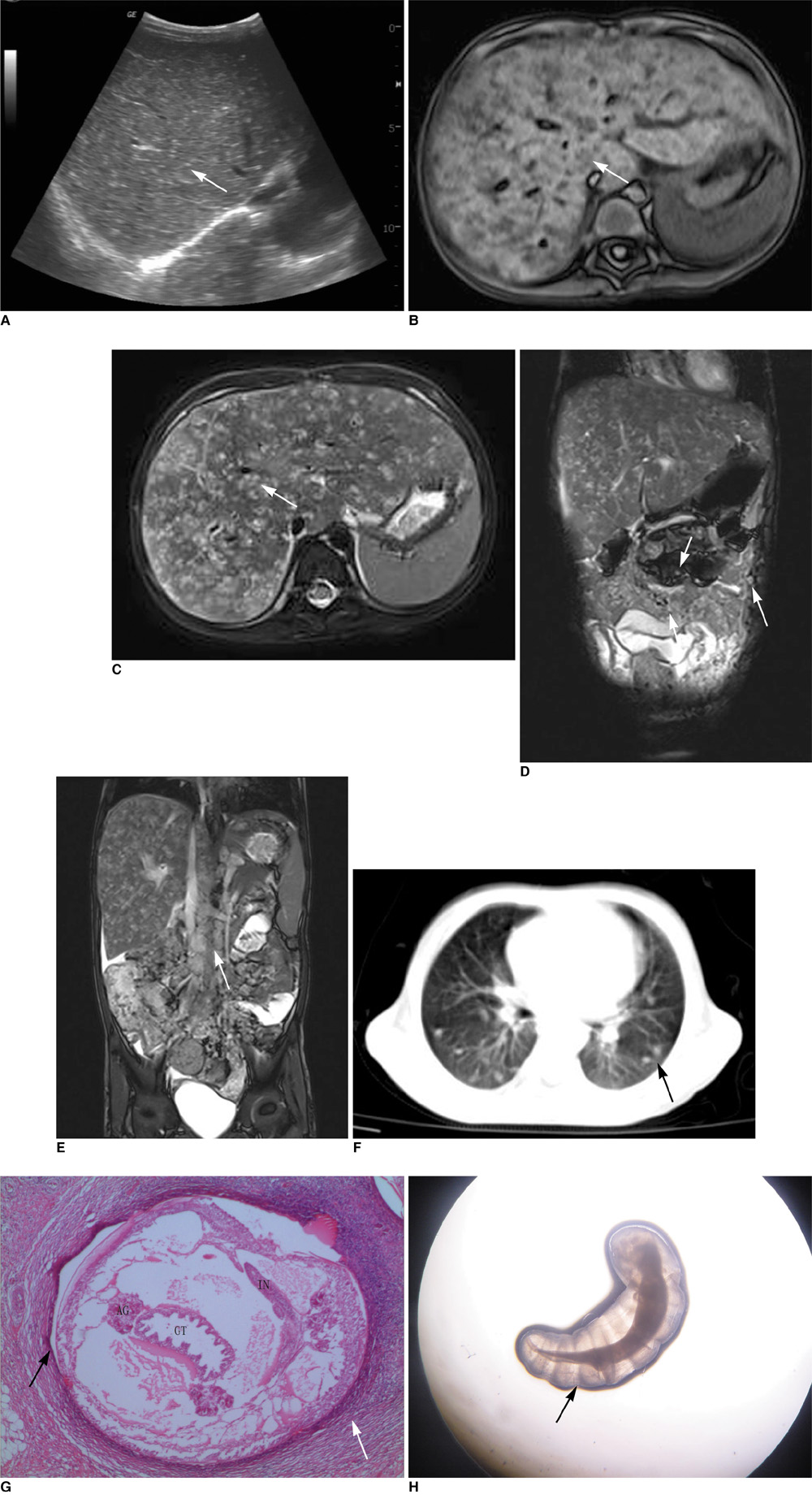
Fig. 1 3-year-old girl with pentastomiasis infection, and she had high fever, abdominal pain, abdominal tension and anemia. A. Abdominal ultrasonography reveals disseminated miliary hyperechoic nodules in liver (arrow). B. T1-weighted transverse MRI (TR, 1,540 ms; TE, 2.3 ms) reveals marked hepatomegaly with miliary nodular lesions (2-4 mm diameter) of low signal intensity throughout hepatic parenchyma (arrow). C. T2-weighted transverse MRI (TR, 4,577 ms; TE, 101 ms) reveals marked hepatomegaly with disseminated nodular lesions of high signal intensity throughout hepatic parenchyma (arrow). D, E. T2-weighted coronal MRI (TR, 3.7 ms; TE, 1.9 ms) shows disseminated miliary mesenteric lymph nodes (short arrows, D), peritoneal nodules (long arrow, D) and diffuse retroperitoneal lymphadenopathy of high signal intensity (arrow, E). F. Chest CT shows multiple small hyperdense nodules in both lungs on lung window (arrow). G. Cross section of pentastomid worm in peritoneum shows primordial genital tract (GT), red acidophilic gland (AG), intestine (IN) and worm wall (black arrow), which are surrounded by group of macrophages and inflammatory cells and fibrosis (white arrow) (Hematoxylin & Eosin stain, × 40). H. Vital well-preserved pentastomid worm from patient feces. Worm is cylindrical, translucent and has prominent spiral rings (arrow). We can see its chest, abdomen and blunt round tail, which is gradually shrinking.
Reference
-
1. Zhang Q, Wang B, Huang M. Armillifer agkistrodontis disease: report of case. Zhonghua Nei Ke Za Zhi. 1996. 35:747–749. [Chinese].2. Lavrov DV, Brown WM, Boore JL. Phylogenetic position of the Pentastomida and (pan)crustacean relationships. Proc Biol Sci. 2004. 271:537–544.3. Qiu MH. Pentastomiasis. Tropical medicine. 2004. 2nd ed. Beijing: People's Health Press;1169–1175.4. Liu JD, Cheng NY, Chu SY. Parasitic disease in the gastroenterological field. Jap J Gastrointest Endosc. 1993. 5:1553–1559.5. Qiu MH, Jiang YY. Advances in studies of human pentastomiasis. Int J Med Parasit Dis. 2006. 33:281–287.6. Tappe D, Büttner DW. Diagnosis of human visceral pentastomiasis. PLoS Negl Trop Dis. 2009. 3:E320.7. Baird JK, Kassebaum LJ, Ludwig GK. Hepatic granuloma in a man from North America caused by a nymph of Linguatula serrata. Pathology. 1988. 20:198–199.8. Smith JA, Oladiran B, Lagundoye SB. Pentastomiasis and malignancy. Ann Trop Med Parasitol. 1975. 69:503–512.9. Qiu MH, Cheng ML. Armilliferiosis agkistrodontis is a visceral larva migrans echinococcosis. Chin J Parasitol Parasite Dis. 1997. 15:117–118.10. Berenji F, Fata A, Hosseininejad Z. A case of Moniliformis moniliformis (Acanthocephala) infection in Iran. Korean J Parasitol. 2007. 45:145–148.11. Pan CM, Tang HF, Qiu MH, Xiong QX. A case with severe infestation with Armilliferiasis moniliformis. Zhonghua Er Ke Za Zhi. 2005. 43:73–74.12. Ihara N, Yashiro N, Kinoshita T, Yoshigi J, Ouchi T, Narita M, et al. Diffuse intrasinusoidal liver metastasis of small cell lung cancer causing fulminant hepatic failure: CT findings-a case report. Radiat Med. 2001. 19:275–277.13. Yao MH, Wu F, Tang LF. Human pentastomiasis in China: case report and literature review. J Parasitol. 2008. 94:1295–1298.
- Full Text Links
-
- Actions
-
Cited
- CITED
-
- Close
- Share
-
- Similar articles
-
- A Case of Pentastomiasis at the Left Maxilla Bone in a Patient with Thyroid Cancer
- Rotavirus infection-associated central nervous system complications: clinicoradiological features and potential mechanisms
- Atypical beta-Catenin Activated Child Hepatocellular Tumor
- Intraductal Papillary Mucinous Neoplasm of the Pancreas with Worrisome Features in a 12-Year-Old Boy
- Radiogenomics Based on PET Imaging
