Utility of Postmortem Autopsy via Whole-Body Imaging: Initial Observations Comparing MDCT and 3.0T MRI Findings with Autopsy Findings
- Affiliations
-
- 1Department of Radiology, Soonchunhyang University Bucheon Hospital, Gyunggi-do 420-020, Korea.
- 2Department of Radiology, Konyang University Hospital, Daejeon 302-718, Korea.
- 3Department of Radiology, Soonchunhyang University Hospital, Seoul 140-743, Korea.
- 4Department of Forensic Medicine, National Institute of Scientific Investigation, Seoul 158-707, Korea. rudany@korea.kr
- KMID: 984893
- DOI: http://doi.org/10.3348/kjr.2010.11.4.395
Abstract
OBJECTIVE
We prospectively compared whole-body multidetector computed tomography (MDCT) and 3.0T magnetic resonance (MR) images with autopsy findings.
MATERIALS AND METHODS
Five cadavers were subjected to whole-body, 16-channel MDCT and 3.0T MR imaging within two hours before an autopsy. A radiologist classified the MDCT and 3.0T MRI findings into major and minor findings, which were compared with autopsy findings.
RESULTS
Most of the imaging findings, pertaining to head and neck, heart and vascular, chest, abdomen, spine, and musculoskeletal lesions, corresponded to autopsy findings. The causes of death that were determined on the bases of MDCT and 3.0T MRI findings were consistent with the autopsy findings in four of five cases. CT was useful in diagnosing fatal hemorrhage and pneumothorax, as well as determining the shapes and characteristics of the fractures and the direction of external force. MRI was effective in evaluating and tracing the route of a metallic object, soft tissue lesions, chronicity of hemorrhage, and bone bruises.
CONCLUSION
A postmortem MDCT combined with MRI is a potentially powerful tool, providing noninvasive and objective measurements for forensic investigations.
MeSH Terms
Figure
-

Fig. 1 40-year-old man (pedestrian) who died in motor vehicle accident.A. Coronal short tau inversion recovery whole-body MR image shows collapse of right lung (asterisk), low signal intensity lesion in right lobe of liver (curved arrow), high signal intensity lesion in L1 vertebral body (dashed arrow), and posterior dislocation of left hip joint (arrowheads).B. Coronal reformatted whole-body CT image showing mediastinal shifting to left side (arrow) and posterior dislocation of left hip joint (arrowheads).C. Axial thoracic CT with lung setting showing subcutaneous emphysema and right rib fracture with inferior displacement (solid arrow). Bilateral pleural effusions are also seen (dashed arrows). Mediastinal shifting (arrowheads) to left side caused by pneumothorax (asterisk), which could not be detected after opening thoracic cavity in conventional autopsy.D. Magnified image of liver from (A) shows low signal lesion with intermediate signal intensity margin in right lobe of liver, suggesting traumatic rupture of liver (arrows).E. Corresponding pathological photograph confirms rupture of right lobe of liver (arrows).F. Volume-rendered CT image shows posterior dislocation of left hip joint, suggesting that external force was applied in anteroposterior direction (arrow).
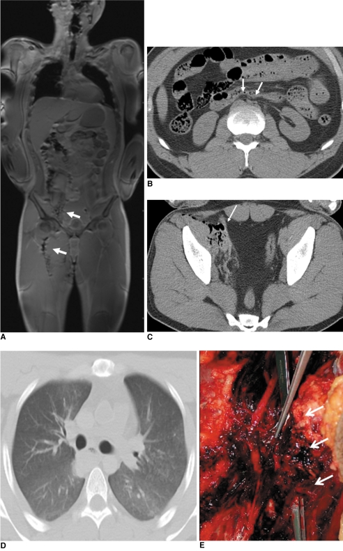
Fig. 2 23-year-old man whose right thigh was pierced by iron bar.A. Coronal T1 fast spoiled gradient-echo image shows curvilinear streaky lesion (arrows) along right thigh and psoas muscle caused by susceptibility artifact, which can help determine path of iron bar in body.B. Axial CT image of pelvis demonstrates collapse of abdominal aorta and inferior vena cava (arrows).C. Axial CT image of pelvis at acetabular level shows loculated air collection along right iliac vessels (arrow).D. Lung window view of transverse CT scan shows absence of any increased density in lung parenchyma.E. Autopsy photography reveals forceps putting through pathway of iron bar (arrows) and ruptured right femoral artery (holded by forceps) located within this pathway.
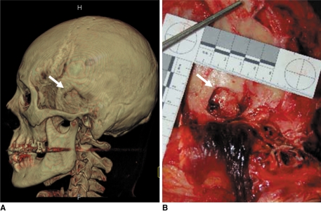
Fig. 3 57-year-old man who died due to head injuries from hammer.A. Volume-rendered CT image of skull shows round depressive fracture in left temporal bone (arrow).B. Autopsy photograph of left temporal bone shows similar size and shape of depressed fracture (arrow) which was observed in CT image.
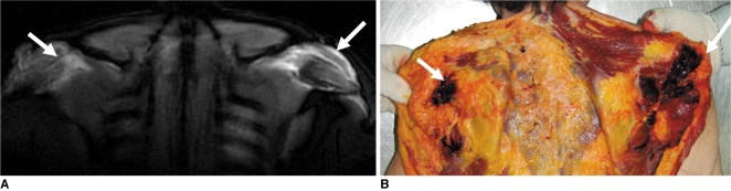
Fig. 4 71-year-old woman who fell from chair after spontaneous intracranial hemorrhage.A. Coronal STIR images show increased signal intensity along posterior subcutaneous area of both scapular regions (arrows).B. Autopsy photography of upper back reveals fresh soft tissue hemorrhage in corresponding areas (arrows).
Reference
-
1. Bolliger SA, Thali MJ, Ross S, Buck U, Naether S, Vock P. Virtual autopsy using imaging: bridging radiologic and forensic sciences. A review of the Virtopsy and similar projects. Eur Radiol. 2008; 18:273–282. PMID: 17705044.
Article2. Lundberg GD. Low-tech autopsies in the era of high-tech medicine: continued value for quality assurance and patient safety. JAMA. 1998; 280:1273–1274. PMID: 9786381.3. Yen K, Lövblad KO, Scheurer E, Ozdoba C, Thali MJ, Aghayev E, et al. Post-mortem forensic neuroimaging: correlation of MDCT and MRI findings with autopsy results. Forensic Sci Int. 2007; 173:21–35. PMID: 17336008.4. Dirnhofer R, Jackowski C, Vock P, Potter K, Thali MJ. VIRTOPSY: minimally invasive, imaging-guided virtual autopsy. Radiographics. 2006; 26:1305–1333. PMID: 16973767.
Article5. Berry PJ, Keeling JW, Wigglesworth JS. Perinatal necropsy by magnetic resonance imaging. Lancet. 1997; 349:55. PMID: 8988133.
Article6. Brookes JA, Hall-Craggs MA, Sams VR, Lees WR. Non-invasive perinatal necropsy by magnetic resonance imaging. Lancet. 1996; 348:1139–1141. PMID: 8888168.
Article7. Woodward PJ, Sohaey R, Harris DP, Jackson GM, Klatt EC, Alexander AL, et al. Postmortem fetal MR imaging: comparison with findings at autopsy. AJR Am J Roentgenol. 1997; 168:41–46. PMID: 8976917.
Article8. Patriquin L, Kassarjian A, Barish M, Casserley L, O'Brien M, Andry C, et al. Postmortem whole-body magnetic resonance imaging as an adjunct to autopsy: preliminary clinical experience. J Magn Reson Imaging. 2001; 13:277–287. PMID: 11169835.
Article9. Ros PR, Li KC, Vo P, Baer H, Staab EV. Preautopsy magnetic resonance imaging: initial experience. Magn Reson Imaging. 1990; 8:303–308. PMID: 2366642.
Article10. Weustink AC, Hunink MG, van Dijke CF, Renken NS, Krestin GP, Oosterhuis JW. Minimally invasive autopsy: an alternative to conventional autopsy? Radiology. 2009; 250:897–904. PMID: 19244053.
Article11. Ross S, Spendlove D, Bolliger S, Christe A, Oesterhelweg L, Grabherr S, et al. Postmortem whole-body CT angiography: evaluation of two contrast media solutions. AJR Am J Roentgenol. 2008; 190:1380–1389. PMID: 18430859.
Article12. Grabherr S, Gygax E, Sollberger B, Ross S, Oesterhelweg L, Bolliger S, et al. Two-step postmortem angiography with a modified heart-lung machine: preliminary results. AJR Am J Roentgenol. 2008; 190:345–351. PMID: 18212219.
Article13. Wüllenweber R, Schneider V, Grumme T. A computer-tomographical examination of cranial bullet wounds (author's transl). Z Rechtsmed. 1977; 80:227–246. [German]. PMID: 602451.14. Dalrymple NC, Prasad SR, El-Merhi FM, Chintapalli KN. Price of isotropy in multidetector CT. Radiographics. 2007; 27:49–62. PMID: 17234998.
Article15. Groves AM, Beadsmoore CJ, Cheow HK, Balan KK, Courtney HM, Kaptoge S, et al. Can 16-detector multislice CT exclude skeletal lesions during tumour staging? Implications for the cancer patient. Eur Radiol. 2006; 16:1066–1073. PMID: 16402253.
Article16. Thali MJ, Yen K, Schweitzer W, Vock P, Boesch C, Ozdoba C, et al. Virtopsy, a new imaging horizon in forensic pathology: virtual autopsy by postmortem multislice computed tomography (MSCT) and magnetic resonance imaging (MRI)--a feasibility study. J Forensic Sci. 2003; 48:386–403. PMID: 12665000.
Article17. Durlacher SH, Banfield WG Jr, Bergner AD. Post-mortem pulmonary edema. Yale J Biol Med. 1950; 22:565–572. PMID: 15431685.18. Schmidt GP, Reiser MF, Baur-Melnyk A. Whole-body imaging of the musculoskeletal system: the value of MR imaging. Skeletal Radiol. 2007; 36:1109–1119. PMID: 17554538.
Article19. Schmidt GP, Kramer H, Reiser MF, Glaser C. Whole-body magnetic resonance imaging and positron emission tomography-computed tomography in oncology. Top Magn Reson Imaging. 2007; 18:193–202. PMID: 17762383.
Article20. Tosaka M, Sato N, Hirato J, Fujimaki H, Yamaguchi R, Kohga H, et al. Assessment of hemorrhage in pituitary macroadenoma by T2*-weighted gradient-echo MR imaging. AJNR Am J Neuroradiol. 2007; 28:2023–2029. PMID: 17898201.
Article21. Yen K, Thali MJ, Aghayev E, Jackowski C, Schweitzer W, Boesch C, et al. Strangulation signs: initial correlation of MRI, MSCT, and forensic neck findings. J Magn Reson Imaging. 2005; 22:501–510. PMID: 16142698.
Article22. Yen K, Vock P, Tiefenthaler B, Ranner G, Scheurer E, Thali MJ, et al. Virtopsy: forensic traumatology of the subcutaneous fatty tissue; multislice computed tomography (MSCT) and magnetic resonance imaging (MRI) as diagnostic tools. J Forensic Sci. 2004; 49:799–806. PMID: 15317198.
Article23. Fidelman NA, Wilson MW, Bloom AI, Kerlan RK Jr, LaBerge JM, Gordon RL. Orthopedic spinal and hip prostheses: effects of magnetic susceptibility artifacts during MR arteriography and venography of abdomen and pelvis. Radiology. 2006; 240:894–899. PMID: 16926332.
Article24. Winkler ML, Olsen WL, Mills TC, Kaufman L. Hemorrhagic and nonhemorrhagic brain lesions: evaluation with 0.35-T fast MR imaging. Radiology. 1987; 165:203–207. PMID: 3628772.
Article25. Newberg AH, Wetzner SM. Bone bruises: their patterns and significance. Semin Ultrasound CT MR. 1994; 15:396–409. PMID: 7803074.
Article
- Full Text Links
-
- Actions
-
Cited
- CITED
-
- Close
- Share
-
- Similar articles
-
- Can Postmortem Fetal MR Imaging Replace Autopsy?
- About reform of autopsy system: focus into the limited autopsy
- Selection of Autopsy Method Depending on Situation
- Differences in the Determination of Cause and Manner of 127 Natural Death Cases by Postmortem Inspection and Autopsy
- Common Postmortem Computed Tomography Findings Following Atraumatic Death: Differentiation between Normal Postmortem Changes and Pathologic Lesions
