Korean J Ophthalmol.
2010 Oct;24(5):318-321. 10.3341/kjo.2010.24.5.318.
A Case of Pantoea Endophthalmitis
- Affiliations
-
- 1Department of Ophthalmology, Gyeongsang National University School of Medicine, Jinju, Korea. parkjm@gnu.ac.kr
- 2Institute of Health Science, Gyeongsang National University, Jinju, Korea.
- KMID: 974329
- DOI: http://doi.org/10.3341/kjo.2010.24.5.318
Abstract
- A previously healthy 50-year-old man was transferred to our hospital for evaluation of acute inflammation in his right eye after ocular trauma while using a grass mower. Slit lamp examination showed 1 mm-length full thickness corneal laceration without leakage, 4+ cells and inflammatory membrane in the anterior chamber, 10% hypopyon, posterior synechiae formation, and cataract change. Upon orbital computerized tomography, a metallic intraocular foreign body in the lens was indentified. Vitrectomy, phacoemulsification, foreign body removal, anterior chamber irrigation, and intravitreal antibiotics injections of vancomycin and ceftazidime were performed. In a culture of humor from the anterior chamber grew Pantoea species. More procedures were performed, including intravitreal antibiotics injection of ceftazidime. Upon administering a course of intravenous ceftazidime, fortified ceftazidime and moxifloxacin eye drops, and oral prednisolone, the patient improved.
Keyword
MeSH Terms
Figure
-
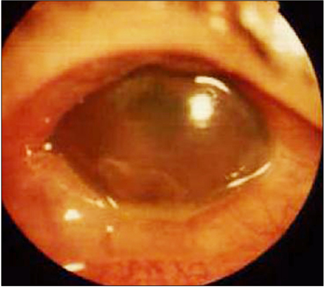
Fig. 1 Slit lamp photography at initial presentation shows conjunctival injection, corneal edema, hypopyon, exudative membrane, and mid-dilated pupil.
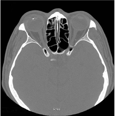
Fig. 2 Orbital computerized tomography shows a metallic intraocular foreign body in the lens.
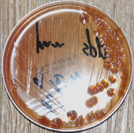
Fig. 3 A culture of aspirated aqueous humor from the anterior chamber shows growth of Pantoea species (culture media: McConkey agar).
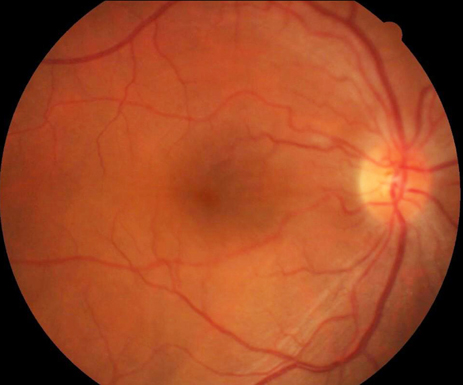
Fig. 4 Fundus photography at two months after trauma shows vitreous and retinal clarity.
Reference
-
1. Jiang CH, Zhang MN. Traumatic endophthalmitis following penetrating ocular injuries with retained intraocular foreign bodies. Chin J Traumatol. 2003. 6:167–170.2. Mieler WF, Ellis MK, Williams DF, Han DP. Retained intraocular foreign bodies and endophthalmitis. Ophthalmology. 1990. 97:1532–1538.3. Thompson JT, Parver LM, Enger CL, et al. Infectious endophthalmitis after penetrating injuries with retained intraocular foreign bodies. National Eye Trauma System. Ophthalmology. 1993. 100:1468–1474.4. Imrie FR, Cox A, Foot B, Macewen CJ. Surveillance of intraocular foreign bodies in the UK. Eye (Lond). 2008. 22:1141–1147.5. Cebulla CM, Flynn HW Jr. Endophthalmitis after open globe injuries. Am J Ophthalmol. 2009. 147:567–568.6. Cruz AT, Cazacu AC, Allen CH. Pantoea agglomerans, a plant pathogen causing human disease. J Clin Microbiol. 2007. 45:1989–1992.7. Deletoile A, Decre D, Courant S, et al. Phylogeny and identification of Pantoea species and typing of Pantoea agglomerans strains by multilocus gene sequencing. J Clin Microbiol. 2009. 47:300–310.8. Brady C, Cleenwerck I, Venter S, et al. Phylogeny and identification of Pantoea species associated with plants, humans and the natural environment based on multilocus sequence analysis (MLSA). Syst Appl Microbiol. 2008. 31:447–460.9. Yeh S, Colyer MH, Weichel ED. Current trends in the management of intraocular foreign bodies. Curr Opin Ophthalmol. 2008. 19:225–233.10. Andreoli CM, Andreoli MT, Kloek CE, et al. Low rate of endophthalmitis in a large series of open globe injuries. Am J Ophthalmol. 2009. 147:601–608.e2.11. Fan JC, Niederer RL, von Lany H, Polkinghorne PJ. Infectious endophthalmitis: clinical features, management and visual outcomes. Clin Experiment Ophthalmol. 2008. 36:631–636.12. Chale-Matsau JR, Snyman HG. The survival of pathogens in soil treated with wastewater sludge and in potatoes grown in such soil. Water Sci Technol. 2006. 54:163–168.13. Van Rostenberghe H, Noraida R, Wan Pauzi WI, et al. The clinical picture of neonatal infection with Pantoea species. Jpn J Infect Dis. 2006. 59:120–121.14. Gora A, Mackiewicz B, Krawczyk P, et al. Occupational exposure to organic dust, microorganisms, endotoxin and peptidoglycan among plants processing workers in Poland. Ann Agric Environ Med. 2009. 16:143–150.15. Sanders WE Jr, Sanders CC. Enterobacter spp.: pathogens poised to flourish at the turn of the century. Clin Microbiol Rev. 1997. 10:220–241.16. Kim HO, Lee HJ, Kim DW, et al. A case of peritonitis caused by Pantoea agglomerans in a patient undergoing continuous ambulatory peritoneal dialysis. Korean J Med. 2008. 74:426–429.17. Coutinho TA, Venter SN. Pantoea ananatis: an unconventional plant pathogen. Mol Plant Pathol. 2009. 10:325–335.18. Mason GI, Bottone EJ, Podos SM. Traumatic endophthalmitis caused by an Erwinia species. Am J Ophthalmol. 1976. 82:709–713.19. Zeiter JH, Koch DD, ParkE DW 2nd, Font RL. Endogenous endophthalmitis with lenticular abscess caused by Enterobacter agglomerans (Erwinia species). Ophthalmic Surg. 1989. 20:9–12.
- Full Text Links
-
- Actions
-
Cited
- CITED
-
- Close
- Share
-
- Similar articles
-
- A Case of Bilateral Endogenous Pantoea Agglomerans Endophthalmitis with Interstitial Lung Disease
- A Case of Pantoea species Cholangitis with Bacteremia
- A case of peritonitis caused by Pantoea agglomerans in a patient undergoing continuous ambulatory peritoneal dialysis
- A case of Endophthalmitis Following Trabeculectomy
- A Case of the Bilateral Metastatic Endophthalmitis
