Thin-Section CT Findings of Arc-Welders' Pneumoconiosis
- Affiliations
-
- 1Department of Radiology and the Institute of Radiation Medicine, MRC, Seoul National University College of Medicine, Seoul, Korea.
- KMID: 966477
- DOI: http://doi.org/10.3348/kjr.2000.1.2.79
Abstract
OBJECTIVE
To describe the thin-section CT findings of arc-welders' pneumoco-niosis. MATERIALS AND METHODS: Eighty-five arc-welders with a three to 30 (mean, 15)-year history of exposure underwent thin-section CT scanning. The extent of abnormalities detected was correlated with the severity of dyspnea and pulmonary function tests. For comparison, images of 43 smoking males (mean 25 pack-year) who underwent thin-section CT for other reasons (smokers' group) were also analyzed. RESULTS: Fifty-four welders (63.5%) and six smokers (14.0%) showed positive findings. Predominant thin-section CT findings were poorly-defined centrilobular micronodules (30/54, 55.6%), branching linear structure (18/54, 33.3%), and ground-glass attenuation (6/54, 11.1%). In the smokers' group, poorly-defined micronodules were found in four patients, branching linear structures in one, and ground-glass attenuation in one. In welders, the extent of abnormalities seen on thin-section CT showed no significant correlation with the severity of dyspnea or the results of pulmonary funotion test. CONCLUSION: Poorly-defined centrilobular micronodules and branching linear structures were the thin-section CT findings most frequently seen in patients with arc-welders' pneumoconiosis. Less commonly, extensive ground-glass attenua-tion was also seen
MeSH Terms
Figure
-

Fig. 1 46-year-old man whose smoking and exposure history were not available. Thin-section CT at the level of the origin of the right middle lobar bronchus shows poorly-defined centrilobular micronodules (arrows). Visual assessment indicated that the extent of abnormalities was 30 - 40%.

Fig. 2 42-year-old man with a 20 pack-year smoking history and 14 years exposure to arc-welding. Pulmonary function tests were normal. Thin-section CT scan obtained at the level of the left lower lobar bronchus shows branching linear structures (arrows). According to visual assessment, the extent of abnormalities was 20-30%.
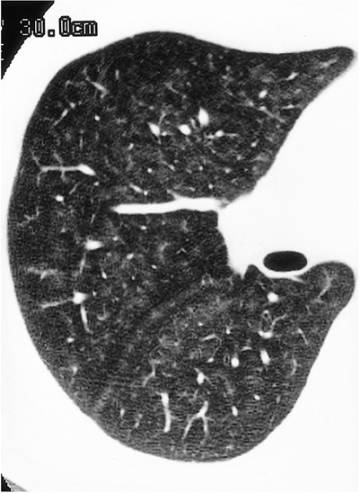
Fig. 3 46-year-old man for whom smoking and exposure history were not available. Thin-section CT scan obtained below the carina shows areas of ground-glass attenuation. Visual assessment revealed that extent of abnormalities was 70-80%.
Reference
-
1. Doig AT, McLaughlin AIG. X-ray appearance of the lungs of electric welders. Lancet. 1936. 1:771–775.2. Attfield MD, Ross DS. Radiological abnormalities in electric-arc welders. Br J Ind Med. 1978. 35:117–122.3. Lee JB, Kim KI, Chang HS, et al. Radiologic findings of chest radiograph for the mass survey in shipyard welders, Pusan. J Korean Radiol Soc. 1990. 26:82–90.4. Akira M. Uncommon pneumoconiosis: CT and pathologic findings. Radiology. 1995. 197:403–409.5. Austin JH, Muller NL, Friedman PJ, et al. Glossary of terms for CT of the lungs: Recommendations of the Nomenclature Committee of the Fleischner Society. Radiology. 1996. 200:327–331.6. Charr R. Respiratory disorders among welders. Am Rev Tuberc Pulm Dis. 1955. 71:877–884.7. Man BT, Lecutier ER. Arc-welders' lung. Br Med J. 1957. 2:921–922.8. Friede E, Rachow DO. Symptomatic pulmonary disease in arc welders. Ann Intern Med. 1961. 54:121.9. Morgan WK. Arc-welders' lung complicated by conglomeration. Amer Rev Resp Dis. 1962. 85:570–575.10. Morgan WK, Kerr HD. Pathologic and physiologic studies of welders' siderosis. Ann Int Med. 1963. 58:293–304.11. Harding HE, Grout JLA, Lloyd Davies TH. The experimental production of x-ray shadows in the lungs by inhalation of industrial dusts: I. Iron oxide. Br J Ind Med. 1947. 4:223.12. Kalliomaki PL, Sutinen S, Kelha V, Lakomaa E, Sortti V, Sutinen S. Amount and distribution of fume contaminants in the lungs of an arc welder post mortem. Br J Ind Med. 1979. 36:224–230.13. Harding HE, McLaughlin AIG, Doig AT. Clinical, radiographic, and pathological studies of the lungs of electric-arc and oxyacetylene welders. Lancet. 1958. 23:394–398.14. Remy-Jardin M, Remy J, Gosselin B, Becette V, Edme JL. Lung parenchymal changes secondary to cigarette smoking: pathologic-CT correlation. Radiology. 1993. 186:643–651.15. Churg A, Wright JL. Small-airway lesions in patients exposed to nonasbestos mineral dusts. Hum Pathol. 1983. 14:688–693.16. Kleinerman J, Green F, Harley RA, et al. Pathology standards for coal workers' pneumoconiosis. Arch Pathol Lab Med. 1979. 103:375–432.
- Full Text Links
-
- Actions
-
Cited
- CITED
-
- Close
- Share
-
- Similar articles
-
- A Study on the Size of Dust in Workplaces of a Shipyard
- Welder' Pneumoconiosis of Shipyard and related Factors
- High-resolution CT Findings of Welders' Pneumoconiosis
- Chest Radiological Changes after Cessation and Decrease of Exposure to Welding Fume in Shipyard Welders
- Comparision of Chest Radiographs and Pulmonary Function in Coal Workers' Pneumoconiosis and Welders' Lung
