Korean J Ophthalmol.
2009 Jun;23(2):121-123. 10.3341/kjo.2009.23.2.121.
A Case of Myotonic Dystrophy With Pigmentary Retinal Changes
- Affiliations
-
- 1Department of Ophthalmology, Kim's Eye Hospital, Konyang University College of Medicine, Seoul, Korea.
- 2Department of Neurology, Seoul National University Bundang Hospital, Seoul National University College of Medicine, Seongnam, Korea.
- 3Department of Ophthalmology, Seoul National University Bundang Hospital, Seoul National University College of Medicine, Seongnam, Korea. hjm@snu.ac.kr
- KMID: 754731
- DOI: http://doi.org/10.3341/kjo.2009.23.2.121
Abstract
- A 46-year-old man presented with visual disturbances in both eyes. His best corrected visual acuity was 0.7 (both eyes). Ptosis and limitation of ocular movement in every direction were observed. Slit lamp examination showed a bilateral iridescent cataract. Fundus examination showed peripheral depigmentation of the retinal pigment epithelium and pigmentary clumping in both eyes that agreed with blocked fluorescence and widow defects on fluorescein angiography. The amplitude of b-wave was decreased on electroretinography. Fourteen months later, the patient's best corrected visual acuity decreased to 0.3 due to increased lens opacity. Phacoemulsification and intraocular lens implantation were performed on both eyes. At the patient's final visit, retinal findings were stable with a best corrected visual acuity of 0.7 in both eyes. In conclusion, the visual disturbance could have been caused by both cataracts and retinal degeneration, meaning the fundus should be examined carefully in patients with myotonic dystrophy.
MeSH Terms
Figure
-

Fig. 1 Bilateral ptosis and ophthalmoplegia with exotropia in every gaze direction.
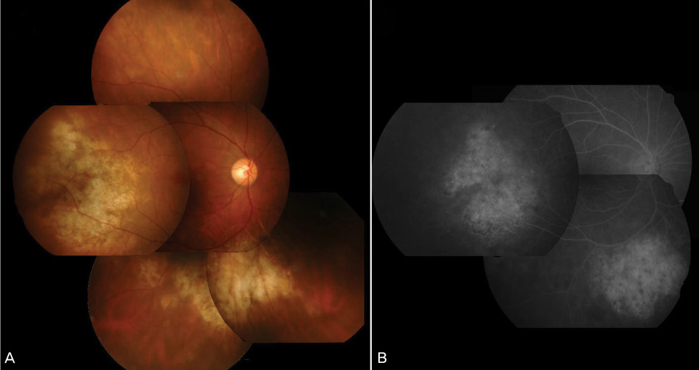
Fig. 2 (A) A fundus photograph demonstrates peripheral geographic depigmentation of the retinal pigment epithelium and pigmentary clumpings. (B) Fluorescein angiographic examination reveals blocked fluorescence due to clumping retinal pigment epithelium and window defects.
Cited by 1 articles
-
Two Cases of Strabismus Surgery in Myotonic Dystrophy
Seonjoo Kim, Mi Ra Park
J Korean Ophthalmol Soc. 2017;58(4):482-487. doi: 10.3341/jkos.2017.58.4.482.
Reference
-
1. Schara U, Schoser BG. Myotonic dystrophies type 1 and 2: a summary on current aspects. Semin Pediatr Neurol. 2006. 13:71–79.2. Meola G, Sansone V. A newly-described myotonic disorder (proximal myotonic myopathy--PROMM): personal experience and review of the literature. Ital J Neurol Sci. 1996. 17:347–353.3. Burian HM, Burns CA. Ocular changes in myotonic dystrophy. Am J Ophthalmol. 1967. 63:22–34.4. Kidd A, Turnpenny P, Kelly K, et al. Ascertainment of myotonic dystrophy through cataract by selective screening. J Med Genet. 1995. 32:519–523.5. Chung YT. Two cases of cataract in two sisters with myotonic dystrophy. J Korean Ophthalmol Soc. 1998. 39:419–423.6. Brook JD, McCurrach ME, Harley HG, et al. Molecular basis of myotonic dystrophy: expansion of a trinucleotide (CTG) repeat at the 3' end of a transcript encoding a protein kinase family member. Cell. 1992. 68:799–808.7. Abe T, Sato M, Kuboki J, et al. Lens epithelial changes and mutated gene expression in patients with myotonic dystrophy. Br J Ophthalmol. 1999. 83:452–457.8. Anastasopoulos D, Kimmig H, Mergner T, Psilas K. Abnormalities of ocular motility in myotonic dystrophy. Brain. 1996. 119:1923–1932.9. Kimizuka Y, Kiyosawa M, Tamai M, Takase S. Retinal changes in myotonic dystrophy. Clinical and follow-up evaluation. Retina. 1993. 13:129–135.10. Lessell S, Coppeto J, Samet S. Ophthalmoplegia in myotonic dystrophy. Am J Ophthalmol. 1971. 71:1231–1235.11. Kuwabara T, Lessel S. Electron microscopic study of extraocular muscles in myotonic dystrophy. Am J Ophthalmol. 1976. 82:303–309.
- Full Text Links
-
- Actions
-
Cited
- CITED
-
- Close
- Share
-
- Similar articles
-
- Two Cases of Cataract in Two Sisters with Myotonic Dystrophy
- Radiofrequency Catheter Ablation of Persistent Atrial Fibrillation with Myotonic Dystrophy and Achalasia-like Esophageal Dilatation
- A Case of Myotonic Dystrophy with Prolonged Atrial Flutter
- Accidentally Diagnosed Myotonic Dystrophy after Cholecystectomy
- Myotonic Dystrophy (A case report)
