Unusual Acute Encephalitis Involving the Thalamus: Imaging Features
- KMID: 754107
- DOI: http://doi.org/10.3348/kjr.2001.2.2.68
Abstract
OBJECTIVE
To describe the brain CT and MR imaging findings of unusual acute encephalitis involving the thalamus. MATERIALS AND METHODS: We retrospectively reviewed the medical records and CT and/or MR imaging findings of six patients with acute encephalitis involving the thalamus. CT (n=6) and MR imaging (n=6) were performed during the acute and/or convalescent stage of the illness. RESULTS: Brain CT showed brain swelling (n=2), low attenuation of both thalami (n=1) or normal findings (n=3). Initial MR imaging indicated that in all patients the thalamus was involved either bilaterally (n=5) or unilaterally (n=1). Lesions were also present in the midbrain (n=5), medial temporal lobe (n=4), pons (n=3), both hippocampi (n=3) the insular cortex (n=2), medulla (n=2), lateral temporal lobe cortex (n=1), both cingulate gyri (n=1), both basal ganglia (n=1), and the left hemispheric cortex (n=1). CONCLUSION: These CT or MR imaging findings of acute encephalitis of unknown etiology were similar to a combination of those of Japanese encephalitis and herpes simplex encephalitis. In order to document the specific causative agents which lead to the appearance of these imaging features, further investigation is required.
Keyword
MeSH Terms
Figure
-
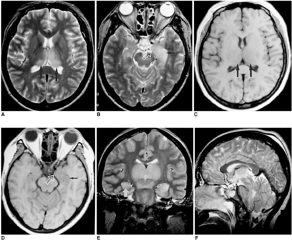
Fig. 1 Case 1. A 40-year-old woman who presented with fever, chill and altered mentality. A-D. T2-weighted (A, B) and T1-weighted (C, D) images obtained 8 days after the onset of illness show abnormal hyperintensity and hypointensity in both thalami (large black arrows), the left hippocampus (small black arrow), and the medial area of the left temporal lobe (open white arrow). E-F. Follow-up T2-weighted images obtained 11 days after onset show marked enlargement of both thalamic lesions and the development of new lesions in the right hippocampus (white arrow) and brain stem.
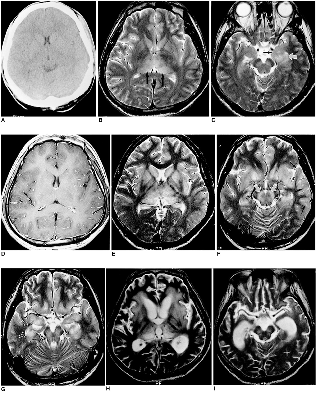
Fig. 2 Case 5: A 32-year-old man who presented with fever and disorientation. A. CT obtained 3 days after the onset of illness reveals subtle diffuse brain swelling. B-D. T2-weighted images (B, C) obtained 6 days after onset reveal abnormal hyperintensity in both thalami (large black arrows), the left hippocampus (white arrow) and medial area of the left temporal lobe (small black arrow). Gd-enhanced T1-weighted image (D) shows no abnormal thalamic enhancement but subtle corticovascular enhancement. E-G. Follow-up T2-weighted images obtained 18 days after onset show the enlargement of previous lesions and development of new lesions in both basal ganglia, both insular cortices, both substantia nigra (white solid arrows), the lateral cortex of the left temporal lobe (black arrows), the right hippocampus (white open arrows) and the medial area of the right temporal lobe. H, I. Follow-up T2-weighted images obtained 48 days after onset show decreased size of previous lesions. Periventricular white matter abnormality around both frontal horns and diffuse brain atrophy are noticed.
Reference
-
1. Jubelt B, Miller JR. Rowland LP, editor. Viral infections. Merritt's Textbook of Neurology. 1995. 9th ed. Philadelphia: Williams & Wilkins;142–179.2. Tien RD, Felsberg GJ, Osumi AK. Herpes virus infection of the CNS: MR findings. AJR. 1993. 161:167–176.3. Abe T, Kojima K, Shoji H, et al. Japanese encephalitis. J Magn Reson Imaging. 1998. 8:755–761.4. Shoji H, Hiraki Y, Kuwasaki N, et al. Japanese encephalitis in the Kurume region of Japan: CT and MRI findings. J Neurol. 1989. 236:255–259.5. Kumar S, Misra UK, Kalita J, et al. MRI in Japanese encephalitis. Neuroradiology. 1997. 39:180–184.6. Misra UK, Kalita J, Jain SK, Mathur A. Radiological and neurophysiological changes in Japanese encephalitis. J Neurol Neurosurg Psychiatry. 1994. 57:1484–1487.7. Shoji H, Murakami T, Murai I, et al. A follow-up study by CT and MRI in 3 cases of Japanese encephalitis. Neuroradiology. 1990. 32:215–219.8. Kimura K, Dosaka A, Hashimoto Y, et al. Single-photon emission CT findings in acute Japanese encephalitis. AJNR. 1997. 18:465–469.9. Ishii K. Virological and serological diagnosis of Japanese encephalitis. Shinkei Kenkyu No Shimpo. 1967. 11:300–311.10. Rowley AH, Whitley RJ, Lakeman FD, Wolinsky SM. Rapid detection of herpes-simplex virus DNA in cerebrospinal fluid of patients with herpes simplex encephalitis. Lancet. 1990. 335:440–441.11. Adams RD, Victor M, Ropper AH. Principles of Neurology. 1997. 6th ed. New York: McGraw-Hill;749–759.12. Fields BN, Knipe DM, Chanock RM, editors. Virology. 1985. New York: Raven Press;967.13. Johnson RT, Burke DS, Elwell M, et al. Japanese encephalitis: immunocytochemical studies of viral antigen and inflammatory cells in fatal cases. Ann Neurol. 1985. 18:567–573.14. Soo MS, Tien RD, Gray L, Andrews PI, Friedman H. Mesenrhombencephalitis: MR findings in nine patients. AJR. 1993. 160:1089–1093.15. Tien RD, Dillon WP. Herpes trigeminal neuritis and rhombencephalitis on Gd-DTPA-enhanced MR imaging. AJNR. 1990. 11:413–414.16. Hayashi K, Arita T. Experimental double infection of Japanese encephalitis virus and herpes simplex virus in mouse brain. Japan J Exp Med. 1977. 47:9–13.17. Tolly TL, Wells RG, Sty JR. MR features of fleeting CNS lesions associated with Epstein-Barr virus infection. J Comput Assist Tomogr. 1989. 13:665–668.18. Gajanana A, Samuel PP, Thenmozhi V, Rajendran R. An appraisal of some recent diagnostic assays for Japanese encephalitis. Southeast Asian J Trop Med Public Health. 1996. 27:673–679.19. Corey L. Fauci AS, Braunwald E, Isselbacher KJ, editors. Herpes simplex virus. Harrison's Principles of Internal Medicine. 1998. 14th ed. New York: McGraw-Hill;1080–1086.20. Fauci AS, Braunwald E, Isselbacher KJ, editors. Harrison's Principles of Internal Medicine. 1998. 14th ed. New York: McGraw-Hill;1137.21. Yagishita A, Nakano I, Ushioda T, Otsuki N, Hasegawa A. Acute encephalopathy with bilateral thalamotegmental involvement in infants and children: imaging and pathology findings. AJNR. 1995. 16:439–447.22. Osborn AG. Diagnostic Neuroradiology. 1994. St. Louis: Mosby;704–706.23. Finkenstaedt M, Szudra A, Zerr I, et al. MR imaging of Creutzfeldt-Jakob disease. Radiology. 1996. 199:793–798.24. Zeidler M, Sellar RJ, Collie DA, et al. The pulvinar sign on magnetic resonance imaging in variant Creutzfeldt-Jakob disease. Lancet. 2000. 355:1412–1418.25. Zeidler M, Stewart GE, Barraclough CR, et al. New variant Creutzfeldt-Jakob disease: neurological features and diagnostic tests. Lancet. 1997. 350:903–907.
- Full Text Links
-
- Actions
-
Cited
- CITED
-
- Close
- Share
-
- Similar articles
-
- An Unusual Case of Japanese Encephalitis Involving Unilateral Deep Gray Matter and Temporal Lobe on Diffusion-Weighted MRI
- Japanese B Encephalitis with Favorable Recovery; Clinical Course, Brain Imaging, and Neuropsychological Findings
- Japanese Encephalitis Associated with Flaccid Quadriplegia
- Astasia and Asterixis after Acute Unilateral Thalamic Infarction
- A Case of Germinoma Involving the Posterior Part of Thalamus and Basal Ganglia
