Complications Arising in Twin Pregnancy: Findings of Prenatal Ultrasonography
- Affiliations
-
- 1Department of Radiology, Samsung Cheil Hospital, Sungkyunkwan University School of Medicine, Seoul Korea. radjycho@samsung.co.kr
- 2Department of Diagnostic Pathology, Samsung Cheil Hospital, Sungkyunkwan University School of Medicine, Seoul Korea.
- KMID: 754043
- DOI: http://doi.org/10.3348/kjr.2003.4.1.54
Abstract
- Multifetal gestations are high-risk pregnancies involving higher perinatal morbidity and mortality, and are subject to unique complications including twin oligohydramnios-polyhydramnios sequence, twin-to-twin transfusion syndrome, acardiac twins, conjoined twins, co-twin demise, and heterotopic pregnancies. The purpose of this study is to describe the prenatal ultrasonographic and pathologic findings of these complications.
Keyword
Figure
-
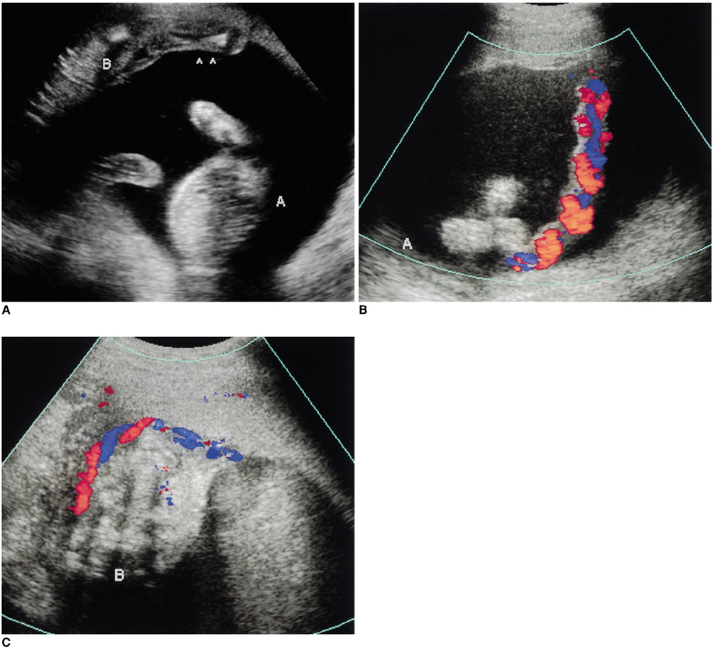
Fig. 1 Prenatal ultrasonographic findings of twin oligohydramnios-polyhydramnios sequence in a monochorionic pregnancy. A. Ultrasonogram obtained at gestational age 22 weeks shows a 20-week sized stuck twin (B) with severe oligohydramnios and a larger 22-week twin (A) with polyhydramnios (arrowheads: thin inter-twin membrane). B, C. Cord insertions of fetus A (B) and fetus B (C) are separated.

Fig. 2 Doppler sonographic findings of twin-to-twin transfusion syndrome in a monochorionic diamniotic twin pregnancy. A. Ultrasonogram obtained at gestational age 21 weeks shows significant discrepancy in fetal sizes. Fetus A is larger than fetus B by more than 2 SD. B. Color Doppler sonogram demonstrates approximate insertions (arrows) of two umbilical cords in a single placenta. C, D. Umbilical arterial Doppler sonogram of the smaller fetus (C) depicts increased vascular resistance and absent diastolic flow, while that of the larger fetus (D) shows normal diastolic flow.
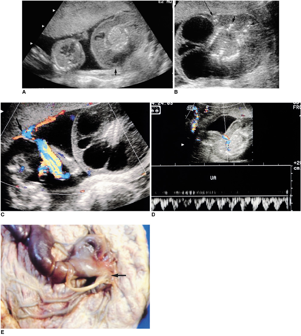
Fig. 3 Sonographic diagnosis of acardiac twins in a monochorionic diamniotic pregnancy. A. Ultrasonogram obtained at gestational age 19 weeks shows significant discrepancy in fetal sizes. The absence of a heart and diffuse soft tissue edema are demonstrated in the larger fetus B (arrow). B. Fetus B has no head (short arrow) and no heart, though tudimentary bony upper extremities (long arrows) are visible. A multiseptated cystic mass suggesting a large cystic hygroma is associated with the acardiac fetus. C. Color Doppler sonogram demonstrates interfetal anastomoses (arrow) of umbilical vessels between the twins. D. Duplex sonogram verifies that in the umbilical artery (UA) of the acardiac fetus B, flow is reversed. E. Placental pathology demonstrates anastomoses (arrow) of the umbilical vessels between the twins in the monochorionic placenta. Dye injection through the umbilical vessels verified interfetal artery-to-artery and vein-to-vein anastomoses (not shown).
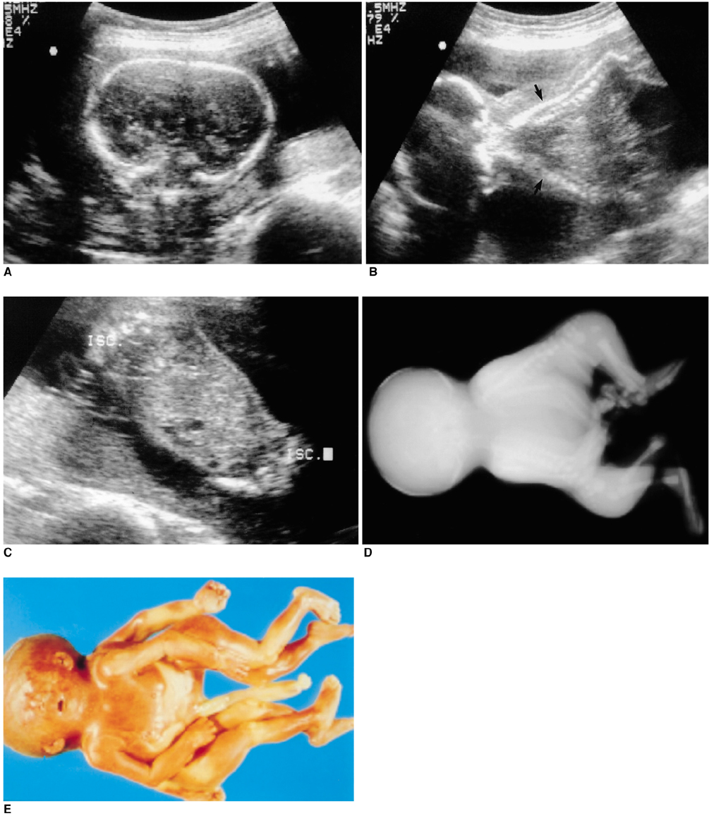
Fig. 4 Ultrasonographic and autopsy findings of conjoined twins. A. Ultrasonogram obtained at gestational age 21 weeks depicts fused cranium and cerebra. B. Fused cranium and anterior chest with two "V"-shaped spines (arrows) are apparent. C. Fused anterior abdomen with two ischia (ISC) is noted. D, E. Specimen radiographs (D) and autopsy specimen (E) demonstrate conjoined twins with fused cranium, anterior chest and abdomen (craniothoracoomphalopagus).
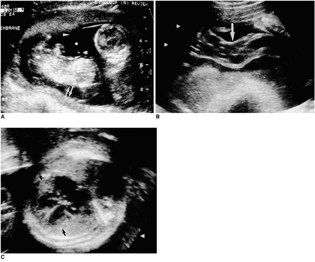
Fig. 5 Sonographic findings of monochorionic diamniotic twins with co-twin demise. A. Ultrasonogram obtained at gestational age 14 weeks shows a fairly-well visualized (short arrow) but preserved (arrowheads) thin intertwin membrane with a single placenta, and intrauterine fetal death of one fetus with diffuse soft tissue edema (arrows). B. Ultrasonogram obtained at gestational age 23 weeks depicts an enlarged umbilical cord (arrow) in the surviving fetus. C. Cardiomegaly with thickened biventricular walls (arrows) developed in the surviving fetus.

Fig. 6 Ultrasonographic findings of a cornual heterotopic pregnancy. A. Ultrasonogram obtained at gestational age 7 weeks shows a gestational sac (black arrow) with an embryo (arrowhead) within the uterine cavity. B. In the cornual portion of the uterus, a coexisting gestational sac (arrows) with an embryo (arrowhead) is visible.
Reference
-
1. Divon MY, Weiner Z. Ultrasound in twin pregnancy. Semin Perinatol. 1995. 19:404–412.2. Mahony BS, Petty CN, Nyberg DA, Luthy DA, Hickok DE, Hirsch JH. The "stuck twin" phenomenon: ultrasonographic findings, pregnancy outcome, and management with serial amniocenteses. Am J Obstet Gynecol. 1990. 163:1513–1522.3. Bruner JP, Rosemond RL. Twin-to-twin transfusion syndrome: a subset of the twin oligohydramnios-polyhydramnios sequence. Am J Obstet Gynecol. 1993. 169:925–930.4. Bruner JP, Anderson TL, Rosemond RL. Placental pathophysiology of the twin oligohydramnios-polyhydramnios sequence and the twin-twin transfusion syndrome. Placenta. 1998. 19:81–86.5. Farley CL, Cox LA, Long BW. Ultrasound and twin-twin transfusion syndrome. Radiol Technol. 2000. 72:95–100.6. Hecher K, Ville Y, Nicolaides KH. Fetal arterial Doppler studies in twin-twin transfusion syndrome. J Ultrasound Med. 1995. 14:101–108.7. Van Allen MI, Smith DW, Shepard TH. Twin reversed arterial perfusion (TRAP) sequence: a study of 14 twin pregnancies with acardius. Semin Perinatol. 1983. 7:285–293.8. Chen CP, Shih SL, Liu FF, Jan SW, Lin YN, Lan CC. Skeletal deformities of acardius anceps: the gross and imaging features. Pediatr Radiol. 1997. 27:221–225.9. Mohanty C, Mishra OP, Singh CP, Das BK, Singla PN. Acardiac anomaly spectrum. Teratology. 2000. 62:356–359.10. Guttmacher AF, Nichols BC. Teratology of conjoined twins. Birth Defects. 1967. 3:3–9.11. Lam YH, Sin SY, Lam C, Lee CP, Tang MH, Tse HY. Prenatal sonographic diagnosis of conjoined twins in the first trimester: two case reports. Ultrasound Obstet Gynecol. 1998. 11:289–291.12. Kilby MD, Govind A, O'Brien PM. Outcome of twin pregnancies complicated by a single intrauterine death: a comparison with viable twin pregnancies. Obstet Gynecol. 1994. 84:107–109.13. Petersen IR, Nyholm HC. Multiple pregnancies with single intrauterine demise: Description of twenty-eight pregnancies. Acta Obstet Gynecol Scand. 1999. 78:202–206.14. Tal J, Haddad S, Gordon N, Timor-Tritsch I. Heterotopic pregnancy after ovulation induction and assisted reproductive technologies: a literature review from 1971 to 1993. Fertil Steril. 1996. 66:1–12.
- Full Text Links
-
- Actions
-
Cited
- CITED
-
- Close
- Share
-
- Similar articles
-
- Rare Complications with Monochorionic Twins: Ultrasonography and Pathology Correlations
- Prenatal Diagnosis of Acardiac Twin : A Case Report
- Ultrasonographic Diagnosis of Conjoined Twins ; A Case Report of Two Cases
- Prenatal diagnosis of epignathus with multiple malformations in one fetus of a twin pregnancy using three-dimensional ultrasonography and magnetic resonance imaging
- Acardiac Twin Presented as a Lower Extremity: Case Report with Serial Prenatal Ultrasonography
