Obstet Gynecol Sci.
2015 Jan;58(1):65-68. 10.5468/ogs.2015.58.1.65.
Prenatal diagnosis of epignathus with multiple malformations in one fetus of a twin pregnancy using three-dimensional ultrasonography and magnetic resonance imaging
- Affiliations
-
- 1Department of Obstetrics and Gynecology, The Catholic University of Korea College of Medicine, Seoul, Korea. ooooobbbbb@catholic.ac.kr
- KMID: 1994229
- DOI: http://doi.org/10.5468/ogs.2015.58.1.65
Abstract
- Epignathus is an extremely rare type of congenital teratoma arising in the oral cavity. Although it is a benign tumor, it is associated with high mortality and morbidity rates because of severe airway obstruction and other malformations. We present a case of epignathus affecting one fetus in a twin pregnancy. The tumor was associated with multiple congenital malformations including cleft palate, bifid tongue, bifid uvula, congenital heart defect, and bilateral inguinal hernias. The diagnostic value of three-dimensional ultrasonography and magnetic resonance imaging was explored with respect to antenatal counseling and peripartum management.
Keyword
MeSH Terms
Figure
-
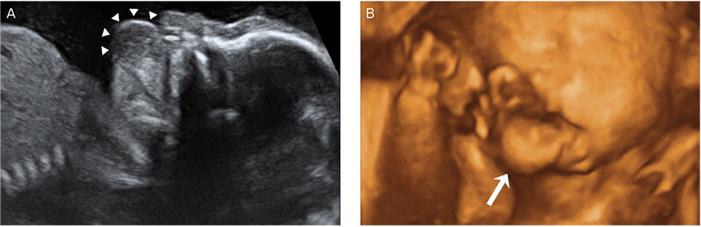
Fig. 1 Antenatal image of the tumor. A round mass (arrow heads) protruding from the oral cavity was found on two-dimensional ultrasonography (A), and three-dimensional image (B).

Fig. 2 Sagittal HASTE T2-weighted magnetic resonance image at 21 weeks 6/7 days of gestation showing a 16×10 mm hyperintense mass (arrows) with a stalk (arrowhead) projecting from the palate and upper lip (UL) (A). A pedunculated tumor originated from the mouth floor in the newborn just after delivery (B). T, tongue; LL, lower lip.
Reference
-
1. Gull I, Wolman I, Har-Toov J, Amster R, Schreiber L, Lessing JB, et al. Antenatal sonographic diagnosis of epignathus at 15 weeks of pregnancy. Ultrasound Obstet Gynecol. 1999; 13:271–273.2. Levine AB, Alvarez M, Wedgwood J, Berkowitz RL, Holzman I. Contemporary management of a potentially lethal fetal anomaly: a successful perinatal approach to epignathus. Obstet Gynecol. 1990; 76:962–966.3. Tonni G, Centini G, Inaudi P, Rosignoli L, Ginanneschi C, De Felice C. Prenatal diagnosis of severe epignathus in a twin: case report and review of the literature. Cleft Palate Craniofac J. 2010; 47:421–425.4. Takagi MM, Bussamra LC, Araujo Junior E, Drummond CL, Herbst SR, Nardozza LM, et al. Prenatal diagnosis of a large epignathus teratoma using two-dimensional and three-dimensional ultrasound: correlation with pathological findings. Cleft Palate Craniofac J. 2014; 51:350–353.5. Dar P, Rosenthal J, Factor S, Dubiosso R, Murthy AS. First-trimester diagnosis of fetal epignathus with 2- and 3-dimensional sonography. J Ultrasound Med. 2009; 28:1743–1746.6. Allen LM. Prenatal 3-dimensional imaging techniques in the sonographic evaluation of an oral mass: comparison with postnatal imaging modalities. J Ultrasound Med. 2011; 30:561–568.7. Tonni G, De Felice C, Centini G, Ginanneschi C. Cervical and oral teratoma in the fetus: a systematic review of etiology, pathology, diagnosis, treatment and prognosis. Arch Gynecol Obstet. 2010; 282:355–361.8. Pasupathy M, Narayanan PV, Mani V, Adenwalla HS. A case report of nasopharyngeal teratoma with a cleft palate and an inguinal hernia. J Plast Reconstr Aesthet Surg. 2011; 64:1525–1527.9. Witters I, Moerman P, Louwagie D, Van Assche FA, Migeon BR, Fryns JP. Second trimester prenatal diagnosis of epignathus teratoma in ring X chromosome mosaicism with inactive ring X chromosome. Ann Genet. 2001; 44:179–182.10. Yapar EG, Ekici E, Gokmen O. Sonographic diagnosis of epignathus (oral teratoma), prosencephaly, meromelia and oligohydramnios in a fetus with trisomy 13. Clin Dysmorphol. 1995; 4:266–271.11. Smart PJ, Schwarz C, Kelsey A. Ultrasonographic and biochemical abnormalities associated with the prenatal diagnosis of epignathus. Prenat Diagn. 1990; 10:327–332.12. Ekici E, Soysal M, Kara S, Dogan M, Gokmen O. Prenatal diagnosis of epignathus causing acute polyhydramnios. Acta Obstet Gynecol Scand. 1996; 75:498–501.13. Abendstein B, Auer A, Pumpel R, Mark E, Desch B, Tscharf J. Epignathus: prenatal diagnosis by sonography and magnetic resonance imaging. Ultraschall Med. 1999; 20:207–211.14. Sichel JY, Eliashar R, Yatsiv I, Moshe Gomori J, Nadjari M, Springer C, et al. A multidisciplinary team approach for management of a giant congenital cervical teratoma. Int J Pediatr Otorhinolaryngol. 2002; 65:241–247.15. Noah MM, Norton ME, Sandberg P, Esakoff T, Farrell J, Albanese CT. Short-term maternal outcomes that are associated with the EXIT procedure, as compared with cesarean delivery. Am J Obstet Gynecol. 2002; 186:773–777.
- Full Text Links
-
- Actions
-
Cited
- CITED
-
- Close
- Share
-
- Similar articles
-
- Prenatal ultrasonography of craniofacial abnormalities
- Ultrasonographic follow-up of involuting placenta in advanced abdominal pregnancy diagnosed by magnetic resonance imaging
- Two Cases of Fetus Papyraceus in Twin Pregnancy
- The Clinical Importance of the Prenatal Diagnosis of Fetal Scalp Hemangioma
- Two Cases of Multicystic Encephalomalacia in a Surviving Co-twin with One Intrauterine Fetal Death
