Characterization of Focal Liver Lesions with Superparamagnetic Iron Oxide-Enhanced MR Imaging: Value of Distributional Phase T1-Weighted Imaging
- Affiliations
-
- 1Department of Radiology, Seoul National University College of Medicine, Seoul, Korea. leejm@radcom.snu.ac.kr
- 2Department of Diagnostic Radiology, Chonbuk National University, Chonju, Korea.
- 3Department of Radiology, Wonkwang University Hospital, Iksan, Korea.
- KMID: 754037
- DOI: http://doi.org/10.3348/kjr.2003.4.1.9
Abstract
OBJECTIVE
To determine the potential value of distributional-phase T1-weighted ferumoxides-enhanced magnetic resonance (MR) imaging for tissue characterization of focal liver lesions. MATERIALS AND METHODS: Ferumoxides-enhanced MR imaging was performed using a 1.5-T system in 46 patients referred for evaluation of known or suspected hepatic malignancies. Seventy-three focal liver lesions (30 hepatocellular carcinomas [HCC], 12 metastases, 15 cysts, 13 hemangiomas, and three cholangiocarcinomas) were evaluated. MR imaging included T1-weighted double-echo gradient-echo (TR/TE: 150/4.2 and 2.1 msec), T2*-weighted gradient-echo (TR/TE: 180/12 msec), and T2-weighted turbo spin-echo MR imaging at 1.5 T before and after intravenous administration of ferumoxides (15 mmol/kg body weight). Postcontrast T1-weighted imaging was performed within eight minutes of infusion of the contrast medium (distributional phase). Both qualitative and quantitative analysis was performed. RESULTS: During the distributional phase after infusion of ferumoxides, unique enhancement patterns of focal liver lesions were observed for hemangiomas, metastases, and hepatocellular carcinomas. On T1-weighted GRE images obtained during the distributional phase, hemangiomas showed a typical positive enhancement pattern of increased signal; metastases showed ring enhancement; and hepatocellar carcinomas showed slight enhancement. Quantitatively, the signal-to-noise ratio of hemangiomas was much higher than that of other tumors (p < .05) and was similar to that of intrahepatic vessels. This finding permitted more effective differentiation between hemangiomas and other malignant tumors. CONCLUSION: T1-weighted double-echo FLASH images obtained soon after the infusion of ferumoxides, show characteristic enhancement patterns and improved the differentiation of focal liver lesions.
Keyword
Figure
-
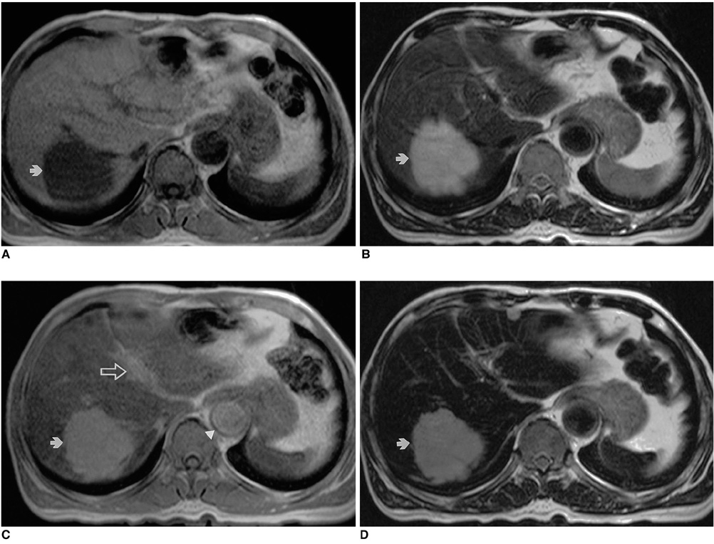
Fig. 1 Liver hemangioma in a 67-year-old woman. A. Precontrast T1-weighted in-phase gradient-echo image depicts a round hypointense lesion (arrow) in the right lobe of the liver. B. Precontrast T2-weighted turbo spin-echo image shows a hyperintense lesion (arrow) in the liver. C. Ferumoxides-enhanced T1-weighted in-phase gradient-echo image obtained during the distributional phase demonstrates positive enhancement of the lesion (arrow), similar to that of the intrahepatic portal vein (open arrow). Note positive enhancement of the abdominal aorta (arrowhead). D. Ferumoxides-enhanced T2-weighted turbo-spin echo image shows reduced intensity of the lesion (arrow) compared to precontrast image (A).

Fig. 2 Liver metastasis in a 67-year-old woman with gastric cancer. A. Precontrast T2-weighted turbo spin-echo image shows a hyperintense nodule (arrow) in the right lobe of the liver. B. Postcontrast T1-weighted out-of-phase image demonstrates rim enhancement (arrow) of the peripheral portion of the lesion, which is hypointense to the liver. Note positive enhancement of the intrahepatic portal vein (small arrowheads) and aorta (arrowhead). C. Postcontrast T1-weighted in-phase image shows that the lesion (arrow) has become hyperintense to the liver. D. Postcontrast T2*-weighted gradient-echo image obtained during the accumulation phase demonstrates markedly improved lesion (arrow)-to-liver contrast compared to precontrast T2-weighted image.
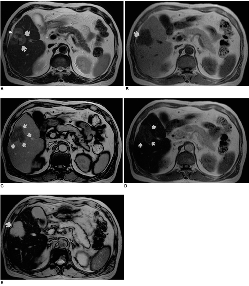
Fig. 3 Surgically-proven cholangiocarcinoma in segment 5 of the liver. A. Precontrast T2-weighted turbo spin-echo image shows a heterogeneously hyperintense lesion (arrows) with mild capsular retraction in the right lobe of the liver (arrowhead). B. Precontrast T1-weighted in-phase gradient-echo image depicts a hypointense mass (arrow). C. On this T1-weighted out-of-phase gradient-echo image obtained after the administration of ferumoxides, the lesion shows peripheral rim enhancement (arrows). D. T1-weighted in-phase gradient-echo image obtained after the administration of ferumoxides shows that the lesion (arrows) has become slightly hyperintense to the liver. E. On this T2*-weighted gradient-echo image obtained after the administration of ferumoxides, the lesion (arrow) has become very hyperintense to the liver. Note that this accumulation phase image provides excellent contrast and lesion conspicuity.
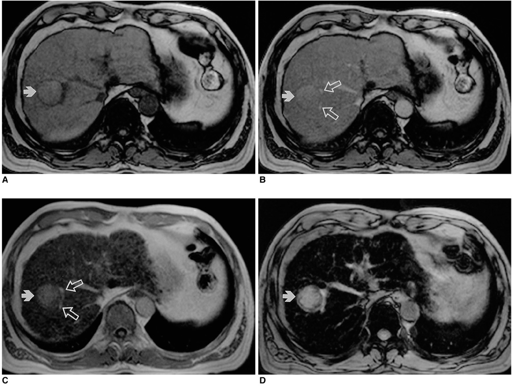
Fig. 4 A 65-year-old man with biopsy-proven hepatocellular carcinoma in the right lobe of the liver. A. Precontrast T1-weighted out-of-phase gradient-echo image depicts a hyperintense tumor (arrow) with a hypointense capsule in the right lobe of the liver. B. Postcontrast T1-weighted out-of-phase gradient-echo image obtained during the distributional phase shows that the lesion (arrow) is hyperintense compared to the liver, but much less hyperintense than the branches of the intrahepatic portal vein (open arrows). C. Postcontrast T1 in-phase gradient-echo image obtained during the distributional phase shows that the lesion (arrow) has become more hyperintense than the liver but is still less hyperintense than the branches of the portal vein (open arrows). Note the presence of multiple hypointense regenerating nodules in the liver parenchyma. D. Postcontrast T2*-weighted gradient echo image shows markedly improved lesion (arrow)-to-liver contrast due to the substantially decreased liver parenchymal signal.
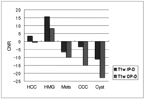
Fig. 5 Lesion-to-liver contrast-to-noise ratio of focal hepatic lesions seen on T1-weighted in-phase and out-of-phase gradient-echo images obtained during the distributional phase
Reference
-
1. Hagspiel KD, Neidel KFW, Eichenberger AC, Weder W, Marincek B. Detection of liver metastases: comparison of superparamagnetic iron-oxide-enhanced MR imaging at 1.5 T with dynamic CT, intraoperative US, and percutaneous US. Radiology. 1995. 196:471–478.2. Pena CS, Saini S, Baron BL, et al. Detection of malignant primary hepatic neoplasms with gadobenate dimeglumine (Gd-BOPTA)-enhanced T1-weighted hepatocyte phase MR imaging: results of off-site blinded review in a phase-II multicenter trial. Korean J Radiol. 2001. 2:210–215.3. Kim SH, Choi D, Lim JH, et al. Optimal pulse sequence for ferumoxides-enhanced MR imaging used in the detection of hepatocellular carcinoma: a comparative study using seven pulse sequences. Korean J Radiol. 2002. 3:87–97.4. Bluemke DA, Paulson EK, Choti MA, DeSena S, Clavien PA. Detection of hepatic lesions in candidates for surgery: comparison of ferumoxides-enhanced MR imaging and dual-phase helical CT. AJR Am J Roentgenol. 2000. 175:1653–1658.5. Kanematsu M, Itoh K, Matsuo M, et al. Malignant hepatic tumor detection with ferumoxides-enhanced MR imaging with a 1.5-T system: comparison of four imaging pulse sequences. J Magn Reson Imaging. 2001. 13:249–257.6. Choi DI, Kim SH, Lim JH, et al. Preoperative detection of hepatocellular carcinoma: ferumoxides-enhanced MR imaging versus combined helical CT during arterial portography and CT hepatic arteriography. AJR Am J Roentgenol. 2001. 176:475–482.7. Karhunen PJ. Benign hepatic tumors and tumor-like conditions in men. J Clin Pathol. 1986. 39:183–189.8. Grangier C, Tourniaire J, Mentha G, et al. Enhancement of liver hemangiomas on T1-weighted MR SE images by superparamagnetic iron oxide particles. J Comput Assist Tomogr. 1994. 18:888–896.9. van Gansbeke D, Metens TM, Matos C, et al. Effects of AMI-25 on liver vessels and tumors on T1-weighted turbo-field-echo images: implications for tumor characterization. J Magn Reson Imaging. 1997. 7:482–489.10. Mergo PJ, Helmberger T, Nicolas AI, Ros PR. Ring enhancement in ultrasmall superparamagnetic iron oxide MR imaging: a potential new sign for characterization of liver lesions. AJR Am J Roentgenol. 1996. 166:379–384.11. Parley M, Mergo PJ, Torres GM, Ros PR. Characterization of focal hepatic lesions with ferumoxides-enhanced T2-weighted MR imaging. AJR Am J Roentgenol. 2000. 175:159–163.12. Nakayama M, Yamashita Y, Mitsuzaki K, et al. Improved tissue characterization of focal liver lesions with ferumoxide-enhanced T1- and T2-weighted MR imaging. J Magn Reson Imaging. 2000. 11:647–654.13. Kim JH, Kim MJ, Suh SH, Chung JJ, You HS, Lee JT. Characterization of focal hepatic lesions with ferumoxides-enhanced MR imaging: utility of T1-weighted spoiled gradient- recalled echo images using different echo times. J Magn Reson Imaging. 2002. 15:573–583.14. Oswald P, Clement O, Chambon C, Schouman-Claeys E, Frija G. Liver-positive enhancement after injection of superparamagnetic nanoparticles: respective role of circulating and uptaken particles. Magn Reson Imaging. 1997. 15:1025–1031.15. Hamm B, Thoeni RF, Gould RG, et al. Focal liver lesions: characterization with nonenhanced and dynamic contrast material-enhanced MR imaging. Radiology. 1994. 190:417–423.16. Yamashita Y, Hatanaka Y, Yamamoto H, et al. Differential diagnosis of focal liver lesions: role of spin-echo and contrast-enhanced dynamic MR imaging. Radiology. 1994. 193:59–65.17. Yoshida H, Itai Y, Ohtomo K, et al. Small hepatocellular carcinoma and cavernous hemangioma: differentiation with dynamic FLASH MR imaging with Gd-DTPA. Radiology. 1989. 171:339–342.18. McFarland EG, Mayo SW, Saini S, et al. Hepatic hemangiomas and malignant tumors: improved differentiation with heavily T2- weighted conventional spin-echo MR imaging. Radiology. 1994. 193:43–47.19. Oudkerk M, van den Heuvel AG, Wielopolski PA, Schmitz PI, Borel Rinkes IH, Wiggers T. Hepatic lesions: detection with ferumoxides-enhanced T1-weighted MR imaging. Radiology. 1997. 203:449–456.20. Reimer P, Müller M, Marx C, et al. T1 effects of a bolus-injectable superparamagnetic iron oxide, SH U 555 A: Dependence on field strength and plasma concentration - preliminary clinical experience with dynamic T1-weighted MR imaging. Radiology. 1998. 209:831–836.21. van Beers B, Gallez B, Pringot J. Contrast-enhanced MR imaging of the liver. Radiology. 1997. 203:297–306.22. Petersein J, Saini S, Weissleder R. Liver. II: Iron oxide-based reticuloendothelial contrast agents for MR imaging. Clinical review. Magn Reson Imaging Clin N Am. 1996. 4:53–60.
- Full Text Links
-
- Actions
-
Cited
- CITED
-
- Close
- Share
-
- Similar articles
-
- Focal Nodular Hyperplasia of the Liver: Imaging Findings with Emphasis on the Findings of Superparamagnetic Iron Oxide-enhanced MR Imaging
- Detectability of Hepatocellular Carcinoma: Comparison of Gd-DT PA-Enhanced and SPIO-Enhanced MR Imaging
- Hemangioma and Hepatocellular Carcinoma: Distinction with Superparamagnetic Iron Oxide-Enhanced MR Imaging
- Detection of Small Hypervascular Hepatocellular Carcinomas in Cirrhotic Patients: Comparison of Superparamagnetic Iron Oxide-Enhanced MR Imaging with Dual-Phase Spiral CT
- Nontumorous Focal Low Attenuated Areas in the Left Lobe around the Falciform Ligament on Contrast Enhanced CTScan: MR Correlation
