Korean J Radiol.
2004 Jun;5(2):96-101. 10.3348/kjr.2004.5.2.96.
Age-Related Changes in Conventional and Magnetization Transfer MR Imaging in Elderly People: Comparison with Neurocognitive Performance
- Affiliations
-
- 1Department of Diagnostic Radiology, Korea University College of Medicine, Seoul, Korea. tkkim@kumc.or.kr
- 2Department of Neurology, Korea University College of Medicine, Seoul, Korea.
- 3Geriatric Health Clinic, Korea University College of Medicine, Ansan Hospital, Ansan City, Kyungki-Do, Korea.
- 4Department of Radiology, Seoul National University Hospital, Seoul, Korea.
- 5Department of Electronic Engineering, College of Engineering, Inha University, Incheon, Korea.
- KMID: 753988
- DOI: http://doi.org/10.3348/kjr.2004.5.2.96
Abstract
OBJECTIVE
This study was designed to compare three different measures of the elderly human brain; the magnetization transfer ratio (MTR) histogram, the percentage of brain parenchymal volume, and the volume of T2 hyperintense areas in terms of correlations with the study subjects' neurocognitive performance. MATERIALS AND METHODS: Thirty-five healthy community-dwelling elderly volunteers aged 60-82 years underwent dual fast spin-echo (FSE) imaging and magnetization transfer imaging. A semi-automated technique was used to generate the MTR histogram, the brain parenchymal volume, and the T2 lesion volume. The subjects' neurocognitive performance was assessed by using the Korean-Mini Mental State Examination (K-MMSE) and additional tests. The peak height of the MTR (PHMTR), the percentage of brain parenchymal volume (PBV), and the normalized T2 lesion volume (T2LV) were compared between the normal group (Z score on the K-MMSE > or = -2, n=23) and the mild cognitive impairment group (Z score on the K-MMSE < -2, n=12), and these parameters were correlated with age and various neurocognitive performance scores. RESULTS: The PHMTR was significantly lower in the cognitively impaired subjects than the PHMTR in the normal subjects (p = 0.005). The PBV scores were lower in the cognitively impaired subjects than in the normal subjects (p = 0.02). The T2LV scores were significantly higher in the cognitively impaired subjects (p = 0.01). An inverse correlation was found between the PHMTR and T2LV (r = -0.747, p < ; 0.001), and also between the PBV and T2LV (r = -0.823, p < ; 0.001). A positive correlation was observed between the PHMTR and the PBV (r = 0.846, p < 0.001). Scores on the various neurocognitive tests were positively correlated with the PHMTR (6 of 7 items) and the PBV (5 of 7 items), and they were negatively correlated with the T2LV (5 of 7 items). CONCLUSION: Our findings of a correlation among the PBV, the T2LV, and the PHMTR suggest that MTR histograms and the PBV and T2LV can be used as a reliable method and valid statistical tool, respectively, for quantifying the total lesion burden in an aging brain.
MeSH Terms
Figure
-

Fig. 1 Segmented volume image of the intracranium (A), cerebrospinal fluid (B), and brain parenchyma (C) from proton density-weighted and T2-weighted images.
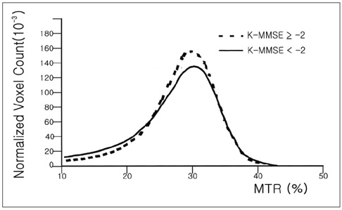
Fig. 2 Magnetization transfer ratio (MTR) histograms from normal subjects (dotted line) and cognitive impaired subjects (solid line). Normalized peak height of the MTR Histogram is lower for the cognitive impairment group compared with that of the normal groups.
Reference
-
1. Salonen O, Autti T, Raininko R, Ylikoski A, Erkinjuntti T. MRI of the brain in neurologically healthy middle-aged and elderly individuals. Neuroradiology. 1997. 39:537–545.2. DeCarli C, Murphy DGM, Trahn M, et al. The effect of white matter hyperintensity volume on brain structure, cognitive performance and cerebral metabolism of glucose in 51 healthy adults. Neurology. 1995. 45:2077–2084.3. Smith CD, Snowdon DA, Wang H, Markesberry WR. White matter volumes and periventricular white matter hyperintensities in aging and dementia. Neurology. 2000. 54:838–842.4. Rossor MN, Freeborough PA, Roques PK. Slowing the progression of Alzheimer's disease: Monitoring progression. Alzheimer Dis Assoc Disord. 1997. 11:Suppl. 5. S6–S9.5. Golomb J, de Leon MJ, Kluger A, George AE, Tarshish C, Ferris SH. Hippocampal atrophy in normal aging. An association with recent memory impairment. Arch Neurol. 1993. 50:967–973.6. Fein G, van Dyke C, Davenport L, et al. Preservation of normal cognitive functioning in elderly subjects with extensive white-matter lesions of long duration. Arch Gen Psychiatry. 1990. 47:220–223.7. Filippi M, Grossman RI, Comi G. Magnetization transfer in multiple sclerosis. Neurology. 1999. 53:Suppl 3. S18–S22.8. Brochet B, Dousset V. Pathological correlates of magnetization transfer imaging abnormalities in animal models and humans with multiple sclerosis. Neurology. 1999. 53:Suppl 3. S12–S17.9. Kabani NJ, Sled JG, Chertkow H. Magnetization transfer ratio in mild cognitive impairment and dementia of Alzheimer's type. Neuroimage. 2002. 15:604–610.10. Kabani NJ, Sled JG, Shuper A, Chertkow H. Regional magnetization transfer ratio changes in mild cognitive impairment. Magn Reson Med. 2002. 47:143–148.11. Tanabe JL, Ezekiel F, Jagusta WJ, et al. Magnetization transfer ratio of white matter hyperintensities in subcortical ischemic vascular dementia. AJNR Am J Neuroradiol. 1999. 20:839–844.12. Udupa JK, Samarasekera S. Fuzzy connectedness and object definition: Theory, algorithms and applications in image segmentation. Graph Models Image Process. 1996. 58:246–261.13. Udupa J, Wei L, Samarasekera S, Miki Y, van Buchem MA, Grossman RI. Multiple sclerosis lesion quantification using fuzzy connectedness principles. IEEE Trans Med Imaging. 1997. 16:598–609.14. Phillips MD, Grossman RI, Miki Y, et al. Comparison of T2 lesion volume and magnetization transfer ratio histogram analysis and of atrophy and measures of lesion burden in patients with multiple sclerosis. AJNR Am J Neuroradiol. 1998. 19:1055–1060.15. Selim SZ, Ismail MA. K-means-type algorithms. IEEE Trans Pattern Anal Mach Intell. 1984. 6:81–87.16. Van Buchem MA, Udupa JK, McGowan JC, et al. Global volumetric estimation of disease burden in multiple sclerosis based on magnetization transfer imaging. AJNR Am J Neuroradiol. 1997. 18:1287–1290.17. Folstein MF, Folstein SE, Mc Hugh PR. Mini-mental state - a practical method grading the cognitive state of patients for the clinician. J Psychiatr Res. 1975. 12:189–198.18. Kang YW, Na DL, Hahn SH. A validity study on the Korean mini-mental state examination (K-MMSE) in dementia patients. J Korean Neurol Assoc. 1997. 15:300–307.19. Kang YW, Kim HJ, Na DL. Parallel short form for the Korean-Boston Naming Test (K-BNT). Korean J Clinical Psy. 2000. 18:144–150.20. Kang YW, Kim JK. Korean-California Verbal Learning Test (K-CVLT): A normative study. Korean J Clinical Psy. 1997. 16:379–396.21. Lee JH, Kang YW, Jin JH, Na DL, Park JS. A normative study of the Korean version of Controlled Oral Word Association Test (COWAT) in the elderly. Korean J clinical Psy. 2000. 19:385–392.22. Hofman PAM, Kemerink GJ, Jolles J, Wilmonk JT. Quantitative analysis of magnetization transfer images of the brain: effect of closed head injury, age and sex on white matter. Magn Reson Med. 1999. 42:803–806.23. Ge Y, Grossman RI, Babb JS, Rabin ML, Mannon LJ, Kolson DL. Age-related total gray matter and white matter changes in normal adult brain II: Quantitative magnetization transfer ratio histogram analysis. AJNR Am J Neuroradiol. 2002. 23:1334–1341.24. Rovaris M, Iannucci G, Cercignani M, et al. Age-related changes in conventional, magnetization transfer, and diffusion-tensor MR imaging findings: study with whole-brain tissue histogram analysis. Radiology. 2003. 227:731–738.25. Ge Y, Kolson DL, Babb JS, Mannon LJ, Grossman RI. Whole brain imaging of HIV-infected patients: quantitative analysis of magnetization transfer ratio histogram and fractional brain volume. AJNR Am J Neuroradiol. 2003. 24:82–87.26. Ge Y, Grossman RI, Udupa JK, Babb JS, Mannon LJ, McGowan JC. Magnetization transfer ratio histogram analysis of normal-appearing gray matter and normal-appearing white matter in multiple sclerosis. J Comput Assist Tomogr. 2002. 26:62–68.27. Hofman PA, Verhey FR, Wilmink JT, Rozendaal N, Jolles J. Brain lesions in patients visiting a memory clinic with postconcussional sequelae after mild to moderate brain injury. J Neuropsychiatry Clin Neurosci. 2002. 14:176–184.28. Dehmeshki J, Van Buchem MA, Bosma GP, Huizinga TW, Tofts PS. Systemic lupus erythematosus: diagnostic application of magnetization transfer ratio histograms in patients with neuropsychiatric symptoms--initial results. Radiology. 2002. 222:722–728.29. Dehmeshki J, Barker GJ, Tofts PS. Classification of disease subgroup and correlation with disease severity using magnetic resonance imaging whole-brain histograms: application to magnetization transfer ratios and multiple sclerosis. IEEE Trans Med Imaging. 2002. 21:320–331.30. Van Der Flier WM, Van Den Heuvel DM, Weverling-Rijnsburger AW, et al. Magnetization transfer imaging in normal aging, mild cognitive impairment, and Alzheimer's disease. Ann Neurol. 2002. 52:62–67.
- Full Text Links
-
- Actions
-
Cited
- CITED
-
- Close
- Share
-
- Similar articles
-
- Postcontrast T1-weighted Brain Magnetic Resonance Imaging in Pediatric Patients: Comparison Between Postcontrast Fat-suppression Imaging and Conventional T1-weighted or Magnetization Transfer Imaging
- Imaging with Magnetization Transfer Technique on the Intracranial Tumors
- Comparison of Magnetization Transfer Ratios of Various Cerebral Edemas
- Magnetization Transfer Contrast Angiography for Organized Thrombosed Intracranial Aneurysm in TOF MR Angiography: a Case Report
- Magnetization Transfer Imaging for Detection of Mesial Temporal Sclerosis in Temporal Lobe Seizure
