Clinical Impact of Microbiome Characteristics in Treatment-Naïve Extranodal NK/T-Cell Lymphoma Patients
- Affiliations
-
- 1Division of Hematology-Oncology, Department of Medicine, Samsung Medical Center, Sungkyunkwan University School of Medicine, Seoul, Korea
- 2CJ Bioscience Inc., Seoul, Korea
- 3Department of Pathology, Samsung Medical Center, Sungkyunkwan University School of Medicine, Seoul, Korea
- KMID: 2566876
- DOI: http://doi.org/10.4143/crt.2024.675
Abstract
- Purpose
Extranodal natural killer/T-cell lymphoma (ENKTL) predominantly manifests in East Asia and Latin America. Despite shared intrinsic factors, such as ethnic and genetic backgrounds, the progression of ENKTL can be influenced by extrinsic factors related to changing lifestyle patterns.
Materials and Methods
This study collected stool samples from newly diagnosed (ND)–ENKTL patients (n=40) and conducted whole genome shotgun sequencing.
Results
ND-ENKTL revealed reduced alpha diversity in ND-ENKTL compared to healthy controls (HCs) (p=0.008), with Enterobacteriaceae abundance significantly contributing to the beta diversity difference between ENKTL and HCs (p < 0.001). Functional analysis indicated upregulated aerobic metabolism and degradation of aromatic compounds in ND-ENKTL. Enterobacteriaceae were associated not only with clinical data explaining disease status (serum C-reactive protein, stage, prognosis index of natural killer cell lymphoma [PINK], and PINK-E) but also with clinical outcomes (early relapse and short progression-free survival). The relative abundance of Enterobacteriaceae at the family level was similar between ENKTL and diffuse large B-cell lymphoma (DLBCL) (p=0.140). However, the ENKTL exhibited a higher abundance of Escherichia, in contrast to the prevalence of Enterobacter and Citrobacter in DLBCL. Linear regression analysis demonstrated a significant association between Escherichia abundance and programmed cell death-ligand-1 (PD-L1) levels in tissue samples (p=0.025), whereas no correlation with PD-L1 was observed for Enterobacteriaceae at the family level (p=0.571).
Conclusion
ND-ENKTL exhibited an abundance of Enterobacteriaceae and a dominant presence of Escherichia. These microbial characteristics correlated with disease status, treatment outcomes, and PD-L1 expression, suggesting the potential of the ENKTL microbiome as a biomarker and cause of lymphomagenesis, which warrants further exploration.
Keyword
Figure
-

Fig. 1. (A) Predominant microbial phyla in healthy control (HC) and extranodal natural killer/T-cell lymphoma (ENKTL), including Firmicutes, Bacteroidetes, Actinobacteria, and Proteobacteria. Phyla with less than 1% abundance are grouped as ‘Others’. (B) Alpha diversity assessed using the Shannon index in HC and ENKTL cohorts. (C) Beta diversity through Bray-Curtis dissimilarity between the groups. (D) Enterobacteriaceae abundance comparison in HC and ENKTL. (E) ROC curves analysis from a random forest model for patient stratification criteria, depicting true positive rate against false positive rate. (F) Identification of top 30 taxa as key features in the random forest model relevant to HC and ENKTL differentiation. AUC, area under curve; SD, standard deviation. ns, not significant; **p < 0.01, ***p < 0.001.
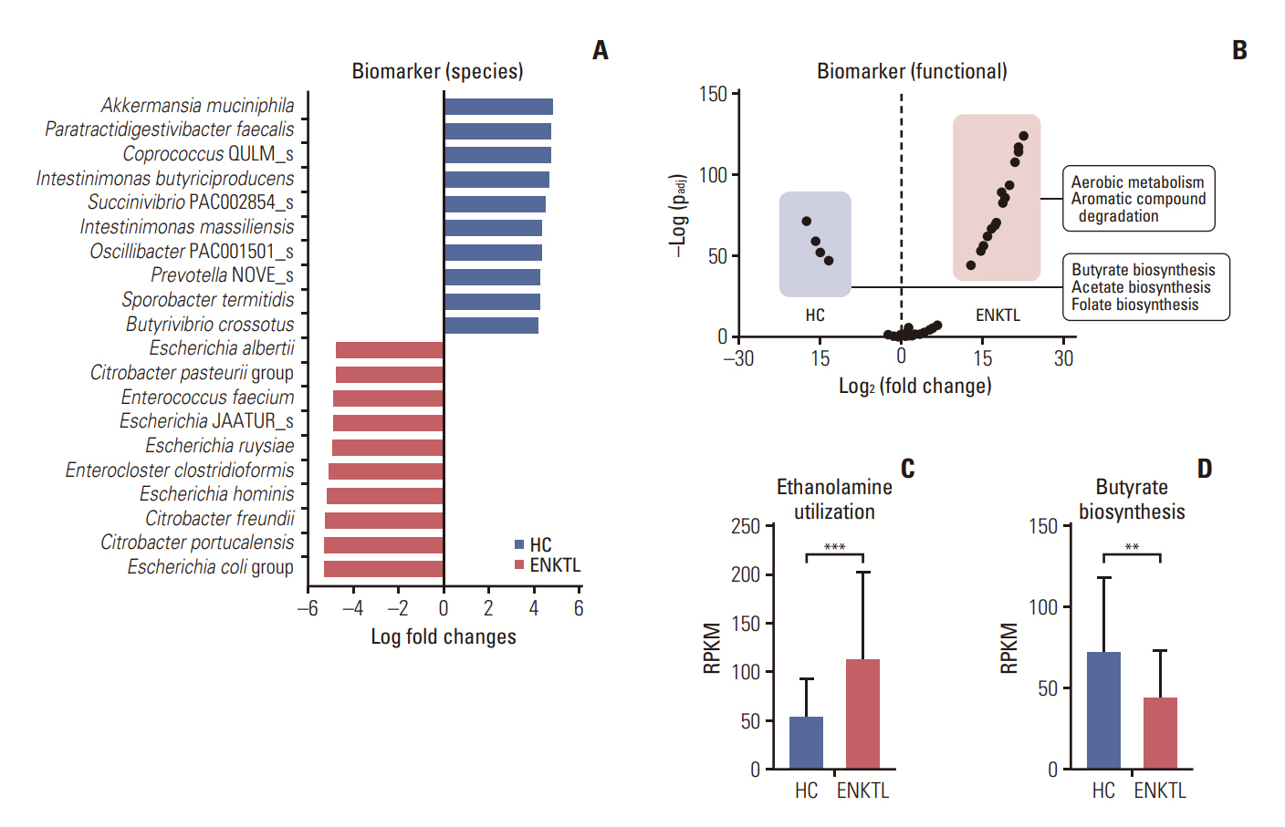
Fig. 2. (A) Species-level gut microbial taxonomic biomarkers for extranodal natural killer/T-cell lymphoma (ENKTL) and healthy control (HC). The top 10 biomarkers for each group are displayed, with ENKTL markers in red and HC markers in blue. Biomarkers identified by using ANCOM-BC analysis. (B) Volcano plot illustrating pathway-level functional biomarkers. Positive x-axis values represent biomarkers for ENKTL (marked in red), whereas negative values indicate HC biomarkers (marked in blue). Statistically significant markers are highlighted. Functional biomarkers analyzed using MaAsLin 2. (C, D) Statistical analysis of the cumulative reads per kilobase million (RPKM) of genes constituting major pathways representative of ENKTL and HC. (C) Ethanolamine utilization pathway. (D) Butyrate biosynthesis pathway. **p < 0.01, ***p < 0.001.

Fig. 3. (A) Redundancy analysis (RDA) on the top 10 families with high feature importance from random forest analysis, in correlation with key clinical features of extranodal natural killer/T-cell lymphoma (ENKTL). Green dots represent individual ENKTL patients, blue dots denote families, and red arrows indicate clinical features. (B) Comparative analysis of Enterobacteriaceae abundance among healthy control (HC), maintained response, and early relapse groups. (C) Beta diversity assessment (Bray-Curtis) for HC, maintained response, and early relapse groups. (D, E) Progression-free survival (PFS) comparison in ENKTL patients divided into top 50% and bottom 50% based on Enterobacteriaceae abundance. The upper panel displays overall PFS (D); the lower panel shows PFS further stratified into stage I/II and III/IV within each subgroup (E). (F) Linear regression plots demonstrating the relationship between Enterobacteriaceae abundance and clinical biomarkers C-reactive protein (CRP), prognosis index of natural killer cell lymphoma (PINK), and PINK-E. BMI, body mass index; EBV, Epstein-Barr virus; LDH, lactate dehydrogenase; OS, overall survival; PFS, progression-free survival. ns, not significant; **p < 0.01, ***p < 0.001.

Fig. 4. (A) Comparative analysis of alpha diversity (Shannon index) among healthy control (HC), diffuse large B-cell lymphoma (DLBCL), and extranodal natural killer/T-cell lymphoma (ENKTL) groups. (B) Beta diversity comparison (Bray-Curtis) across HC, DLBCL, and ENKTL groups. (C) Relative abundance of Enterobacteriaceae in DLBCL and ENKTL groups. (D) Comparison of the genus composition within the Enterobacteriaceae family between DLBCL and ENKTL groups. (E) Progression-free survival (PFS) analysis in ENKTL patients categorized into top 50% and bottom 50% based on Escherichia abundance. (F) PFS comparison in ENKTL patients stratified by relative abundance of Escherichia into top 50% and bottom 50%, further subdivided into stage I/II and III/IV. (G, H) Linear regression analysis illustrating the relationship between Escherichia relative abundance and serum programmed cell death-ligand-1 (PD-L1) levels (G), and between Enterobacteriaceae relative abundance and serum PD-L1 levels (H).
Reference
-
References
1. Damania B, Kenney SC, Raab-Traub N. Epstein-Barr virus: biology and clinical disease. Cell. 2022; 185:3652–70.
Article2. Tse E, Fox CP, Glover A, Yoon SE, Kim WS, Kwong YL. Extranodal natural killer/T-cell lymphoma: an overview on pathology and clinical management. Semin Hematol. 2022; 59:198–209.
Article3. Lin GW, Xu C, Chen K, Huang HQ, Chen J, Song B, et al. Genetic risk of extranodal natural killer T-cell lymphoma: a genome-wide association study in multiple populations. Lancet Oncol. 2020; 21:306–16.4. Li Z, Xia Y, Feng LN, Chen JR, Li HM, Cui J, et al. Genetic risk of extranodal natural killer T-cell lymphoma: a genome-wide association study. Lancet Oncol. 2016; 17:1240–7.
Article5. Vose J, Armitage J, Weisenburger D; International T-Cell Lymphoma Project. International peripheral T-cell and natural killer/T-cell lymphoma study: pathology findings and clinical outcomes. J Clin Oncol. 2008; 26:4124–30.
Article6. Yoon SE, Song Y, Kim SJ, Yoon DH, Chen TY, Koh Y, et al. Comprehensive analysis of peripheral T-cell and natural killer/T-cell lymphoma in Asian patients: a multinational, multicenter, prospective registry study in Asia. Lancet Reg Health West Pac. 2021; 10:100126.
Article7. Gupta VK, Paul S, Dutta C. Geography, ethnicity or subsistence-specific variations in human microbiome composition and diversity. Front Microbiol. 2017; 8:1162.
Article8. Shi Z, Li X, Wang X, Zhang L, Li L, Fu X, et al. Characteristics and clinical implications of the nasal microbiota in extranodal NK/T-cell lymphoma, nasal type. Front Cell Infect Microbiol. 2021; 11:686595.
Article9. Shi Z, Hu G, Li MW, Zhang L, Li X, Li L, et al. Gut microbiota as non-invasive diagnostic and prognostic biomarkers for natural killer/T-cell lymphoma. Gut. 2023; 72:1999–2002.
Article10. Yoon SE, Kang W, Choi S, Park Y, Chalita M, Kim H, et al. The influence of microbial dysbiosis on immunochemotherapy-related efficacy and safety in diffuse large B-cell lymphoma. Blood. 2023; 141:2224–38.
Article11. Kim SJ, Yoon DH, Jaccard A, Chng WJ, Lim ST, Hong H, et al. A prognostic index for natural killer cell lymphoma after non-anthracycline-based treatment: a multicentre, retrospective analysis. Lancet Oncol. 2016; 17:389–400.
Article12. Cho J, Kim SJ, Park WY, Kim J, Woo J, Kim G, et al. Immune subtyping of extranodal NK/T-cell lymphoma: a new biomarker and an immune shift during disease progression. Mod Pathol. 2020; 33:603–15.
Article13. Cheson BD, Pfistner B, Juweid ME, Gascoyne RD, Specht L, Horning SJ, et al. Revised response criteria for malignant lymphoma. J Clin Oncol. 2007; 25:579–86.
Article14. Sepich-Poore GD, Zitvogel L, Straussman R, Hasty J, Wargo JA, Knight R. The microbiome and human cancer. Science. 2021; 371:eabc4552.
Article15. Park EM, Chelvanambi M, Bhutiani N, Kroemer G, Zitvogel L, Wargo JA. Targeting the gut and tumor microbiota in cancer. Nat Med. 2022; 28:690–703.
Article16. Upadhyay Banskota S, Skupa SA, El-Gamal D, D’Angelo CR. Defining the role of the gut microbiome in the pathogenesis and treatment of lymphoid malignancies. Int J Mol Sci. 2023; 24:2309.
Article17. Hamerman JA, Ogasawara K, Lanier LL. NK cells in innate immunity. Curr Opin Immunol. 2005; 17:29–35.
Article18. Bajor-Dattilo EB, Pittaluga S, Jaffe ES. Pathobiology of T-cell and NK-cell lymphomas. Best Pract Res Clin Haematol. 2013; 26:75–87.
Article19. Kers JG, Saccenti E. The power of microbiome studies: some considerations on which alpha and beta metrics to use and how to report results. Front Microbiol. 2021; 12:796025.
Article20. Helmink BA, Khan MA, Hermann A, Gopalakrishnan V, Wargo JA. The microbiome, cancer, and cancer therapy. Nat Med. 2019; 25:377–88.
Article21. Gopalakrishnan V, Spencer CN, Nezi L, Reuben A, Andrews MC, Karpinets TV, et al. Gut microbiome modulates response to anti-PD-1 immunotherapy in melanoma patients. Science. 2018; 359:97–103.22. Yuan L, Wang W, Zhang W, Zhang Y, Wei C, Li J, et al. Gut microbiota in untreated diffuse large B cell lymphoma patients. Front Microbiol. 2021; 12:646361.
Article23. Diefenbach CS, Peters BA, Li H, Raphael B, Moskovits T, Hymes K, et al. Microbial dysbiosis is associated with aggressive histology and adverse clinical outcome in B-cell nonHodgkin lymphoma. Blood Adv. 2021; 5:1194–8.
Article24. Rivera-Chavez F, Lopez CA, Baumler AJ. Oxygen as a driver of gut dysbiosis. Free Radic Biol Med. 2017; 105:93–101.
Article25. Litvak Y, Byndloss MX, Baumler AJ. Colonocyte metabolism shapes the gut microbiota. Science. 2018; 362:eaat9076.
Article26. Zeng MY, Inohara N, Nunez G. Mechanisms of inflammation-driven bacterial dysbiosis in the gut. Mucosal Immunol. 2017; 10:18–26.
Article27. Liu XF, Shao JH, Liao YT, Wang LN, Jia Y, Dong PJ, et al. Reglation of short-chain fatty acids in the immune system. Front Immunol. 2023; 14:1186892.28. Mikkelsen K, Apostolopoulos V. Vitamin B12, folic acid, and the immune system. In : Mahmoudi M, Razaei N, editors. Nutrition and immunity. Springer;2019. p. 103–14.29. Li YJ, Li ZM, Xia Y, Huang JJ, Huang HQ, Xia ZJ, et al. Serum C-reactive protein (CRP) as a simple and independent prognostic factor in extranodal natural killer/T-cell lymphoma, nasal type. PLoS One. 2013; 8:e64158.
Article30. Bao C, Zhou D, Zhu L, Qian W, Ye X. Increased serum level of interleukin-6 correlates with negative prognostic factors in extranodal NK/T-cell lymphoma. Transl Cancer Res. 2020; 9:2378–89.
Article31. Mitsunaga S, Ikeda M, Shimizu S, Ohno I, Takahashi H, Okuyama H, et al. C-reactive protein level is an indicator of the aggressiveness of advanced pancreatic cancer. Pancreas. 2016; 45:110–6.
Article32. Troppan KT, Schlick K, Deutsch A, Melchardt T, Egle A, Stojakovic T, et al. C-reactive protein level is a prognostic indicator for survival and improves the predictive ability of the R-IPI score in diffuse large B-cell lymphoma patients. Br J Cancer. 2014; 111:55–60.
Article33. Song TL, Nairismagi ML, Laurensia Y, Lim JQ, Tan J, Li ZM, et al. Oncogenic activation of the STAT3 pathway drives PD-L1 expression in natural killer/T-cell lymphoma. Blood. 2018; 132:1146–58.34. Li X, Cheng Y, Zhang M, Yan J, Li L, Fu X, et al. Activity of pembrolizumab in relapsed/refractory NK/T-cell lymphoma. J Hematol Oncol. 2018; 11:15.
Article
- Full Text Links
-
- Actions
-
Cited
- CITED
-
- Close
- Share
-
- Similar articles
-
- Recent updates on extranodal NK/T-cell lymphoma
- Extranodal NK/T Cell Lymphoma, Nasal Type that Occurred in Patients with Atrophic Rhinitis
- A Case of Extranodal NK/T-cell Lymphoma at the Base of Tongue
- A Case of Extranodal NK/T Cell Lymphoma, Nasal Type with Cutaneous Involvement
- Extranodal NK/T cell lymphoma
