Korean J Orthod.
2024 Mar;54(2):128-135. 10.4041/kjod23.166.
Is three-piece maxillary segmentation surgery a stable procedure?
- Affiliations
-
- 1Department of Morphology and Pediatric Clinic, School of Dentistry of Araraquara, São Paulo State University, Araraquara, Brazil
- 2Departments of Oral and Maxillofacial Surgery and Orthodontics, Texas A&M University Health Science Center, Baylor College of Dentistry, Dallas, TX, USA
- 3Private Practice, Nova Lima, Brazil
- 4Department of Orthodontics, University of the Pacific, Arthur A. Dugoni School of Dentistry, San Francisco, CA, USA
- KMID: 2554369
- DOI: http://doi.org/10.4041/kjod23.166
Abstract
Objective
The number of three-piece maxillary osteotomies has increased over the years; however, the literature remains controversial. The objective of this study was to evaluate the skeletal stability of this surgical modality compared with that of one-piece maxillary osteotomy.
Methods
This retrospective cohort study included 39 individuals who underwent Le Fort I maxillary osteotomies and were divided into two groups: group 1 (three pieces, n = 22) and group 2 (one piece, n = 17). Three cone-beam computed tomography scans from each patient (T1, pre-surgical; T2, post-surgical; and T3, follow-up) were used to evaluate the three-dimensional skeletal changes.
Results
The differences within groups were statistically significant only for group 1 in terms of surgical changes (T2-T1) with a mean difference in the canine region of 3.09 mm and the posterior region of 3.08 mm. No significant differences in surgical stability were identified between or within the groups. The mean values of the differences between groups were 0.05 mm (posterior region) and –0.39 mm (canine region).
Conclusions
Our findings suggest that one- and three-piece maxillary osteotomies result in similar post-surgical skeletal stability.
Keyword
Figure
-

Figure 1 Landmark points marked on the three-dimensional maxillary surface. PR, posterior right; CR, canine right; AR, anterior right; AL, anterior left; CL, canine left; PL, posterior left.
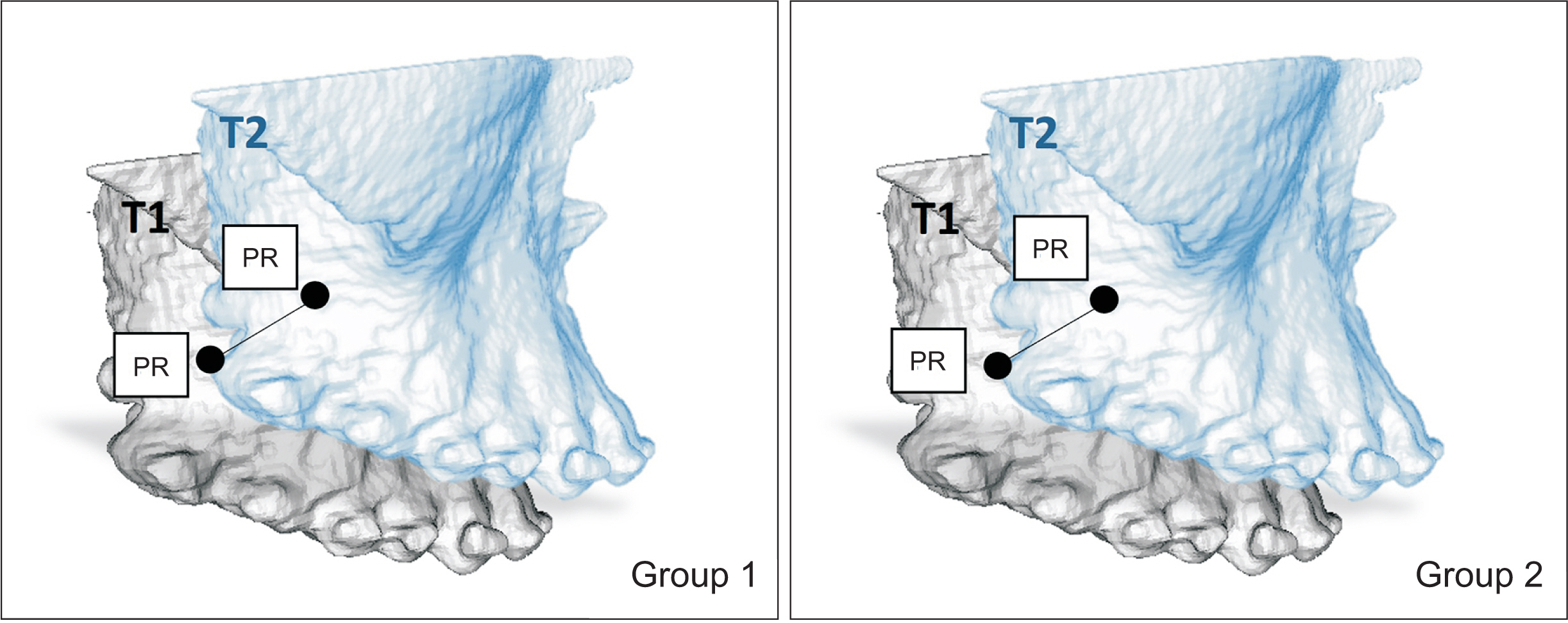
Figure 2 Example of the Euclidean three-dimensional (3D) distance using two landmarks on a 3D surface. PR measurements within groups. Example: PR T2-PR T1. T1, pre-surgical; T2, post-surgical; PR, posterior right.

Figure 3 Euclidean three-dimensional distance obtained using two landmarks within the model. Example: posterior right to posterior left.
Reference
-
1. Bell WH, Fonseca RJ, Kenneky JW, Levy BM. 1975; Bone healing and revascularization after total maxillary osteotomy. J Oral Surg. 33:253–60. https://pubmed.ncbi.nlm.nih.gov/1054396/.2. Proffit WR, Turvey TA, Phillips C. 2007; The hierarchy of stability and predictability in orthognathic surgery with rigid fixation: an update and extension. Head Face Med. 3:21. https://doi.org/10.1186/1746-160X-3-21. DOI: 10.1186/1746-160X-3-21. PMID: 17470277. PMCID: PMC1876453. PMID: 14b2d6631ff64d8b9171769e5bc4cda3.
Article3. Kretschmer WB, Baciut G, Baciut M, Zoder W, Wangerin K. 2010; Stability of Le Fort I osteotomy in bimaxillary osteotomies: single-piece versus 3-piece maxilla. J Oral Maxillofac Surg. 68:372–80. https://doi.org/10.1016/j.joms.2009.09.053. DOI: 10.1016/j.joms.2009.09.053. PMID: 20116710.
Article4. Kretschmer WB, Baciut G, Baciut M, Zoder W, Wangerin K. 2011; Transverse stability of 3-piece Le Fort I osteotomies. J Oral Maxillofac Surg. 69:861–9. https://doi.org/10.1016/j.joms.2010.05.024. DOI: 10.1016/j.joms.2010.05.024. PMID: 21050640.
Article5. Blæhr TL, Jensen T, Due KM, Neumann-Jensen B. 2014; Stability of the anterior maxillary segment and teeth after segmental Le Fort I osteotomy and postoperative skeletal elastic fixation with or without occlusal splint. J Oral Maxillofac Res. 5:e4. https://doi.org/10.5037/jomr.2014.5304. DOI: 10.5037/jomr.2014.5304. PMID: 25386231. PMCID: PMC4219863. PMID: d1cc23106fc24e82a92e31be2885783e.
Article6. Tai W, Leung YY, Li DTS. 2022; Le Fort I osteotomy with segmentation for the treatment of maxillary dentoalveolar protrusion: a single-centre, 10-year outcome study. Int J Oral Maxillofac Surg. 51:1197–204. https://doi.org/10.1016/j.ijom.2022.01.012. DOI: 10.1016/j.ijom.2022.01.012. PMID: 35101320.
Article7. Kahnberg KE, Hagberg C. 2007; The approach to dentofacial skeletal deformities using a multisegmentation technique. Clin Plast Surg. 34:477–84. https://doi.org/10.1016/j.cps.2007.05.003. DOI: 10.1016/j.cps.2007.05.003. PMID: 17692705.
Article8. Morgan TA, Fridrich KL. 2001; Effects of the multiple-piece maxillary osteotomy on the periodontium. Int J Adult Orthodon Orthognath Surg. 16:255–65. https://pubmed.ncbi.nlm.nih.gov/12390003/.9. Reyneke JP, Conley RS. 2020; Surgical/orthodontic correction of transverse maxillary discrepancies. Oral Maxillofac Surg Clin North Am. 32:53–69. https://doi.org/10.1016/j.coms.2019.08.007. DOI: 10.1016/j.coms.2019.08.007. PMID: 31699580.
Article10. Posnick JC, Adachie A, Choi E. 2016; Segmental maxillary osteotomies in conjunction with bimaxillary orthognathic surgery: indications - safety - outcome. J Oral Maxillofac Surg. 74:1422–40. https://doi.org/10.1016/j.joms.2016.01.051. DOI: 10.1016/j.joms.2016.01.051. PMID: 26923557.
Article11. Haas Junior OL, Guijarro-Martínez R, de Sousa Gil AP, da Silva Meirelles L, de Oliveira RB, Hernández-Alfaro F. 2017; Stability and surgical complications in segmental Le Fort I osteotomy: a systematic review. Int J Oral Maxillofac Surg. 46:1071–87. https://doi.org/10.1016/j.ijom.2017.05.011. DOI: 10.1016/j.ijom.2017.05.011. PMID: 28601432.
Article12. Kramer FJ, Baethge C, Swennen G, Teltzrow T, Schulze A, Berten J, et al. 2004; Intra- and perioperative complications of the LeFort I osteotomy: a prospective evaluation of 1000 patients. J Craniofac Surg. 15:971–7. discussion 978–9. https://doi.org/10.1097/00001665-200411000-00016. DOI: 10.1097/00001665-200411000-00016. PMID: 15547385.
Article13. Kahnberg KE, Vannas-Löfqvist L, Zellin G. 2005; Complications associated with segmentation of the maxilla: a retrospective radiographic follow up of 82 patients. Int J Oral Maxillofac Surg. 34:840–5. https://doi.org/10.1016/j.ijom.2005.04.016. DOI: 10.1016/j.ijom.2005.04.016. PMID: 16105727.
Article14. Rodrigues DB, Campos PSF, Wolford LM, Ignácio J, Gonçalves JR. 2018; Maxillary interdental osteotomies have low morbidity for alveolar crestal bone and adjacent teeth: a cone beam computed tomography-based study. J Oral Maxillofac Surg. 76:1763–71. https://doi.org/10.1016/j.joms.2018.01.031. DOI: 10.1016/j.joms.2018.01.031. PMID: 29544755.
Article15. Venugoplan SR, Nanda V, Turkistani K, Desai S, Allareddy V. 2012; Discharge patterns of orthognathic surgeries in the United States. J Oral Maxillofac Surg. 70:e77–86. https://doi.org/10.1016/j.joms.2011.09.030. DOI: 10.1016/j.joms.2011.09.030. PMID: 22182664.
Article16. Jodeh DS, Nguyen ATH, Rottgers SA. 2020; Le Fort 1 and bimaxillary osteotomies increase the length of stay but not postoperative morbidity compared to mandibular osteotomies and single jaw procedures. J Craniofac Surg. 31:1734–8. https://doi.org/10.1097/SCS.0000000000006514. DOI: 10.1097/SCS.0000000000006514. PMID: 32371693.
Article17. Perez MM, Sameshima GT, Sinclair PM. 1997; The long-term stability of LeFort I maxillary downgrafts with rigid fixation to correct vertical maxillary deficiency. Am J Orthod Dentofacial Orthop. 112:104–8. https://doi.org/10.1016/s0889-5406(97)70280-4. DOI: 10.1016/S0889-5406(97)70280-4. PMID: 9228848.
Article18. Hoppenreijs TJ, Freihofer HP, Stoelinga PJ, Tuinzing DB, van't Hof MA, van der Linden FP, et al. 1997; Skeletal and dento-alveolar stability of Le Fort I intrusion osteotomies and bimaxillary osteotomies in anterior open bite deformities. A retrospective three-centre study. Int J Oral Maxillofac Surg. 26:161–75. https://doi.org/10.1016/s0901-5027(97)80813-2. DOI: 10.1016/S0901-5027(97)80813-2. PMID: 9180224.
Article19. Bailey LJ, Phillips C, Proffit WR, Turvey TA. 1994; Stability following superior repositioning of the maxilla by Le Fort I osteotomy: five-year follow-up. Int J Adult Orthodon Orthognath Surg. 9:163–73. https://pubmed.ncbi.nlm.nih.gov/7814921/.20. Arpornmaeklong P, Heggie AA, Shand JM. 2003; A comparison of the stability of single-piece and segmental Le Fort I maxillary advancements. J Craniofac Surg. 14:3–9. https://doi.org/10.1097/00001665-200301000-00002. DOI: 10.1097/00001665-200301000-00002. PMID: 12544214.
Article21. Parizotto JOL, Borsato KT, Peixoto AP, Bianchi J, Cassano DS, Gonçalves JR. 2020; Can palatal splint improve stability of segmental Le Fort I osteotomies? Orthod Craniofac Res. 23:486–92. https://doi.org/10.1111/ocr.12399. DOI: 10.1111/ocr.12399. PMID: 32533749.
Article22. Bailey L', Cevidanes LH, Proffit WR. 2004; Stability and predictability of orthognathic surgery. Am J Orthod Dentofacial Orthop. 126:273–7. https://pubmed.ncbi.nlm.nih.gov/15356484/. DOI: 10.1016/j.ajodo.2004.06.003. PMID: 15356484. PMCID: PMC3681098.
Article23. Proffit WR, Turvey TA, Phillips C. 1996; Orthognathic surgery: a hierarchy of stability. Int J Adult Orthodon Orthognath Surg. 11:191–204. https://pubmed.ncbi.nlm.nih.gov/9456622/.24. Bailey LJ, White RP Jr, Proffit WR, Turvey TA. 1997; Segmental LeFort I osteotomy for management of transverse maxillary deficiency. J Oral Maxillofac Surg. 55:728–31. https://doi.org/10.1016/s0278-2391(97)90588-7. DOI: 10.1016/S0278-2391(97)90588-7. PMID: 9216506.
Article25. Lele S, Richtsmeier JT. 1991; Euclidean distance matrix analysis: a coordinate-free approach for comparing biological shapes using landmark data. Am J Phys Anthropol. 86:415–27. https://doi.org/10.1002/ajpa.1330860307. DOI: 10.1002/ajpa.1330860307. PMID: 1746646.
Article26. Wolford LM, Bennett MA, Rafferty CG. 1987; Modification of the mandibular ramus sagittal split osteotomy. Oral Surg Oral Med Oral Pathol. 64:146–55. https://doi.org/10.1016/0030-4220(87)90080-6. DOI: 10.1016/0030-4220(87)90080-6. PMID: 3476891.
Article27. Bennett MA, Wolford LM. 1985; The maxillary step osteotomy and Steinmann pin stabilization. J Oral Maxillofac Surg. 43:307–11. https://doi.org/10.1016/0278-2391(85)90297-6. DOI: 10.1016/0278-2391(85)90297-6. PMID: 3884756.
Article28. Yushkevich PA, Piven J, Hazlett HC, Smith RG, Ho S, Gee JC, et al. 2006; User-guided 3D active contour segmentation of anatomical structures: significantly improved efficiency and reliability. Neuroimage. 31:1116–28. https://doi.org/10.1016/j.neuroimage.2006.01.015. DOI: 10.1016/j.neuroimage.2006.01.015. PMID: 16545965.
Article29. Cevidanes LH, Bailey LJ, Tucker GR Jr, Styner MA, Mol A, Phillips CL, et al. 2005; Superimposition of 3D cone-beam CT models of orthognathic surgery patients. Dentomaxillofac Radiol. 34:369–75. https://doi.org/10.1259/dmfr/17102411. DOI: 10.1259/dmfr/17102411. PMID: 16227481. PMCID: PMC3552302.
Article30. Ruellas AC, Tonello C, Gomes LR, Yatabe MS, Macron L, Lopinto J, et al. 2016; Common 3-dimensional coordinate system for assessment of directional changes. Am J Orthod Dentofacial Orthop. 149:645–56. https://doi.org/10.1016/j.ajodo.2015.10.021. DOI: 10.1016/j.ajodo.2015.10.021. PMID: 27131246. PMCID: PMC4959834.
Article31. Ruellas AC, Yatabe MS, Souki BQ, Benavides E, Nguyen T, Luiz RR, et al. 2016; 3D mandibular superimposition: comparison of regions of reference for voxel-based registration. PLoS One. 11:e0157625. https://doi.org/10.1371/journal.pone.0157625. DOI: 10.1371/journal.pone.0157625. PMID: 27336366. PMCID: PMC4919005. PMID: e2a528cc49064bfdb9b578f71a498dba.
Article32. Casko JS, Vaden JL, Kokich VG, Damone J, James RD, Cangialosi TJ, et al. 1998; Objective grading system for dental casts and panoramic radiographs. American Board of Orthodontics. Am J Orthod Dentofacial Orthop. 114:589–99. https://doi.org/10.1016/s0889-5406(98)70179-9. DOI: 10.1016/S0889-5406(98)70179-9. PMID: 9810056.
Article33. Wolford LM, Rieche-Fischel O, Mehra P. 2002; Soft tissue healing after parasagittal palatal incisions in segmental maxillary surgery: a review of 311 patients. J Oral Maxillofac Surg. 60:20–5. discussion 26https://doi.org/10.1053/joms.2002.29068. DOI: 10.1053/joms.2002.29068. PMID: 11757000.
Article34. Haas Junior OL, Guijarro-Martínez R, de Sousa Gil AP, da Silva Meirelles L, Scolari N, Muñoz-Pereira ME, et al. 2019; Hierarchy of surgical stability in orthognathic surgery: overview of systematic reviews. Int J Oral Maxillofac Surg. 48:1415–33. https://doi.org/10.1016/j.ijom.2019.03.003. DOI: 10.1016/j.ijom.2019.03.003. PMID: 30910409.
Article35. Starch-Jensen T, Blæhr TL. 2016; Transverse expansion and stability after segmental Le Fort I osteotomy versus surgically assisted rapid maxillary expansion: a systematic review. J Oral Maxillofac Res. 7:e1. https://doi.org/10.5037/jomr.2016.7401. DOI: 10.5037/jomr.2016.7401. PMID: 28154745. PMCID: PMC5279767. PMID: cbbbc465f5b94ea39757381d3341a2d7.
Article36. Marchetti C, Pironi M, Bianchi A, Musci A. 2009; Surgically assisted rapid palatal expansion vs. segmental Le Fort I osteotomy: transverse stability over a 2-year period. J Craniomaxillofac Surg. 37:74–8. https://doi.org/10.1016/j.jcms.2008.08.006. DOI: 10.1016/j.jcms.2008.08.006. PMID: 19062299.
Article
- Full Text Links
-
- Actions
-
Cited
- CITED
-
- Close
- Share
-
- Similar articles
-
- Implant Surgery with Both Sinus Bone Graft in the Maxillary edentulous patient: A Case Report
- Comparison of Anterior Chamber Parameter and Refractive Change between Three-Piece and Single-Piece Aspheric Intraocular Lenses
- Delayed Occurrence of Maxillary Sinusitis after Simultaneous Maxillary Sinus Augmentation and Implant: A Case Report and Literature Review
- Volumetric growth analysis of maxillary sinus using computed tomography scan segmentation: a pilot study of Indonesian population
- THE SKELETAL STABILITY OF LE FORT I MAXILLARY ADVANCEMENT
