Anat Cell Biol.
2021 Dec;54(4):431-435. 10.5115/acb.21.051.
Volumetric growth analysis of maxillary sinus using computed tomography scan segmentation: a pilot study of Indonesian population
- Affiliations
-
- 1Department of Oral Biology (Anatomy Laboratory), Faculty of Dentistry, Universitas Padjadjaran, West Java, Indonesia.
- 2Department of Oral Maxillofacial Radiology, Faculty of Dentistry, Universitas Padjadjaran, West Java, Indonesia.
- 3Department of Radiology, Faculty of Medicine, Universitas Padjadjaran, West Java, Indonesia.
- 4Department of Oral Maxillofacial Surgery, Faculty of Dentistry, Universitas Padjadjaran, West Java, Indonesia.
- KMID: 2523573
- DOI: http://doi.org/10.5115/acb.21.051
Abstract
- The aim of this study is to investigate the volumetric measurements of the maxillary sinus among Indonesian population through computed tomography (CT) scan semi-automated segmentation. This project collected 802 retrospective head CT scan archives from Department of Radiology, Hasan Sadikin Hospital, Bandung, Indonesia between 2019–2020. Patients with craniofacial anomalies/pathology fracture in proximity of the maxillary sinuses, and mediocre image quality were excluded from this study resulting only 97 CT scan archives (194 maxillary sinuses; 52 males; 45 females; age range 0–25 years old). Three-dimensional craniofacial structures were reconstructed and volumetric measurements of the maxillary sinus were computed through semi-automated segmentation using ITK-SNAP. This study recorded the initial phase of maxillary sinus pneumatization during infancy. The maxillary sinus developed until reaching the maximum of average maxillary sinus volume at 13,278.73 mm 3 in 16 to 20 years old group in which afterwards fell to 12,325.21 mm 3 . There was no difference found between right and left maxillary sinus volume. This study revealed that the pneumatization of maxillary sinus begin during infancy and climb until reaching the second decade of life, in which after that slowly decrease. Moreover, no difference between right and left maxillary sinus volume was detected. The volumetric dimension of maxillary sinus presented in this study may serve as the basis knowledge surgical intervention of maxillary sinus and its related structures.
Figure
-
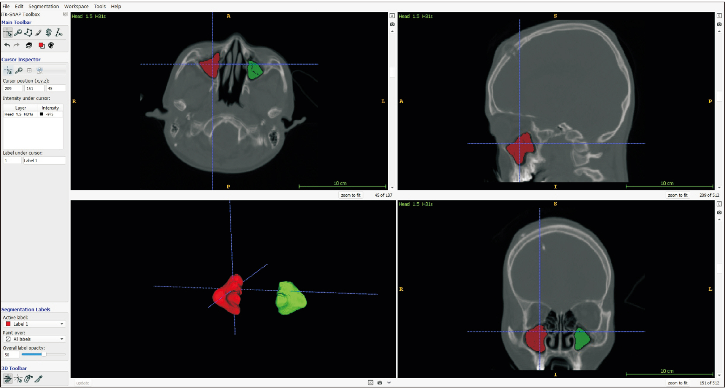
Fig. 1 Segmentation procedure of maxillary sinus using ITK-SNAP.
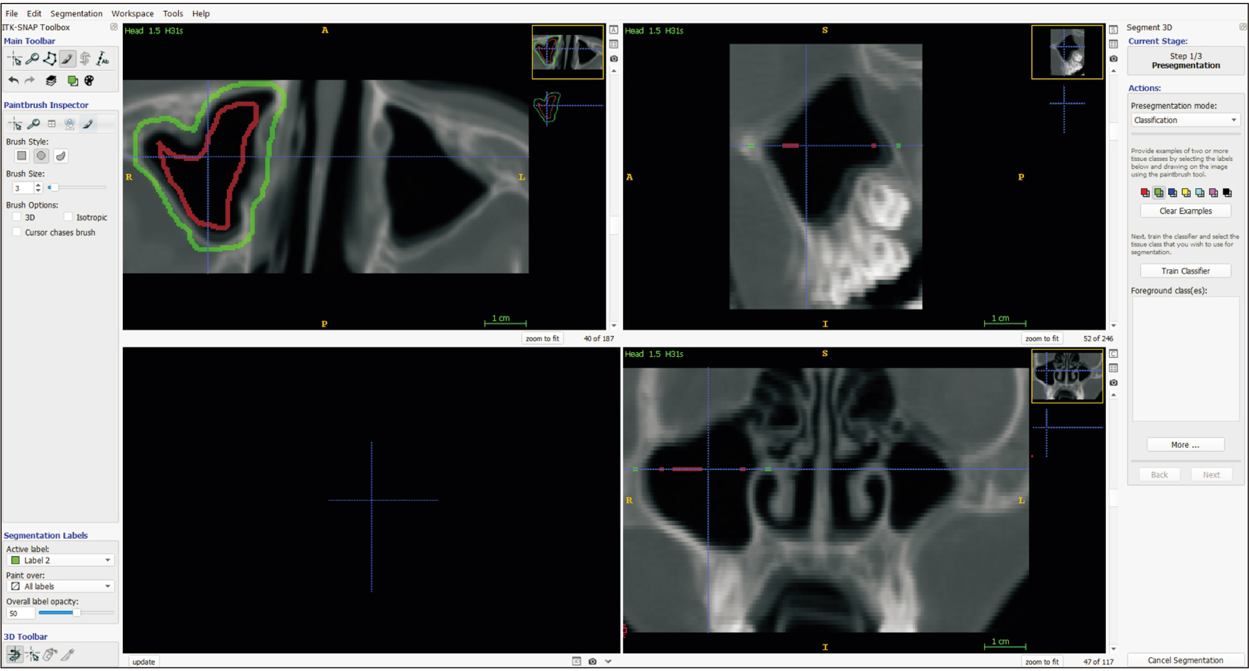
Fig. 2 Maxillary sinus classification of image regions.
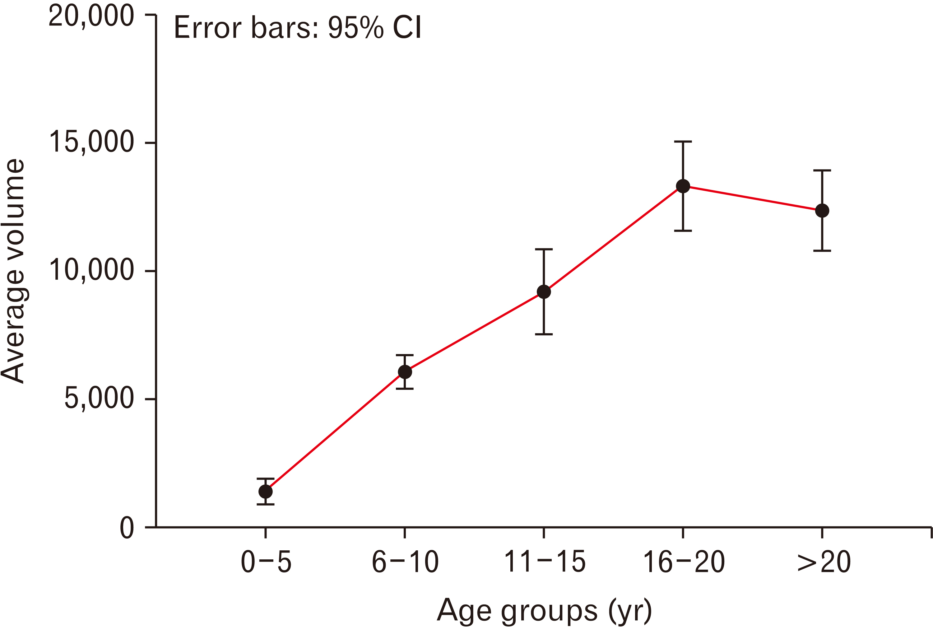
Fig. 3 Distribution of mean maxillary sinus volume by age groups (mm3). CI, confidence interval.
Reference
-
References
1. Maroldi R, Ravanelli M, Borghesi A, Farina D. 2008; Paranasal sinus imaging. Eur J Radiol. 66:372–86. DOI: 10.1016/j.ejrad.2008.01.059. PMID: 18375083.
Article2. Cellina M, Gibelli D, Cappella A, Toluian T, Pittino CV, Carlo M, Oliva G. 2021; Segmentation procedures for the assessment of paranasal sinuses volumes. Neuroradiol J. 34:13–20. DOI: 10.1177/1971400920946635. PMID: 32757847. PMCID: PMC7868593.
Article3. Natsis K, Karabatakis V, Tsikaras P, Chatzibalis T, Stangos A, Stangos N. 2004; Frontal sinus anatomical variations with potential consequences for the orbit. Study on cadavers. Morphologie. 88:35–8. DOI: 10.1016/S1286-0115(04)97997-0. PMID: 15208811.
Article4. Emirzeoglu M, Sahin B, Bilgic S, Celebi M, Uzun A. 2007; Volumetric evaluation of the paranasal sinuses in normal subjects using computer tomography images: a stereological study. Auris Nasus Larynx. 34:191–5. DOI: 10.1016/j.anl.2006.09.003. PMID: 17084569.
Article5. Aktuna Belgin C, Colak M, Adiguzel O, Akkus Z, Orhan K. 2019; Three-dimensional evaluation of maxillary sinus volume in different age and sex groups using CBCT. Eur Arch Otorhinolaryngol. 276:1493–9. DOI: 10.1007/s00405-019-05383-y. PMID: 30879193.
Article6. Pirimoglu B, Sade R, Sakat MS, Ogul H, Eren S, Kantarci M. 2017; Comparing subjective and objective image quality at two different radiation exposure ranges of the paranasal sinus CT examinations using a volumetric 320-row detector CT system. Dentomaxillofac Radiol. 46:20170212. DOI: 10.1259/dmfr.20170212. PMID: 28937286. PMCID: PMC5965943.
Article7. Sahlstrand-Johnson P, Jannert M, Strömbeck A, Abul-Kasim K. 2011; Computed tomography measurements of different dimensions of maxillary and frontal sinuses. BMC Med Imaging. 11:8. DOI: 10.1186/1471-2342-11-8. PMID: 21466703. PMCID: PMC3080316.
Article8. Stutzki M, Jahns E, Mandapathil MM, Diogo I, Werner JA, Güldner C. 2015; Indications of cone beam CT in head and neck imaging. Acta Otolaryngol. 135:1337–43. DOI: 10.3109/00016489.2015.1076172. PMID: 26313160.
Article9. Kirmeier R, Arnetzl C, Robl T, Payer M, Lorenzoni M, Jakse N. 2011; Reproducibility of volumetric measurements on maxillary sinuses. Int J Oral Maxillofac Surg. 40:195–9. DOI: 10.1016/j.ijom.2010.10.008. PMID: 21074367.
Article10. Bui NL, Ong SH, Foong KW. 2015; Automatic segmentation of the nasal cavity and paranasal sinuses from cone-beam CT images. Int J Comput Assist Radiol Surg. 10:1269–77. DOI: 10.1007/s11548-014-1134-5. PMID: 25503593.
Article11. Humphries SM, Centeno JP, Notary AM, Gerow J, Cicchetti G, Katial RK, Beswick DM, Ramakrishnan VR, Alam R, Lynch DA. 2020; Volumetric assessment of paranasal sinus opacification on computed tomography can be automated using a convolutional neural network. Int Forum Allergy Rhinol. 10:1218–25. DOI: 10.1002/alr.22588. PMID: 32306522.
Article12. Giacomini G, Pavan ALM, Altemani JMC, Duarte SB, Fortaleza CMCB, Miranda JRA, de Pina DR. 2018; Computed tomography-based volumetric tool for standardized measurement of the maxillary sinus. PLoS One. 13:e0190770. DOI: 10.1371/journal.pone.0190770. PMID: 29304130. PMCID: PMC5755892.
Article13. Spaeth J, Krügelstein U, Schlöndorff G. 1997; The paranasal sinuses in CT-imaging: development from birth to age 25. Int J Pediatr Otorhinolaryngol. 39:25–40. DOI: 10.1016/S0165-5876(96)01458-9. PMID: 9051437.
Article14. Park IH, Song JS, Choi H, Kim TH, Hoon S, Lee SH, Lee HM. 2010; Volumetric study in the development of paranasal sinuses by CT imaging in Asian: a pilot study. Int J Pediatr Otorhinolaryngol. 74:1347–50. DOI: 10.1016/j.ijporl.2010.08.018. PMID: 20863577.
Article15. Yushkevich PA, Piven J, Hazlett HC, Smith RG, Ho S, Gee JC, Gerig G. 2006; User-guided 3D active contour segmentation of anatomical structures: significantly improved efficiency and reliability. Neuroimage. 31:1116–28. DOI: 10.1016/j.neuroimage.2006.01.015. PMID: 16545965.
Article16. Ariji Y, Kuroki T, Moriguchi S, Ariji E, Kanda S. 1994; Age changes in the volume of the human maxillary sinus: a study using computed tomography. Dentomaxillofac Radiol. 23:163–8. DOI: 10.1259/dmfr.23.3.7835518. PMID: 7835518.
Article17. Ikeda A, Ikeda M, Komatsuzaki A. 1998; A CT study of the course of growth of the maxillary sinus: normal subjects and subjects with chronic sinusitis. ORL J Otorhinolaryngol Relat Spec. 60:147–52. DOI: 10.1159/000027584. PMID: 9579359.
Article18. Kawarai Y, Fukushima K, Ogawa T, Nishizaki K, Gunduz M, Fujimoto M, Masuda Y. 1999; Volume quantification of healthy paranasal cavity by three-dimensional CT imaging. Acta Otolaryngol Suppl. 540:45–9. DOI: 10.1080/00016489950181198. PMID: 10445079.19. Sarilita E, Rynn C, Mossey PA, Black S, Oscandar F. 2018; Nose profile morphology and accuracy study of nose profile estimation method in Scottish subadult and Indonesian adult populations. Int J Legal Med. 132:923–31. DOI: 10.1007/s00414-017-1758-4. PMID: 29260392. PMCID: PMC5919985.
Article20. Adibelli ZH, Songu M, Adibelli H. 2011; Paranasal sinus development in children: a magnetic resonance imaging analysis. Am J Rhinol Allergy. 25:30–5. DOI: 10.2500/ajra.2011.25.3552. PMID: 21711972.
Article21. Jun BC, Song SW, Park CS, Lee DH, Cho KJ, Cho JH. 2005; The analysis of maxillary sinus aeration according to aging process; volume assessment by 3-dimensional reconstruction by high-resolutional CT scanning. Otolaryngol Head Neck Surg. 132:429–34. DOI: 10.1016/j.otohns.2004.11.012. PMID: 15746857.
Article22. Waluyo RF, Priaminiarti M, Yuniastuti M, Soedarsono N, Susilo BT. 2020; Measurements of sex-related differences in maxillary sinus and mandibular canal characteristic using cone beam computed tomography. Forensic Imaging. 21:200371. DOI: 10.1016/j.fri.2020.200371.
Article
- Full Text Links
-
- Actions
-
Cited
- CITED
-
- Close
- Share
-
- Similar articles
-
- Maxillary sinus volumetric changes in jet aircraft pilots: A multislice computed tomography pilot study
- A case report of incidental finding of fungus ball on CBCT of maxillary sinus in treatment planning of dental implant
- Effect of Pediatric Endoscopic Sinus Surgery with Antrochoanal Polyps on Sinus Growth
- Comparison of panoramic radiography and cone beam computed tomography for assessing the relationship between the maxillary sinus floor and maxillary molars
- Three-dimensional linear and volumetric computed tomography analysis of the frontal sinus
