A novel histologic description of the fibrous networks in the lid-cheek junction and infraorbital region
- Affiliations
-
- 1Division in Anatomy and Developmental Biology, Department of Oral Biology, Human Identification Research Institute, BK21 FOUR Project, Yonsei University College of Dentistry, Seoul, Korea
- 2Maylin Clinic, Seoul, Korea
- 3Yonsei Severance Dental Clinic, Seoul, Korea
- 4Department of Anatomy, College of Medicine, Konkuk University, Chungju, Korea
- 5Department of Anatomy, Chungbuk National University, Cheongju, Korea
- 6Department of Plastic and Reconstructive Surgery, Royal Free Hospital, London, UK
- KMID: 2554238
- DOI: http://doi.org/10.5115/acb.23.275
Abstract
- The aim of this study was to identify the anatomical feature of retaining ligament and fat compartment on the lower eyelid and infraorbital region using a histological method, and to investigate clear definitions for them which could be used generally in the clinical area. Eighteen specimens from eight fresh Korean cadavers were stained with Masson trichrome or hematoxylin and eosin. The ligamentous and fascial fibrous tissue were clearly identified. The ligamentous fibrous tissue which traversed in the superficial and deep fat layer was skin ligament and orbicularis retaining ligament (ORL). The fascial fibrous tissue enclosed the orbicularis oculi muscle (OOc) and circumferencial adipose tissue. Based on the ligamentous and fascial structure, three fat compartments, septal, suborbicularis oculi and infraorbital fat compartment, could be identified. The OOc attached to orbital rim and dermis by ORL and skin ligament, and the muscle fascicle and fat fascicle provided the connection point to the ORL and skin ligament as enclosing all muscle and fat tissue. The combination of the force made by the skin ligament in the lower eyelid and ORL may decide the level and form of the infraorbital grooves.
Figure
-
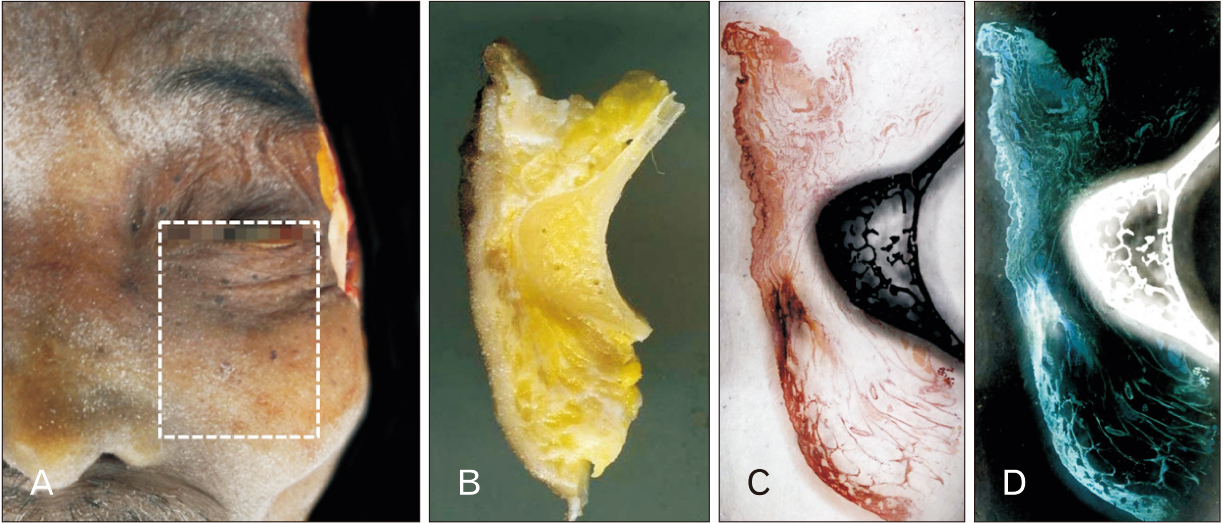
Fig. 1 Full thickness specimens with a bony structure. (A) The boundaries of the specimens were lower eyelid (superior), medial canthus (medial), lateral canthus (lateral), and alare of nose (inferior). (B) The sectional planes of the specimens were scanned. (C) The specimens were stained with Masson trichrome or hematoxylin and eosin. (D) The color of original image was inverted by the photo editing program.

Fig. 2 The histological features of the infraorbital specimen (H&E staining, 8×). The superficial and deep fat layer was composed of fibrous tissue and adipose tissue (blue star), and there were two types of the fibrous tissue: ligamentous (red arrowhead) and fascial fibrous tissue (green arrowhead). (A) The layers of the facial skin: (a) skin, (b) superficial fat, (c) orbicularis oculi muscle (OOc), (d) deep fat, and (e) periosteum. (B) The skin ligament was observed thoroughly in the superficial fat layer. (C) The orbicularis retaining ligament (ORL, black arrowheads) bundled the deep aspect of the OOc, and the OOc of the attachment site was observed to be tugged inwards. The number of ORL strands and the tugged site of the OOc were equivalent (Fig. 2C). Adapted from Clinical anatomy of the face for filler and botulinum toxin injection. Springer, 2016, with permission [16].

Fig. 3 The fascicular system of the fat tissue. Many adipose cells constituted a fat unit, and a few fat units constituted again the fat compartment, such as inferior orbital fat and the sub orbicularis oculi fat. The fascicle encircles an adipose cell, a fat unit, and the fat compartment could be named the endoadiposium (blue arrowhead), peiadiposium (red arrowhead), and epiadiposium (black arrowhead), respectively.
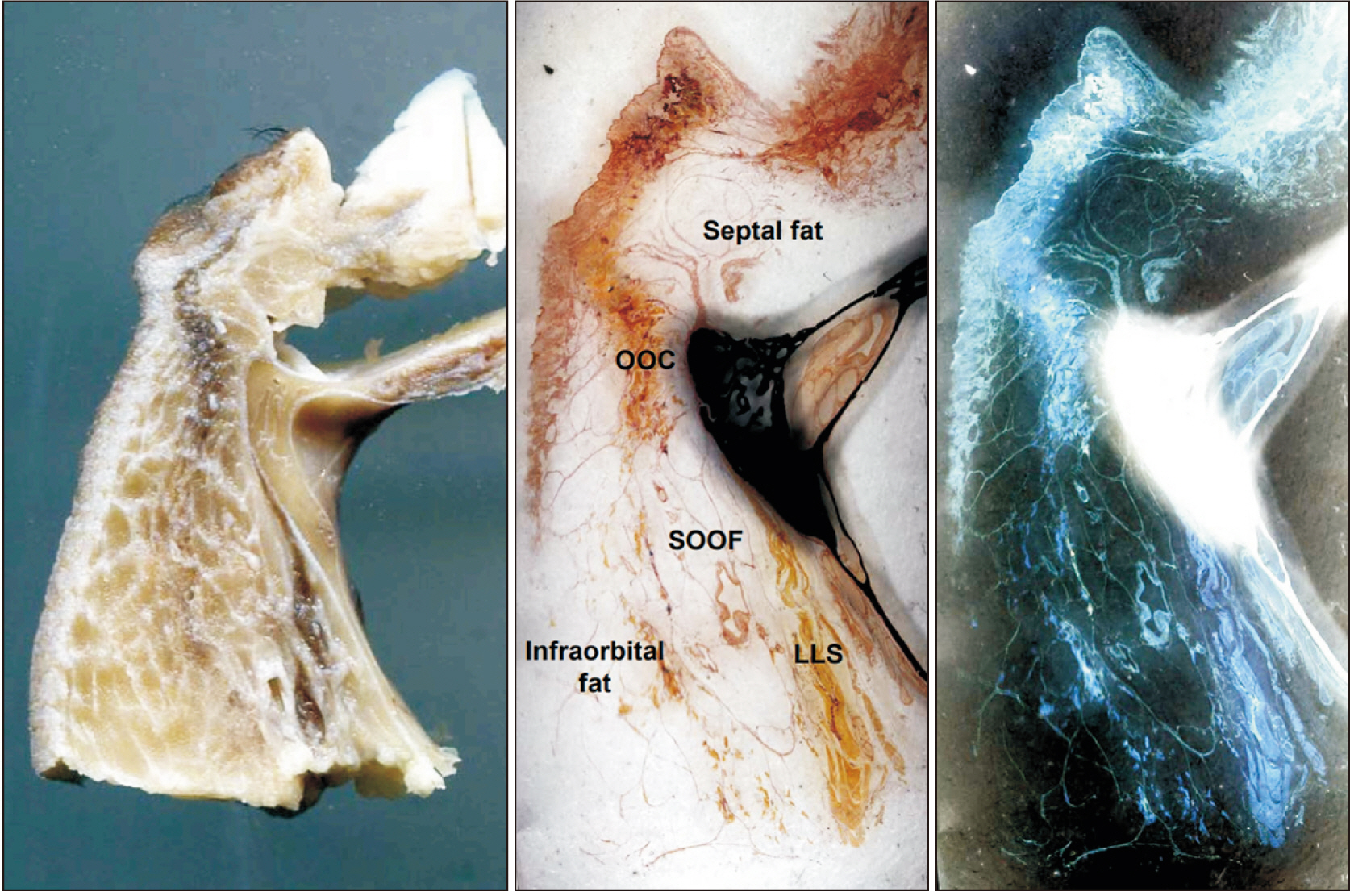
Fig. 4 Three fat compartments in the infraorbital area: septal, suborbicularis oculi and infraorbital fat compartment. OOc, orbicularis oculi muscle; SOOF, sub orbicularis oculi fat; LLS, levator labii superioris.

Fig. 5 Various number of the orbicularis retaining ligament (ORL, arrowheads). The number of ORL different among specimens, actually, it was impossible to count the number of ORL because it was condensed fibrous structure. OOc, orbicularis oculi muscle.
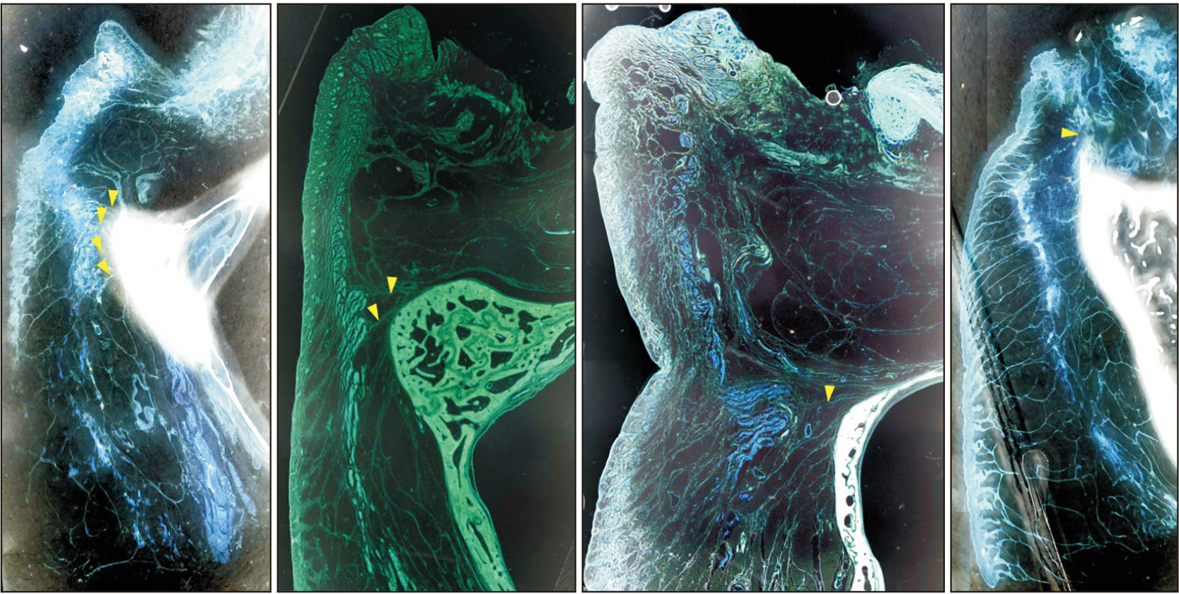
Fig. 6 The histologic features of the infraorbital skin showing baggy lower lid. The septal fat compartment protruded anteriorly as the deep fatty layer thickened, and the more-inwardly tugged orbicularis oculi muscle (OOc) compensated for the increased space. In other words, while there was no difference in the length of the orbicularis retaining ligament (ORL, yellow arrowheads), the OOc was tugged considerably more inwards. And, the level of the groove was not the same with the level of ORL but rather equivalent to the level of the orbital rim or the inferior border of the septal fat compartment. Also, the superficial fatty layer as well as the deep fatty layer was thickened. In other words, the laxity of skin ligament was increased.
Reference
-
References
1. Haddock NT, Saadeh PB, Boutros S, Thorne CH. 2009; The tear trough and lid/cheek junction: anatomy and implications for surgical correction. Plast Reconstr Surg. 123:1332–40. DOI: 10.1097/PRS.0b013e31819f2b36. PMID: 19337101.
Article2. Stuzin JM, Baker TJ, Gordon HL. 1992; The relationship of the superficial and deep facial fascias: relevance to rhytidectomy and aging. Plast Reconstr Surg. 89:441–9. discussion 450–1. DOI: 10.1097/00006534-199203000-00008.3. Wong CH, Hsieh MKH, Mendelson B. 2012; The tear trough ligament: anatomical basis for the tear trough deformity. Plast Reconstr Surg. 129:1392–402. DOI: 10.1097/PRS.0b013e31824ecd77. PMID: 22634656.4. Furnas DW. 1989; The retaining ligaments of the cheek. Plast Reconstr Surg. 83:11–6. DOI: 10.1097/00006534-198901000-00003. PMID: 2909050.
Article5. Nash LG, Phillips MN, Nicholson H, Barnett R, Zhang M. 2004; Skin ligaments: regional distribution and variation in morphology. Clin Anat. 17:287–93. DOI: 10.1002/ca.10203. PMID: 15108331.6. Lambros V. 2007; Observations on periorbital and midface aging. Plast Reconstr Surg. 120:1367–76. DOI: 10.1097/01.prs.0000279348.09156.c3. PMID: 17898614.
Article7. Moss CJ, Mendelson BC, Taylor GI. 2000; Surgical anatomy of the ligamentous attachments in the temple and periorbital regions. Plast Reconstr Surg. 105:1475–90. discussion 1491–8. DOI: 10.1097/00006534-200004000-00034.
Article8. Donofrio LM. 2000; Fat distribution: a morphologic study of the aging face. Dermatol Surg. 26:1107–12. DOI: 10.1046/j.1524-4725.2000.00270.x. PMID: 11134986.
Article9. Rohrich RJ, Pessa JE. 2008; The retaining system of the face: histologic evaluation of the septal boundaries of the subcutaneous fat compartments. Plast Reconstr Surg. 121:1804–9. DOI: 10.1097/PRS.0b013e31816c3c1a. PMID: 18454006.
Article10. Ghavami A, Pessa JE, Janis J, Khosla R, Reece EM, Rohrich RJ. 2008; The orbicularis retaining ligament of the medial orbit: closing the circle. Plast Reconstr Surg. 121:994–1001. DOI: 10.1097/01.prs.0000299941.62645.4e. PMID: 18317148.
Article11. Hwang K, Nam YS, Kim DJ, Han SH. 2008; Surgical anatomy of retaining ligaments in the periorbital area. J Craniofac Surg. 19:800–4. DOI: 10.1097/SCS.0b013e31816b6c5e. PMID: 18520402.
Article12. Byrd HS, Burt JD. 2002; Achieving aesthetic balance in the brow, eyelids, and midface. Plast Reconstr Surg. 110:926–33. discussion 934–9. DOI: 10.1097/01.PRS.0000019877.41086.EB.
Article13. Mendelson BC, Muzaffar AR, Adams WP Jr. 2002; Surgical anatomy of the midcheek and malar mounds. Plast Reconstr Surg. 110:885–96. discussion 897–911. DOI: 10.1097/01.PRS.0000019706.34235.E5.
Article14. Muzaffar AR, Mendelson BC, Adams WP Jr. 2002; Surgical anatomy of the ligamentous attachments of the lower lid and lateral canthus. Plast Reconstr Surg. 110:873–84. discussion 897–911. DOI: 10.1097/00006534-200209010-00025. PMID: 12172154.
Article15. Yang C, Zhang P, Xing X. 2013; Tear trough and palpebromalar groove in young versus elderly adults: a sectional anatomy study. Plast Reconstr Surg. 132:796–808. DOI: 10.1097/PRS.0b0133182a0539e. PMID: 24076672.16. Kim HJ, Seo K.K, Lee HK, Kim J. Clinical anatomy of the face for filler and botulinum toxin injection. Springer;2016. DOI: 10.1007/978-981-10-0240-3.17. Standring S, Gray H. Gray's anatomy: the anatomical basis of clinical practice. 40th ed. Churchill Livingstone/Elsevier;2008.18. Davison SP, Iorio ML, Oh C. 2015; Transconjunctival lower lid blepharoplasty with and without fat repositioning. Clin Plast Surg. 42:51–6. Erratum in: Clin Plast Surg 2017;44:xv. DOI: 10.1016/j.cps.2014.09.001. PMID: 25440740.
Article19. Furnas DW. 1981; The orbicularis oculi muscle. Management in blepharoplasty. Clin Plast Surg. 8:687–715. DOI: 10.1016/S0094-1298(20)30398-9. PMID: 7338003.
Article20. Mendelson BC. 2013; Anatomic study of the retaining ligaments of the face and applications for facial rejuvenation. Aesthetic Plast Surg. 37:513–5. DOI: 10.1007/s00266-013-0066-8. PMID: 23494030. PMCID: PMC3657075.
Article21. Nassif PS. 2005; Lower blepharoplasty: transconjunctival fat repositioning. Facial Plast Surg Clin North Am. 13:553–9. viDOI: 10.1016/j.fsc.2005.06.006. PMID: 16253842.
Article22. Zarem HA, Resnick JI. 1991; Expanded applications for transconjunctival lower lid blepharoplasty. Plast Reconstr Surg. 88:215–20. discussion 221DOI: 10.1097/00006534-199108000-00006.
Article23. Rohrich RJ, Ghavami A, Mojallal A. 2011; The five-step lower blepharoplasty: blending the eyelid-cheek junction. Plast Reconstr Surg. 128:775–83. DOI: 10.1097/PRS.0b013e3182121618. PMID: 21278622.24. Yoo DB, Peng GL, Massry GG. 2013; Transconjunctival lower blepharoplasty with fat repositioning: a retrospective comparison of transposing fat to the subperiosteal vs supraperiosteal planes. JAMA Facial Plast Surg. 15:176–81. DOI: 10.1001/jamafacial.2013.749. PMID: 23471339.
Article
