Black esophagus: a life-threatening adverse event associated with endoscopic retrograde cholangiopancreatography
- Affiliations
-
- 1Department of Gastroenterology, Nara Medical University, Nara, Japan
- 2Division of Endoscopy, Nara Medical University, Nara, Japan
- KMID: 2553766
- DOI: http://doi.org/10.5946/ce.2023.047
Figure
-
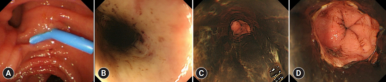
Fig. 1. (A) Endoscopy showing normal findings of the duodenal mucosa and papilla during endoscopic retrograde cholangiopancreatography. A plastic stent was placed into the bile duct. (B–D) Endoscopic findings the day after endoscopic retrograde cholangiopancreatography (B, upper esophagus; C, middle esophagus; D, distal esophagus). Black esophageal mucosa with ulcers present mainly in the distal esophagus.

Fig. 2. (A) Forty-five days after endoscopic retrograde cholangiopancreatography, the necrosis of the esophagus appears to have healed, but stricture formation in the distal esophagus was observed. (B) Photomicrograph of esophageal biopsy specimen showing degenerated esophageal epithelium, granulation tissue extending into the submucosa (hematoxylin &eosin stain, ×100). (C) The fluoroscopic image of balloon dilation.

Fig. 3. (A, B) A second endoscopic dilatation was performed for restenosis of the esophagus. (C) Computed tomography findings after endoscopic balloon dilation. Massive free air was noted in the abdominal cavity. (D) Gross pathologic image of the resected esophagus specimen. Circumferential erosion and ulcers were observed in the distal esophagus. (E) Photomicrograph of resected esophagus specimen. The esophageal wall was infiltrated with numerous neutrophils and inflammatory cells (hematoxylin & eosin stain, ×40).
Reference
-
1. Gurvits GE. Black esophagus: acute esophageal necrosis syndrome. World J Gastroenterol. 2010; 16:3219–3225.2. Lamers CR, Mares WGN, Bac DJ. Black esophagus: a case series and literature review of acute esophageal necrosis. Scand J Gastroenterol. 2018; 53:1421–1424.3. Jinushi R, Ishii N, Yano T, et al. Endoscopic balloon dilation for the prevention of severe strictures caused by acute esophageal necrosis. DEN Open. 2021; 2:e43.4. Forman R, Gopal K, Huysman AM, et al. Black esophagus: a case series. Dig Endosc. 2007; 19:142–146.5. Kabaçam G, Yakut M, Soykan I. Acute esophageal necrosis: a rare cause of gastrointestinal bleeding. Dig Endosc. 2012; 24:283.6. Augusto F, Fernandes V, Cremers MI, et al. Acute necrotizing esophagitis: a large retrospective case series. Endoscopy. 2004; 36:411–415.7. Gurvits GE, Cherian K, Shami MN, et al. Black esophagus: new insights and multicenter international experience in 2014. Dig Dis Sci. 2015; 60:444–453.8. Goldenberg SP, Wain SL, Marignani P. Acute necrotizing esophagitis. Gastroenterology. 1990; 98:493–496.9. Gurvits GE, Shapsis A, Lau N, et al. Acute esophageal necrosis: a rare syndrome. J Gastroenterol. 2007; 42:29–38.10. Day A, Sayegh M. Acute oesophageal necrosis: a case report and review of the literature. Int J Surg. 2010; 8:6–14.
- Full Text Links
-
- Actions
-
Cited
- CITED
-
- Close
- Share
-
- Similar articles
-
- Visceral Artery Pseudoaneurysm Rupture after Endoscopic Retrograde Cholangiopancreatography
- Prevention of Post-endoscopic Retrograde Cholangiopancreatography Pancreatitis: An Endoscopic Perspective
- Portal cavernography during endoscopic retrograde cholangiopancreatography: from bilhemia to hemobilia
- A Road Less Traveled: Endoscopic Retrograde Cholangiopancreatography in a Patient with Long-standing Achalasia and Sigmoid Esophagus
- Endoscopic Retrograde Cholangiopancreatography during Pregnancy: Really Guarantee to Safety?
