Rehabilitation using endocrown for fracture of maxillary anterior teeth due to trauma in adolescence: a case report
- Affiliations
-
- 1Department of Conservative Dentistry, School of Dentistry, Pusan National University, Yangsan, Republic of Korea
- KMID: 2553628
- DOI: http://doi.org/10.14368/jdras.2024.40.1.24
Abstract
- Complicated crown fractures of maxillary anterior teeth caused by trauma in adolescence can cause functional and aesthetic problems. For crown fractures with pulp exposure, various restorative methods can be considered depending on the amount of remaining tooth structure. Direct resin restorations are the most traditional and effective method, but they are likely to discolor and break over time. Fixed prosthesis have a high possibility of re-restoration due to marginal disharmony due to tooth movement during the growth period, and restorations using post which are mainly performed for extensive crown fractures increase the risk of root perforation and root fracture. However, endocrown is an integrated structure that gains retention force from the pulp space, enabling effective reconstruction from a biomechanical perspective and providing advantages in restoring function and aesthetics. Therefore, endocrown can be considered as a restoration method for complicated crown fractures caused by trauma in adolescence.
Keyword
Figure
-
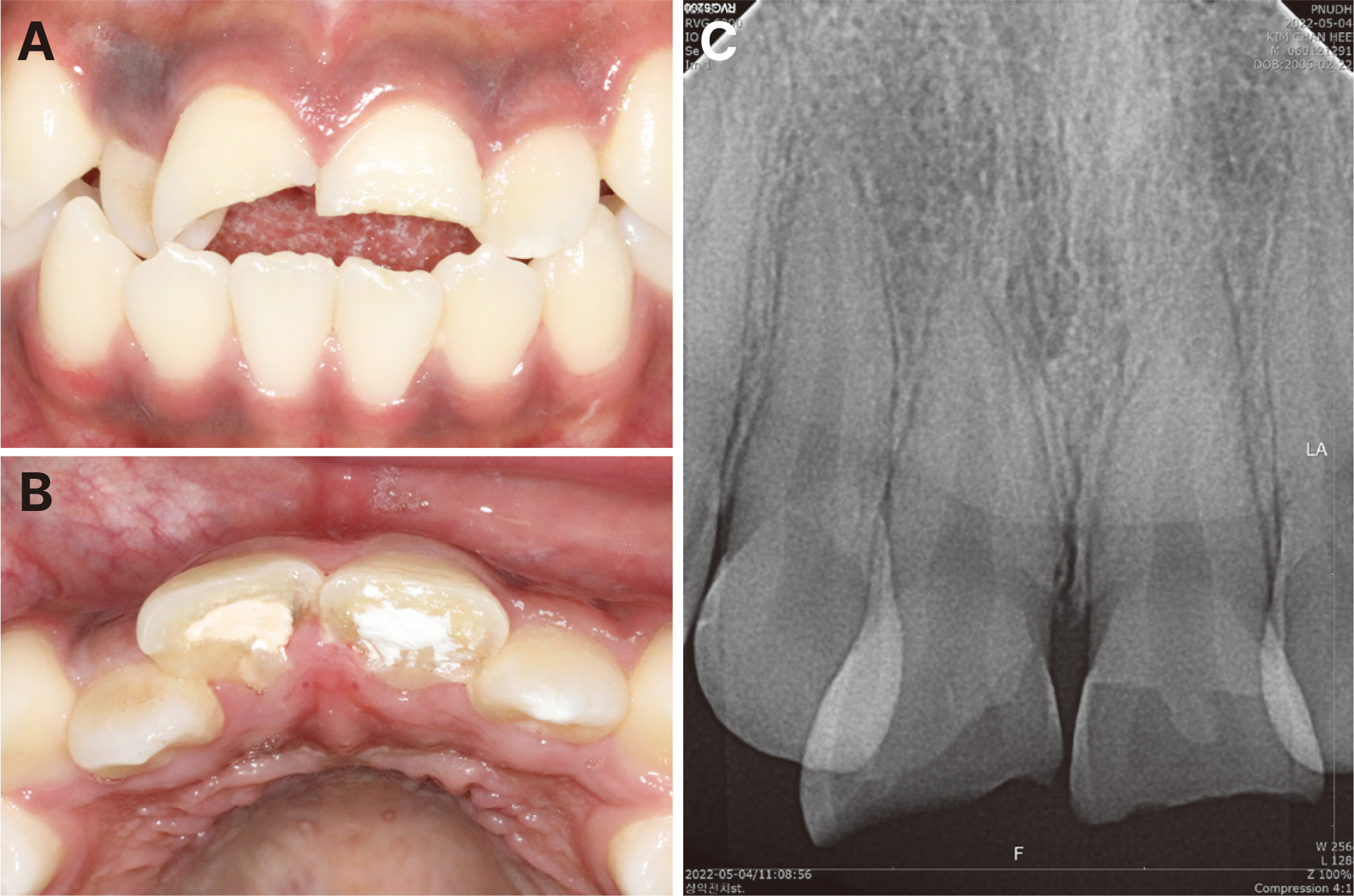
Fig. 1 Initial examination of maxillary right central incisor and maxillary left central incisor. (A) Preoperative photograph: frontal view, (B) Preoperative photograph: occlusal view, (C) Preoperative radiograph.
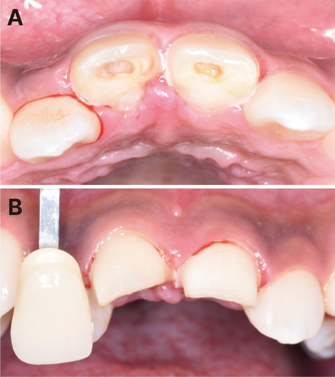
Fig. 2 The preparation design of Endocrown and selection of tooth shade with VITASPAN. (A) Tooth preparation, (B) Shade selection.
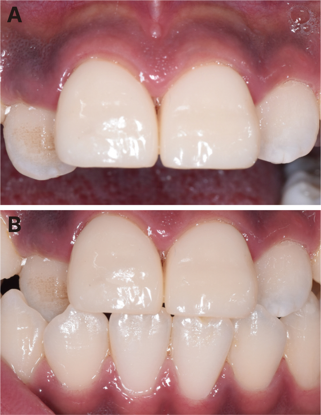
Fig. 3 Conventional resin cement was used to bond the Endocrown to the prepared tooth. (A) Postoperative photograph: frontal view of upper anterior teeth, (B) Postoperative photograph: frontal view.
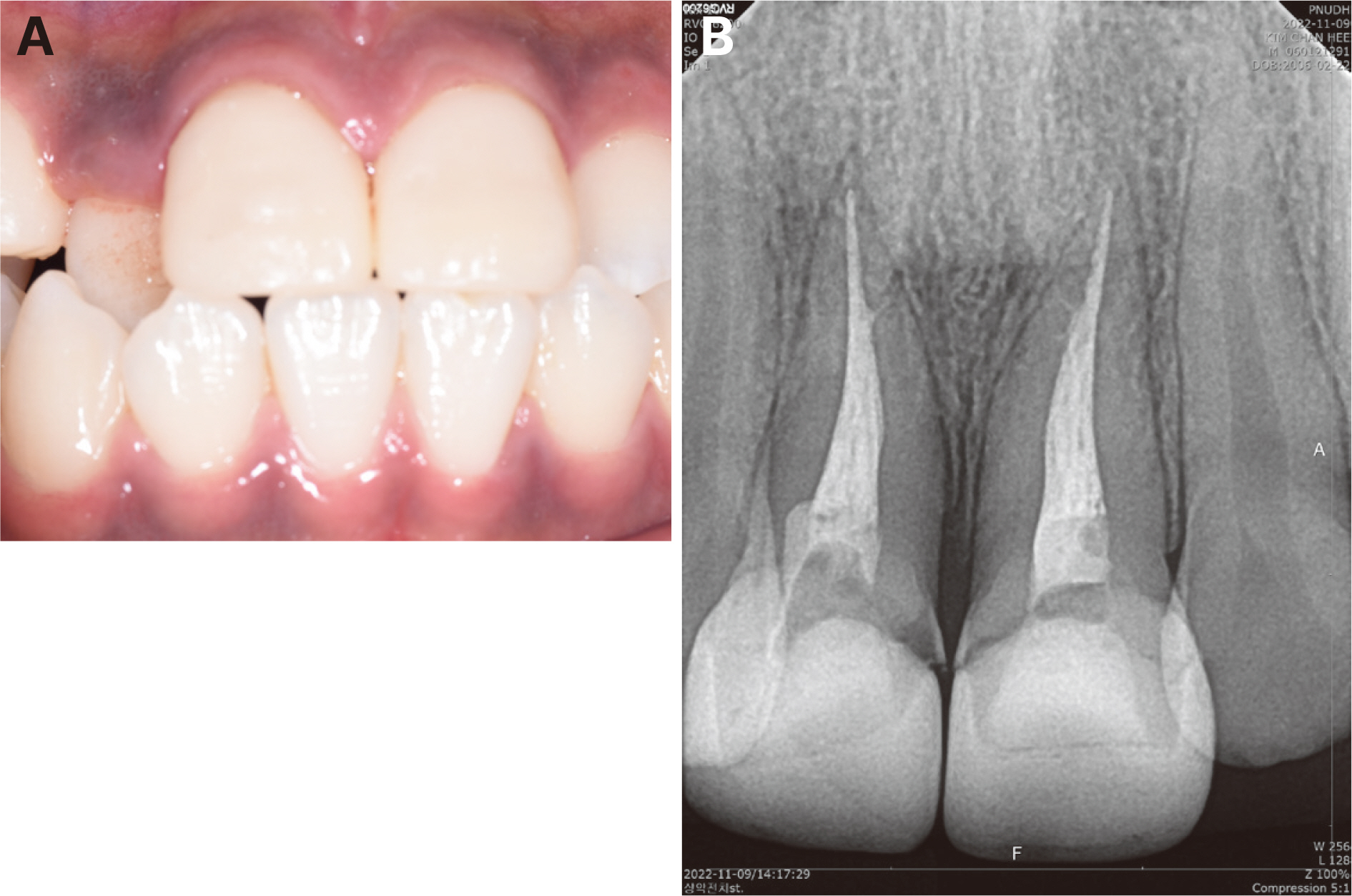
Fig. 4 3-month follow-up. (A) 3-month follow-up photograph, (B) 3-month follow-up radiograph.

Fig. 5 Initial examination of maxillary right central incisor and maxillary left central incisor. (A) preoperative photograph: facial view, (B) fractured fragment reattachment to maxillary right central incisor.

Fig. 6 The preparation design of Endocrown. (A) Tooth preparation, (B) Gingival retraction.
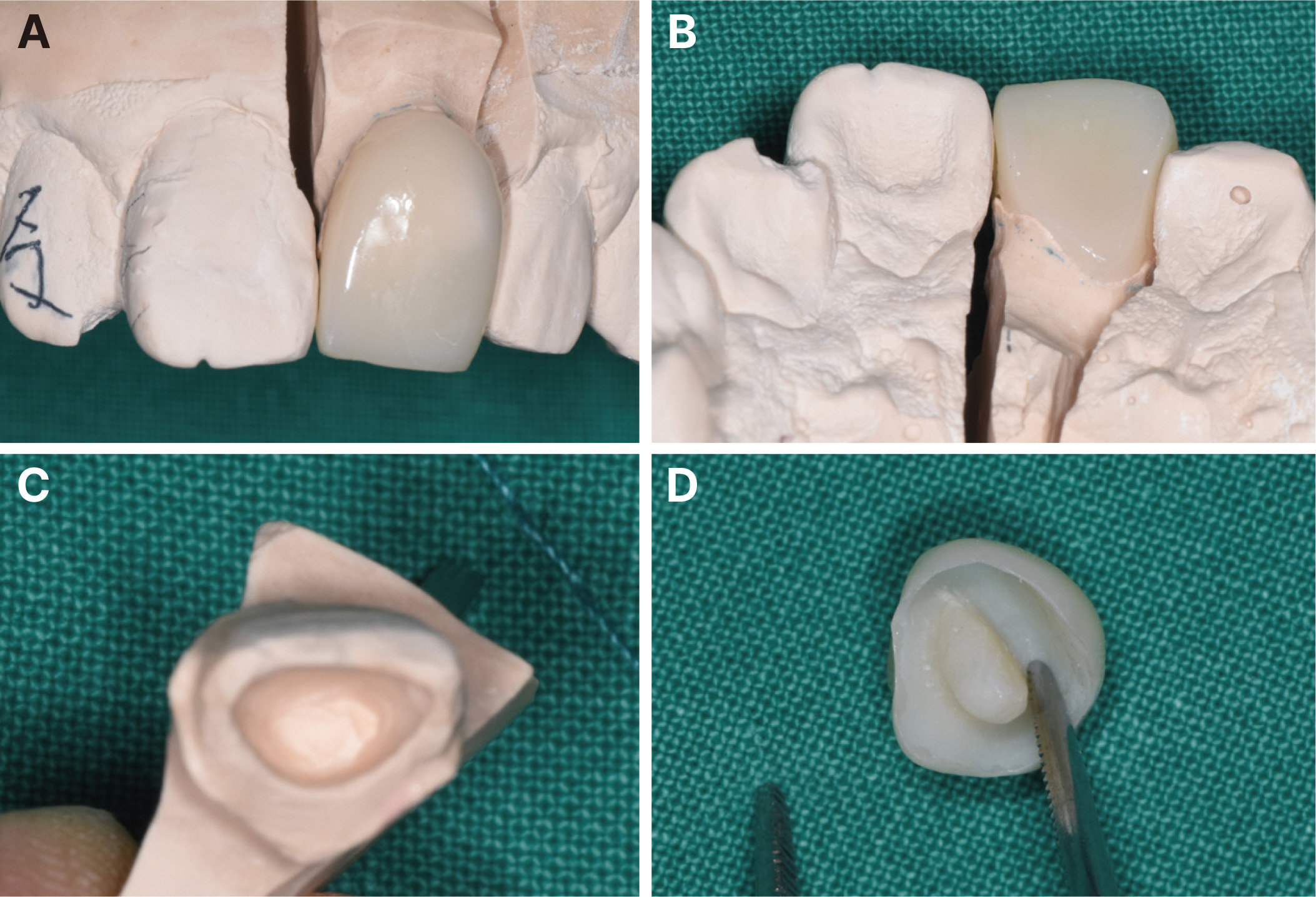
Fig. 7 The endocrown was fabricated with Tescera in laboratory. (A) Fabricated endocrown on master cast: frontal view, (B) Fabricated endocrown on master cast: occlusal view, (C) Master cast: internal view, (D) Fabricated endocrown.

Fig. 8 Clinical and radiograph examination during follow up. (A) Postoperative photograph, (B) 2-month follow-up photograph, (C) 4-month follow-up photograph, (D) Postoperative radiograph, (E) 2-month follow-up radiograph, (F) 4-month follow-up radiograph.
Reference
-
References
1. Oz IA, Haytaç MC, Toroglu MS. 2006; Multidisciplinary approach to the rehabilitation of a crown-root fracture with original fragment for immediate esthetics: a case report with 4-year follow-up. Dent Traumatol. 22:48–52. DOI: 10.1111/j.1600-9657.2006.00335.x. PMID: 16422760.2. Bissinger R, Müller DD, Reymus M, Khazaei Y, Hickel R, Bücher K, Kühnisch J. 2021; Treatment outcomes after uncomplicated and complicated crown fractures in permanent teeth. Clin Oral Investig. 25:133–43. DOI: 10.1007/s00784-020-03344-y. PMID: 32705398. PMCID: PMC7785561.
Article3. Aggarwal V, Logani A, Shah N. 2009; Complicated crown fractures - management and treatment options. Int Endod J. 42:740–53. DOI: 10.1111/j.1365-2591.2009.01588.x. PMID: 19548932.
Article4. Sedgley CM, Messer HH. 1992; Are endodontically treated teeth more brittle? J Endod. 18:332–5. DOI: 10.1016/S0099-2399(06)80483-8. PMID: 1402595.
Article5. Elfadil S, Nassar HI, Elbeshbeishy RA, Annamma LM. 2023; Esthetic Rehabilitation of Pediatric Patients Using Direct Bonding Technique - A Case Series Report. Children (Basel). 10:546. DOI: 10.3390/children10030546. PMID: 36980104. PMCID: PMC10047593.6. Shah YR, Shiraguppi VL, Deosarkar BA, Shelke UR. 2021; Long-term survival and reasons for failure in direct anterior composite restorations: A systematic review. J Conserv Dent. 24:415–20. DOI: 10.4103/jcd.jcd_527_21. PMID: 35399771. PMCID: PMC8989165.
Article7. Riverwalk Dental. What age do people get crowns? Available from: https://www.riverwalkdentalpotranco.com/what-age-do-people-get-crowns. updated 2023 Nov 11.8. Schwartz RS, Robbins JW. 2004; Post placement and restoration of endodontically treated teeth: a literature review. J Endod. 30:289–301. DOI: 10.1097/00004770-200405000-00001. PMID: 15107639.
Article9. Biacchi GR, Mello B, Basting RT. 2013; The endocrown: an alternative approach for restoring extensively damaged molars. J Esthet Restor Dent. 25:383–90. DOI: 10.1111/jerd.12065. PMID: 24148141.
Article10. Sevimli G, Cengiz S, Oruc MS. 2015; Endocrowns: review. J Istanb Univ Fac Dent. 49:57–63. DOI: 10.17096/jiufd.71363. PMID: 28955538. PMCID: PMC5573486.
Article11. Bindl A, Mörmann WH. 1999; Clinical evaluation of adhesively placed Cerec endo-crowns after 2 years -preliminary results. J Adhes Dent. 1:255–65.12. Soares CJ, Versluis A, Tantbirojn D, Kim HC, Veríssimo C, Santos-Filho PCF. Perdigão J, editor. Biomechanical Principles of Root Canal-Treated-Teeth Restored with Fiber-Reinforced Resin Posts. Restoration of Root Canal-Treated Teeth. Zürich: Springer;2016. p. 87–100. DOI: 10.1007/978-3-319-15401-5_5.
Article13. Krishna A, Malur MH, Swapna DV, Benjamin S, Deepak CA. 2012; Traumatic dental injury-an enigma for adolescents: a series of case reports. Case Rep Dent. 2012:756526. DOI: 10.1155/2012/756526. PMID: 23198166. PMCID: PMC3502796.14. Deulkar PV, Bane SP, Rathi NV, Thosar NR. 2022; Rehabilitation of Traumatised Maxillary Anterior Teeth in Children Using Endocrown: A Case Series. Cureus. 14:e28102. DOI: 10.7759/cureus.28102.15. Bankoğlu Güngör M, Turhan Bal B, Yilmaz H, Aydin C, Karakoca Nemli S. 2017; Fracture strength of CAD/CAM fabricated lithium disilicate and resin nano ceramic restorations used for endodontically treated teeth. Dent Mater J. 3:135–41. DOI: 10.4012/dmj.2016-017. PMID: 28111383.16. Jeong H, Kim S, Kim J, Choi N. 2019; Post-endodontic Restoration on Erupting Permanent First Molars Using Endocrown with a Polyglass Composite Resin: Report of Two Cases. J Korean Acad Pediatr Dent. 46:111–8. DOI: 10.5933/JKAPD.2019.46.1.111.17. Biacchi GR, Basting RT. 2012; Comparison of fracture strength of endocrowns and glass fiber postretained conventional crowns. Oper Dent. 37:130–6. DOI: 10.2341/11-105-L. PMID: 21942234.18. Ozakar-Ilday N, Zorba YO, Yildiz M, Erdem V, Seven N, Demirbuga S. 2013; Three-year clinical performance of two indirect composite inlays compared to direct composite restorations. Med Oral Patol Oral Cir Bucal. 18:e521–8. DOI: 10.4317/medoral.18491. PMID: 23524423. PMCID: PMC3668883.19. Zarone F, Sorrentino R, Apicella D, Valentino B, Ferrari M, Aversa R, Apicella A. 2006; Evaluation of the biomechanical behavior of maxillary central incisors restored by means of endocrowns compared to a natural tooth: a 3D static linear finite elements analysis. Dent Mater. 22:1035–44. DOI: 10.1016/j.dental.2005.11.034. PMID: 16406084.
Article
- Full Text Links
-
- Actions
-
Cited
- CITED
-
- Close
- Share
-
- Similar articles
-
- Anterior esthetic restoration using DSD (digital smile design) for a patient with congenital missing tooth of maxillary central incisor
- Anterior esthetic restoration accompanied by gingivectomy of patient with unesthetic tooth proportion of maxillary anterior teeth: a case report
- Conservative approach for anterior crown-root fractured teeth: forced eruption
- Study of Normative Gingival Proportion in Anterior Maxilla
- Prosthetic reconstruction of maxillary defect resulting from a traumatic fall in an elderly patient: A case report
