Anterior esthetic restoration accompanied by gingivectomy of patient with unesthetic tooth proportion of maxillary anterior teeth: a case report
- Affiliations
-
- 1Department of Prosthodontics, College of Dentistry, School of Health Science, Dankook University, Cheonan, Republic of Korea. hyuk928@chol.com
- 2Jangbaek Dental Laboratory, Seoul, Republic of Korea.
- KMID: 2422639
- DOI: http://doi.org/10.14368/jdras.2018.34.3.208
Abstract
- The maxillary anterior teeth play an important role in esthetics. The esthetic of maxillary anterior teeth is closely related to tooth morphology and also harmony with gingiva. Precise diagnosis and treatment plan are essential to satisfy patient's demand, and sometimes surrounding soft tissue management is involved to achieve the goal. Gingivectomy can be considered as one method to make esthetic restoration possible. As well as esthetics, function has to be considered in maxillary anterior teeth restoration. Definitive cast of abutment and diagnostic cast waxed up labially were superimposed with model scanner, so can provide former comfortable occlusion. This case report demonstrates functional and esthetic improvements of two patients through gingivectomy and the data of superimposed image of casts.
Figure
-
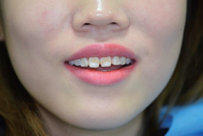
Fig. 1 Preoperative extraoral view.
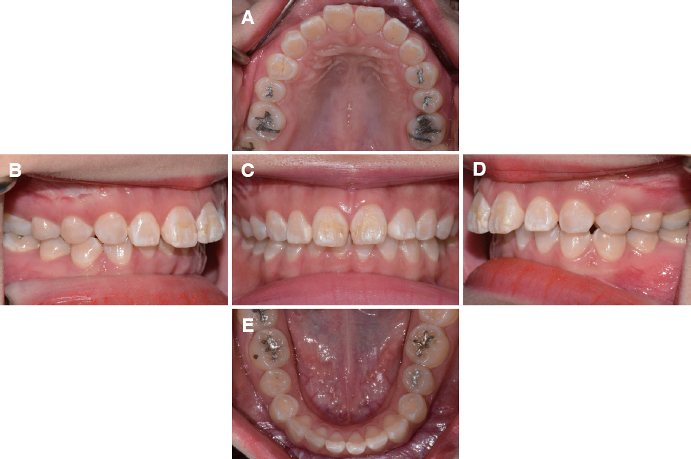
Fig. 2 Preoperative intraoral view. (A) Occlusal view of maxilla, (B) Right lateral view, (C) Frontal view, (D) Left lateral view, (E) Occlusal view of mandible.
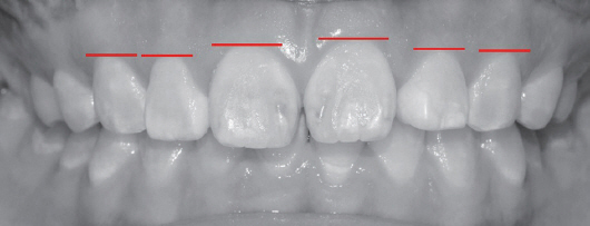
Fig. 3 Zenith of the gingival contour of maxillary anterior teeth.
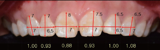
Fig. 4 Analysis of maxillary anterior tooth size and proportion.
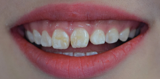
Fig. 5 Extraoral view of smile line.

Fig. 6 Gingivectomy and lip repositioning. (A) Surgical splint for gingivectomy, (B) After gingivectomy and lip repositioning.

Fig. 7 Intraoral and extraoral view 1 month after periodontal surgery. (A) Intraoral view, (B) Extraoral view.

Fig. 8 Diagnostic model fabrication. (A) Face bow transfer, (B) Frontal view of diagnostic model, (C) Lateral view of diagnostic model, (D) Diagnostic model of maxilla, (E) Diagnostic wax-up model of maxilla.

Fig. 9 Intraoral view after tooth preparation and temporary bridge restoration. (A) Tooth preparation, (B) Frontal view of temporary crown, (C) Occlusal view of temporary crown.

Fig. 10 Model scan and coping design. (A) Model scanned image, (B) Superimposed image of definitive model and wax-up diagnostic model, (C) Coping design image.

Fig. 11 Intraoral view of coping and final restoration try in. (A) Intraoral view of pretreatment state, (B) Coping try in, (C) Final restoration try in, (D) 1 year after treatment.
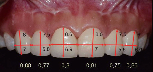
Fig. 12 Analysis of maxillary anterior tooth size and proportion after treatment.

Fig. 13 Analysis of overjet and overbite. (A) Before treatment, (B) After treatment.
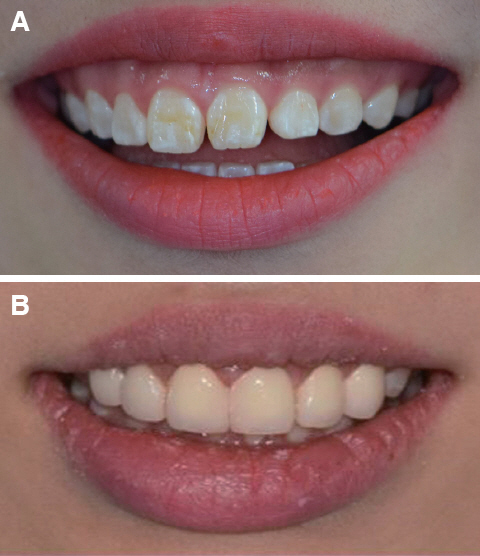
Fig. 14 Analysis of smile line. (A) Before treatment, (B) After treatment.
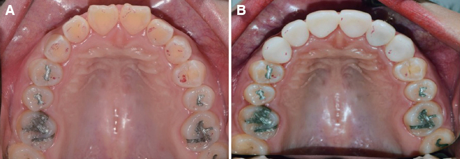
Fig. 15 Intraoral view of occlusion. (A) Occlusion before treatment, (B) Occlusion after treatment.

Fig. 16 Intraoral image of frontal view.
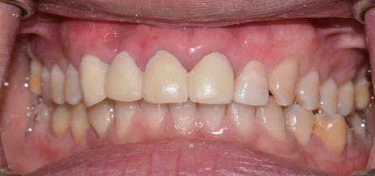
Fig. 17 Intraoral image of frontal view, 1 month after periodontal treatment and temporary restoration.
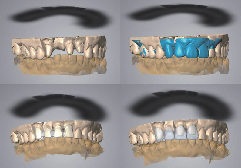
Fig. 18 CAD image of coping design using scanned and superimposed image.
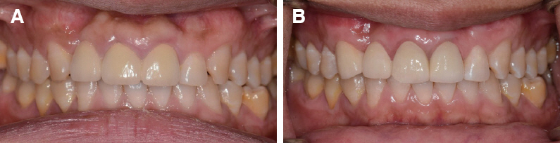
Fig. 19 Intraoral image of frontal view. (A) Before treatment, (B) After treatment.

Fig. 20 Intraoral view of occlusion. (A) Before treatment, (B) After treatment view.
Reference
-
References
1. Magne P, Gallucci GO, Belser UC. Anatomic crown width/length ratios of unworn and worn maxillary teeth in white subjects. J Prosthet Dent. 2003; 89:453–61. DOI: 10.1016/S0022-3913(03)00125-2. PMID: 12806322.2. Dong JK, Jin TH, Cho HW, Oh SC. The esthetics of the smile:a review of some recent studies. Int J Prosthodont. 1999; 12:9–19. PMID: 10196823.3. Radia S, Sherriff M, McDonald F, Naini FB. Relationship between maxillary central incisor proportions and facial proportions. J Prosthet Dent. 2016; 115:741–8. DOI: 10.1016/j.prosdent.2015.10.019. PMID: 26794701.4. Marcuschamer E, Tsukiyama T, Griffin TJ, Arguello E, Gallucci GO, Magne P. Anatomical crown width/length ratios of worn and unworn maxillary teeth in Asian subjects. Int J Periodontics Restorative Dent. 2011; 31:495–503. PMID: 21845244.5. Sherwood IA. Fluorosis varied treatment options. J Conserv Dent. 2010; 13:47–53. DOI: 10.4103/0972-0707.62631. PMID: 20582220. PMCID: PMC2883808.6. Ahmad I. Anterior dental aesthetics:dentofacial perspective. Br Dent J. 2005; 199:81–8. DOI: 10.1038/sj.bdj.4812521. PMID: 16041333.7. Lee EA. Esthetic Crown Lengthening:Contemporary Guidelines for Achieving Ideal Gingival Architecture and Stability. Curr Oral Health Rep. 2017; 4:105–11. DOI: 10.1007/s40496-017-0140-4.8. Al-Harbi F, Ahmad I. A guide to minimally invasive crown lengthening and tooth preparation for rehabilitating pink and white aesthetics. Br Dent J. 2018; 224:228–34. DOI: 10.1038/sj.bdj.2018.121. PMID: 29472662.9. Tjan AH, Miller GD, The JG. Some esthetic factors in a smile. J Prosthet Dent. 1984; 51:24–8. DOI: 10.1016/S0022-3913(84)80097-9. PMID: 6583388.10. Mahn DH. Elimination of a “Gummy Smile” With Crown Lengthening and Lip Repositioning. Compend Contin Educ Dent. 2016; 37:52–5. PMID: 26863221.11. Silva CO, Ribeiro-Júnior NV, Campos TV, Rodrigues JG, Tatakis DN. Excessive gingival display:treatment by a modified lip repositioning technique. J Clin Periodontol. 2013; 40:260–5. DOI: 10.1111/jcpe.12046. PMID: 23278672.12. Miyazaki T, Nakamura T, Matsumura H, Ban S, Kobayashi T. Current status of zirconia restoration. J Prosthodont Res. 2013; 57:236–61. DOI: 10.1016/j.jpor.2013.09.001. PMID: 24140561.
- Full Text Links
-
- Actions
-
Cited
- CITED
-
- Close
- Share
-
- Similar articles
-
- Esthetic improvements through systematic diagnosis and treatment procedures in patients with unesthetic maxillary anterior teeth proportion after orthodontic treatment: Case report
- Anterior esthetic restoration using DSD (digital smile design) for a patient with congenital missing tooth of maxillary central incisor
- Esthetic restoration of malpositioned anterior teeth by tooth shape and gingival contour modification : a clinical report
- Application of Systematic Digital Diagnosis to Create Dental Virtual Patients with Dynamic Occlusion for Esthetic Restoration of Anterior Teeth: A Case Report
- Decoronation and implant restoration of ankylosed tooth resulted from anterior avulsion: A case report
