Case 19: A 65-Year-Old Man With Melena and Hematochezia
- Affiliations
-
- 1Division of Gastroenterology and Hepatology, Department of Internal Medicine, Incheon St. Mary’s Hospital, College of Medicine, The Catholic University of Korea, Seoul, Korea
- 2Department of Internal Medicine, Seoul St. Mary’s Hospital, College of Medicine, The Catholic University of Korea, Seoul, Korea
- 3Division of Gastroenterology and Hepatology, Department of Internal Medicine, Yeouido St. Mary’s Hospital, College of Medicine, The Catholic University of Korea, Seoul, Korea
- 4Division of Gastroenterology and Hepatology, Department of Internal Medicine, Seoul St. Mary’s Hospital, College of Medicine, The Catholic University of Korea, Seoul, Korea
- 5Division of Gastroenterology and Hepatology, Department of Internal Medicine, Bucheon St. Mary’s Hospital, College of Medicine, The Catholic University of Korea, Seoul, Korea
- KMID: 2553309
- DOI: http://doi.org/10.3346/jkms.2024.39.e66
Figure
-

Fig. 1 Endoscopic findings in a patient who came to the emergency room with melena and hematochezia. (A) Normal findings on esophagogastroduodenoscopy, (B) Normal findings on colonoscopy, (C) Eythema and ulcers in the proximal jejunum on capsule endoscopy.
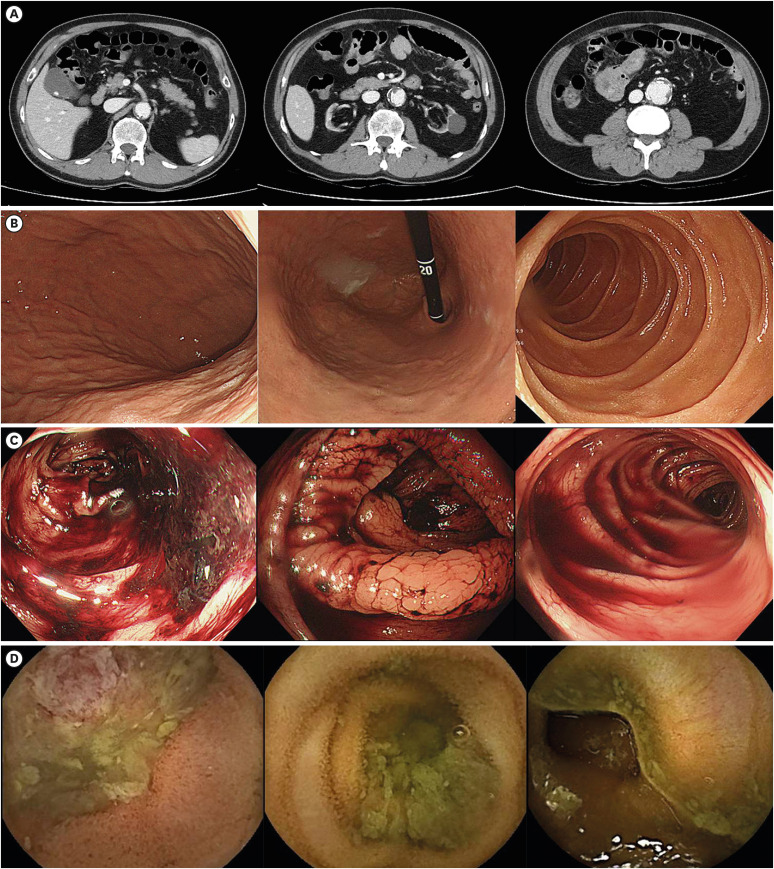
Fig. 2 Imaging and endoscopic findings in a patient admitted with second gastrointestinal bleeding. (A) Abdomen pelvis computed tomography findings showed no lesions that could cause bleeding; gallbladder stone, atrophied both kidneys, and aneurysmal dilatation of abdominal aorta were observed. (B) Normal esophagogastroduodenoscopy findings, (C) Colonoscopy findings covered with hematochezia, (D) Suspected jejunal ulcers on small bowel capsule endoscopy.
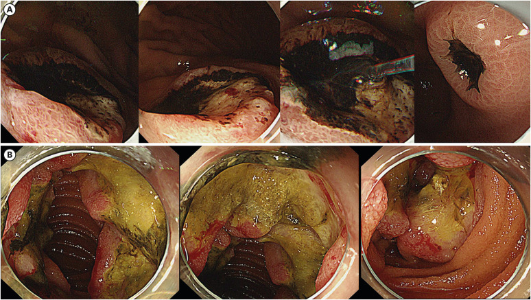
Fig. 3 Ulcerofungating mass observed in the stomach and jejunum. (A) Stomach and (B) proximal jejunum.

Fig. 4 Pathological findings. (A) H&E stain (×200). (B) Positive for CD20, a B cell marker. (C) High Ki-67 proliferation index.H&E = hematoxylin and eosin.
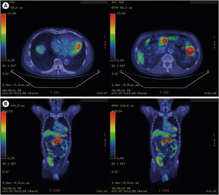
Fig. 5 Lymphoma of the stomach and small bowel on positron emission tomography and computed tomography. (A) Axial view, (B) Coronal view.There is intense multifocal FDG uptake in the stomach (SULmax: 17.9). FDG uptake is also seen in the jejunal loop of the left upper quadrant and the small bowel loop of the right-side abdomen. There is intense nodular FDG uptake in the mesentery. Aneurysmal dilatation is seen in the abdominal aorta.FDG = fluorodeoxyglucose, SULmax = standardized uptake value normalized by lean body mass for maximum.
Reference
-
1. Xie Y, Pittaluga S, Jaffe ES. The histological classification of diffuse large B-cell lymphomas. Semin Hematol. 2015; 52(2):57–66. PMID: 25805585.2. Kim BS, Li BT, Engel A, Samra JS, Clarke S, Norton ID, et al. Diagnosis of gastrointestinal bleeding: a practical guide for clinicians. World J Gastrointest Pathophysiol. 2014; 5(4):467–478. PMID: 25400991.3. Kim SE, Kim HJ, Koh M, Kim MC, Kim JS, Nam JH, et al. A practical approach for small bowel bleeding. Clin Endosc. 2023; 56(3):283–289. PMID: 37165534.4. Teshima CW, Kuipers EJ, van Zanten SV, Mensink PB. Double balloon enteroscopy and capsule endoscopy for obscure gastrointestinal bleeding: an updated meta-analysis. J Gastroenterol Hepatol. 2011; 26(5):796–801. PMID: 21155884.5. Shim KN, Moon JS, Chang DK, Do JH, Kim JH, Min BH, et al. Guideline for capsule endoscopy: obscure gastrointestinal bleeding. Clin Endosc. 2013; 46(1):45–53. PMID: 23423225.6. Ghimire P, Wu GY, Zhu L. Primary gastrointestinal lymphoma. World J Gastroenterol. 2011; 17(6):697–707. PMID: 21390139.7. Ahn JY. Gastrointestinal tract lymphoma. Korean J Helicobacter Up Gastrointest Res. 2022; 22(1):18–28.8. Abbas F, El Kossi M, Shaheen IS, Sharma A, Halawa A. Post-transplantation lymphoproliferative disorders: current concepts and future therapeutic approaches. World J Transplant. 2020; 10(2):29–46. PMID: 32226769.9. Taylor AL, Watson CJ, Bradley JA. Immunosuppressive agents in solid organ transplantation: mechanisms of action and therapeutic efficacy. Crit Rev Oncol Hematol. 2005; 56(1):23–46. PMID: 16039869.10. Francis A, Johnson DW, Teixeira-Pinto A, Craig JC, Wong G. Incidence and predictors of post-transplant lymphoproliferative disease after kidney transplantation during adulthood and childhood: a registry study. Nephrol Dial Transplant. 2018; 33(5):881–889. PMID: 29342279.
- Full Text Links
-
- Actions
-
Cited
- CITED
-
- Close
- Share
-
- Similar articles
-
- A Case of Ischemic Colitis Results from the Bleeding on Gastric Dieulafoy Lesions
- A Case of Arteriovenous Malformation of the Pancreas
- A Case of Jejunal Leiomyoma Presenting with Recurrent Lower Gastrointestinal Bleeding
- Primary Aortoduodenal Fistula Causes Massive Melena: A Case Report
- A Case of traumatic Pancreatic Pseudoaneurysm
