Huge Chondroblastoma of the Talus: A Case Report
- Affiliations
-
- 1Department of Orthopedic Surgery, Dankook University Hospital, Cheonan, Korea
- KMID: 2553009
- DOI: http://doi.org/10.14193/jkfas.2023.27.4.154
Abstract
- Chondroblastoma is a rare benign cartilaginous neoplasm that accounts for 1% of bone tumors and is common in the epiphysis of the long bones. The condition is rarely found in the talus bone (4% of cases). This paper reports a 15-year-old male patient treated for a talus bone lesion discovered incidentally on imaging. Excisional biopsy, curettage, and an autobone and allobone graft were performed, with good results.
Keyword
Figure
-
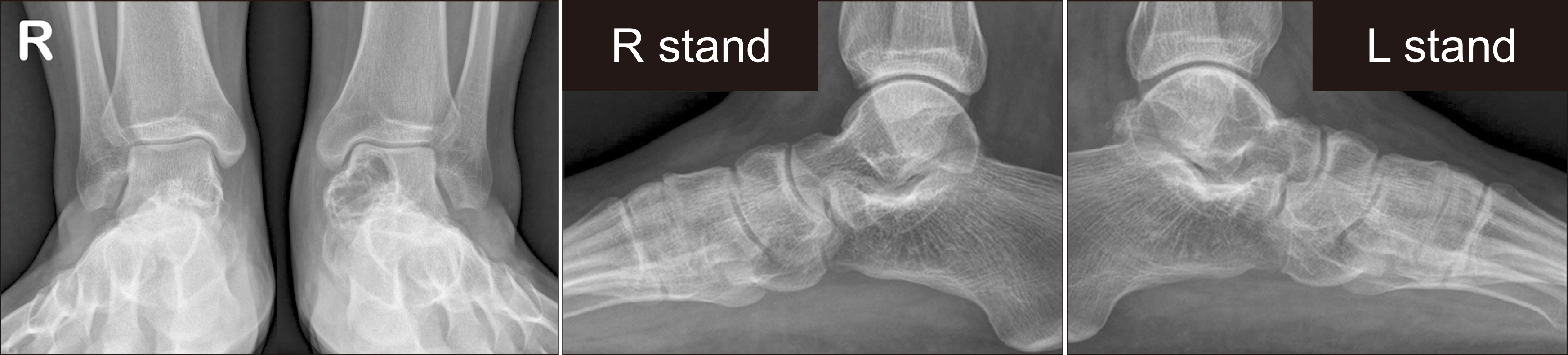
Figure 1 Preoperative plain radiography. Multi-lobulated huge cystic lesion is found on the left talar body and neck.

Figure 2 Preoperative magnetic resonance imaging in the axial (A), sagittal (B) and coronal (C) plane images. Multi-lobulated huge cystic lesion is found on the left talar body and neck. Heterogeneous intensity in the cystic lesion and fluid-fluid level are found, these findings suggest secondary aneurysmal bone cyst change.
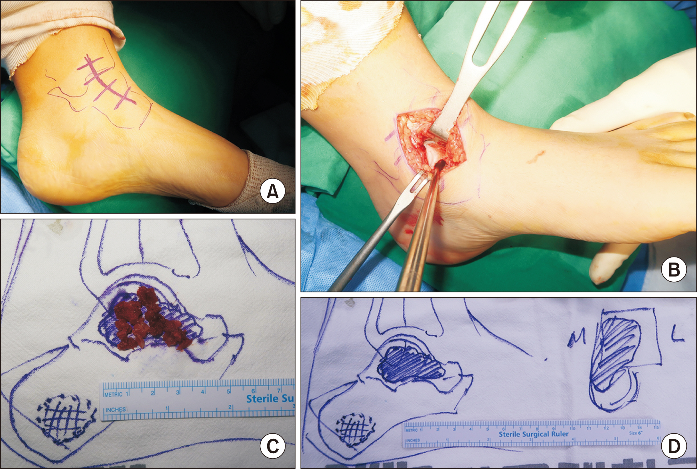
Figure 3 Intraoperative clinical photos. (A) Skin incision. (B) Curettage thru the opened cortex of anterolateral talar body area. (C) Removed mass lesion from the cavity. (D) Schematic drawing of the mass lesion on the sagittal and axial plane.

Figure 4 Specimen pathologic images. Hematoxylin and Eosin (H&E) stain preparation. Multinucleated giant cell with a chondroid matrix, surrounded by dystrophic calcification are revealed as ‘chicken wire appearance’. (A) Magnification ×200, (B) magnification ×400.

Figure 5 Postoperative plain radiography after 7 months from surgery. Radiopacity is increased in the cystic lesion in the lateral (A) and anteroposterior (B) views.

Figure 6 Postoperative three-dimensional computed tomography scan images after 7 months from surgery in the axial (A), sagittal (B) and coronal (C) plane images. The findings indicate that there is newly formed bone tissue within the cyst, and an increase in cortical bone thickness.
Reference
-
References
1. Sl B, A S, J M, R B. 2020; Tumours of the talus - a pictorial review. J Clin Orthop Trauma. 11:410–6. doi: 10.1016/j.jcot.2020.03.021. DOI: 10.1016/j.jcot.2020.03.021. PMID: 32405200. PMCID: PMC7211828.
Article2. Jagiella-Lodise O, McAleese T, Curtin M, Molloy A, Walsh J. 2023; Recurrent chondroblastoma of the talus: a case report and literature review of recurrent lesions in the foot and ankle. Int J Surg Case Rep. 106:108192. doi: 10.1016/j.ijscr.2023.108192. DOI: 10.1016/j.ijscr.2023.108192. PMID: 37105027. PMCID: PMC10164886.
Article3. Angelini A, Arguedas F, Varela A, Ruggieri P. 2018; Chondroblastoma of the foot: 40 cases from a single institution. J Foot Ankle Surg. 57:1105–9. doi: 10.1053/j.jfas.2018.05.005. DOI: 10.1053/j.jfas.2018.05.005. PMID: 30368424.
Article4. Ningegowda RV, Subramanian K, Suresh I. 2013; Chondroblastoma of the talus. J Foot Ankle Surg. 52:673–7. doi: 10.1053/j.jfas.2013.02.020. DOI: 10.1053/j.jfas.2013.02.020. PMID: 23540757.
Article5. Springfield DS, Capanna R, Gherlinzoni F, Picci P, Campanacci M. 1985; Chondroblastoma. A review of seventy cases. J Bone Joint Surg Am. 67:748–55. doi: 10.2106/00004623-198567050-00009. DOI: 10.2106/00004623-198567050-00009. PMID: 3997927.
Article6. Focaccia M, Gambarotti M, Hakim R, Paioli A, Cesari M, Spazzoli B, et al. 2021; Chondroblastoma's lung metastases treated with denosumab in pediatric patient. Cancer Res Treat. 53:279–82. doi: 10.4143/crt.2020.384. DOI: 10.4143/crt.2020.384. PMID: 32777878. PMCID: PMC7812007.
Article7. Park JS, Suh JS, Choi JY. 2019; Chondroblastoma of the talus mimicking an aneurysmal bone cyst: a case report. J Korean Foot Ankle Soc. 23:31–4. doi: 10.14193/jkfas.2019.23.1.31. DOI: 10.14193/jkfas.2019.23.1.31.
Article
