Chondroblastoma of the Talus Mimicking an Aneurysmal Bone Cyst: A Case Report
- Affiliations
-
- 1Department of Orthopedic Surgery, Ilsan Paik Hospital, Inje University College of Medicine, Goyang, Korea. osddr8151@paik.ac.kr
- KMID: 2440582
- DOI: http://doi.org/10.14193/jkfas.2019.23.1.31
Abstract
- Chondroblastoma is a rare benign tumor that produces giant cells and cartilage matrix. The tumor occurs in people between 10 and 25 years with slightly higher incidence in males. The condition occurs in the proximal epiphysis of the tibia and humerus, distal epiphysis of the femur, but its occurrence in the talus is relatively rare, accounting for 4% of the total number of chondroblastoma cases. Chondroblastoma is often misdiagnosed as a primary aneurysmal bone cyst, giant cell tumor, chondromyxoid, and lesion of a secondary aneurysmal bone cyst by fibrous dysplasia. The most commonly used surgical method for chondroblastoma is broad curettage with bone grafting. In general, an aneurysmal bone cyst is associated with a second degree chondroblastoma, which is approximately 20%. Chondroblastoma of the talus and secondary aneurysmal bone cysts can be misdiagnosed as primary aneurysmal bone cysts. This paper reports a case of a young male patient with chondroblastoma of the talus, which was initially misdiagnosed as an aneurysmal bone cyst with involvement of the talo-navicular joint.
Keyword
MeSH Terms
Figure
-
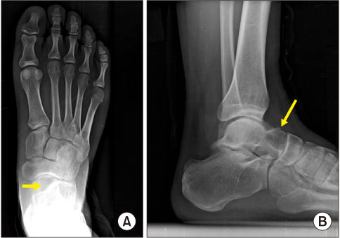
Figure 1 Preoperative standing foot anteroposterior (A) and lateral (B) radiographs present well circumscribed osteolytic lesion (arrows) with a sclerotic rim involving talo-navicular joint in the talar head.
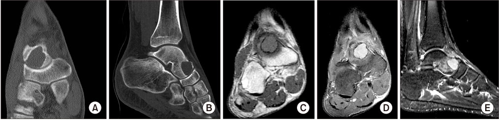
Figure 2 Preoperative computed tomographic images (A, B) show well marginated bony cyst with a preserved articular cartilage at talo-navicular joint. A low on T1-weighted coronal image (C), high signal intensity on T2-weighted coronal image (D), and T2-weighted sagittal image (E) on magnetic resonance imaging were shown.
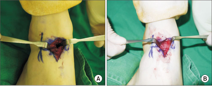
Figure 3 (A) A 1×1 cm2 cortical window was made on the dorsal side of talar head with an osteotome to approach the intraosseous mass lesion. (B) An aspiration of hemorrhagic fluid and removal of sclerotic wall with curette were conducted. Mixed bone graft was filled in the space.

Figure 4 (A) A removed sclerotic wall tissue was sent for histopathologic confirmation. (B) Multinucleated giant cell with a background of chondroid matrix compatible to chondroblastoma was noted. (H&E stain, ×200) Dystrophic calcification surrounding individual cells, giving the classic “chicken wire appearance” was not detected.
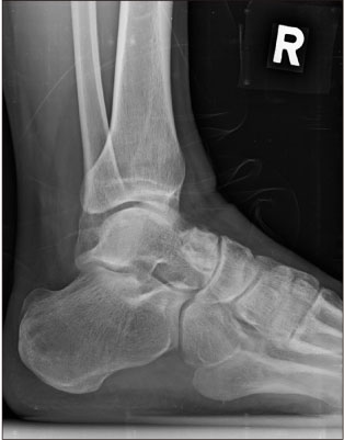
Figure 5 A consolidation of bone graft site was noted on the postoperative 1 year radiography.
Cited by 3 articles
-
Treatment of a Huge Aneurysmal Bone Cyst of the Talus through a Segmental Bone Graft of the Tricortical Bone: A Case Report
Seung-Jin Lee, Hyobeom Lee, Gab-Lae Kim, Donghyeon Kim
J Korean Foot Ankle Soc. 2021;25(4):185-189. doi: 10.14193/jkfas.2021.25.4.185.Surgical Treatment of Talar Chondroblastoma via Partial Posterior Medial Malleolar Osteotomy: A Case Report
Oh Jun Yu, Jin Soo Suh, Han Sung Kim, Jun Young Choi
J Korean Foot Ankle Soc. 2023;27(2):75-78. doi: 10.14193/jkfas.2023.27.2.75.Huge Chondroblastoma of the Talus: A Case Report
Sung Hyun Yoon, Hyun-woo Park
J Korean Foot Ankle Soc. 2023;27(4):154-157. doi: 10.14193/jkfas.2023.27.4.154.
Reference
-
1. Heck RK Jr, Toy PC. Benign/aggressive tumors of bone. In : Azar FM, Beaty JH, Canale ST, editors. Campbell's operative orthopaedics. 13th ed. Philadelphia: Elsevier;2017. p. 925–931.2. Ningegowda RV, Subramanian K, Suresh I. Chondroblastoma of the talus. J Foot Ankle Surg. 2013; 52:673–677.
Article3. Ryu JJ, Kim W, Lee JS, Kim YK, Lee HS, Seo SG. Combined autograft and bone cement for painful chondroblastoma: a case report. J Foot Ankle Surg. 2018; 57:396–400.
Article4. Kudo T, Okada K, Hirano Y, Sageshima M. Chondroblastoma of a metacarpal bone mimicking an aneurysmal bone cyst: a case report and a review of the literature. Tohoku J Exp Med. 2001; 194:251–257.
Article5. Springfield DS, Capanna R, Gherlinzoni F, Picci P, Campanacci M. Chondroblastoma. A review of seventy cases. J Bone Joint Surg Am. 1985; 67:748–755.
Article6. Ramappa AJ, Lee FY, Tang P, Carlson JR, Gebhardt MC, Mankin HJ. Chondroblastoma of bone. J Bone Joint Surg Am. 2000; 82:1140–1145.
Article7. Bloem JL, Mulder JD. Chondroblastoma: a clinical and radiological study of 104 cases. Skeletal Radiol. 1985; 14:1–9.
Article8. Zhang K, Gao Y, Dai H, Zhang S, Li G, Yu B. Chondroblastoma of the talus: a case report and literature review. J Foot Ankle Surg. 2012; 51:262–265.
Article9. Anderson AF, Ramsey JR. Chondroblastoma of the talus treated with osteochondral autograft transfer from the lateral femoral condyle. Foot Ankle Int. 2003; 24:283–287.
Article10. Sterling G, Wilson A. Chondroblastoma of the talus: a case report. J Foot Ankle Surg. 2002; 41:178–182.
Article11. Nolan DJ, Middlemiss H. Chondroblastoma of bone. Clin Radiol. 1975; 26:343–350.
Article12. Huvos AG, Marcove RC. Chondroblastoma of bone. A critical review. Clin Orthop Relat Res. 1973; (95):300–312.
Article13. Sharma S, Gupta P, Sharma S, Singh M, Singh D. Primary aneurysmal bone cyst of talus. J Res Med Sci. 2012; 17:1192–1194.14. Sherman RS, Uzel AR. Benign chondroblastoma of bone; its roentgen diagnosis. Am J Roentgenol Radium Ther Nucl Med. 1956; 76:1132–1140.15. Lehner B, Witte D, Weiss S. Clinical and radiological long-term results after operative treatment of chondroblastoma. Arch Orthop Trauma Surg. 2011; 131:45–52.
Article16. Turcotte RE, Kurt AM, Sim FH, Unni KK, McLeod RA. Chondroblastoma. Hum Pathol. 1993; 24:944–949.
Article17. Sailhan F, Chotel F, Parot R. SOFOP. Chondroblastoma of bone in a pediatric population. J Bone Joint Surg Am. 2009; 91:2159–2168.
Article
- Full Text Links
-
- Actions
-
Cited
- CITED
-
- Close
- Share
-
- Similar articles
-
- Huge Chondroblastoma of the Talus: A Case Report
- Treatment of a Huge Aneurysmal Bone Cyst of the Talus through a Segmental Bone Graft of the Tricortical Bone: A Case Report
- Aneurysmal Bone Cyst in Clavicle: Report of A Case
- Chondroblastoma with Secondary Aneurysmal Bone Cyst in the Sphenoid Sinus: A Case Report
- A Case of Aneurysmal Bone Cyst in the Skull Base
