Distal Soft Tissue Procedure in Hallux Valgus Deformity: Comparison of Modified Mcbride Procedure and Trans-Articular Approach
- Affiliations
-
- 1Department of Orthopedic Surgery, Kangbuk Samsung Hospital, Sungkyunkwan University School of Medicine, Seoul, Korea
- KMID: 2552991
- DOI: http://doi.org/10.14193/jkfas.2023.27.4.123
Abstract
- Purpose
“Hallux valgus” is a common disease encountered in clinical practice and is accompanied by foot deformities. Conservative treatment is commonly used in the early stages of hallux valgus. On the other hand, surgical treatment often becomes necessary as the deformity progresses. Surgical treatments involve various osteotomy methods or joint fusion procedures combined with soft tissue release, and outcomes from these surgical treatments are generally favorable. This study compared two soft tissue release techniques in the hallux region.
Materials and Methods
This study conducted a retrospective cohort study on 48 participants who underwent surgical treatment for hallux valgus at a single institution from March 1, 2018, to March 31, 2023. A scarf osteotomy was performed in all cases, and the “Modified Mcbride procedure” or “Trans-articular approach” was done for soft tissue release. Hallux valgus angle (HVA), intermetatarsal angle (IMA), and the degree of subluxation of the lateral sesamoid were measured through simple foot radiographs taken before surgery and one year after surgery.
Results
In the Modified Mcbride procedure group, HVA, IMA, and the sesamoid position grade decreased from 34.94º to 9.98º, 15.64º to 5.44º, and 2.47 to 0.44, respectively. In the trans-articular approach group, HVA, IMA, and the sesamoid position grade decreased from 33.42º to 7.34º, 15.06º to 6.03º, and 2.17 to 0.58, respectively. There was no significant difference in these changes between the preoperative and one-year postoperative measurements for both techniques (p-value>0.05).
Conclusion
A radiological assessment of soft tissue release through the Modified Mcbride procedure and trans-articular approach in hallux valgus did not show significant differences. Therefore, both surgical techniques can be considered in the distal soft tissue release for a hallux valgus correction.
Figure
-
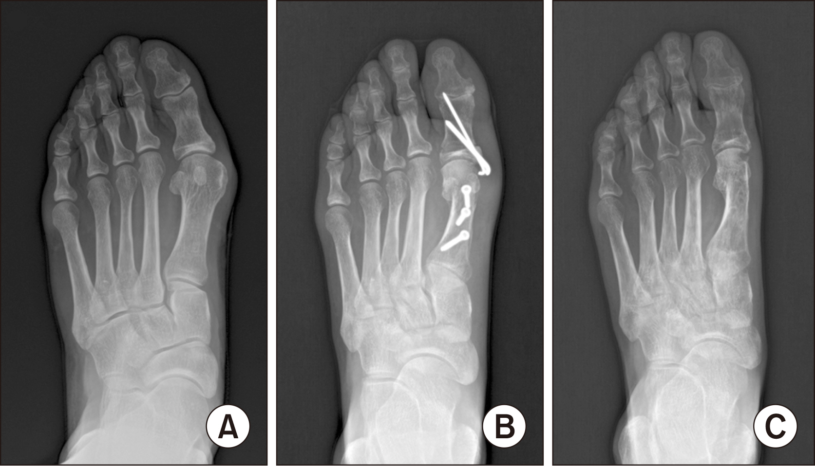
Figure 1 Radiographs showing the pre-operative deformity (A) and the immediate post-operative correction through modified Mcbride procedure and Scarf osteotomy, Akin osteotomy (B). As well as Fig. 1, final follow up radiographs (C) shows well maintained 1st metatarsophalangeal joint alignment.

Figure 2 Radiographs showing the pre-operative deformity (A) and the immediate post-operative correction through the trans-articular distal soft tissue release and Scarf osteotomy (B). Last follow up radiographs (C) shows maintained neutral 1st metatarsophalangeal joint alignment.
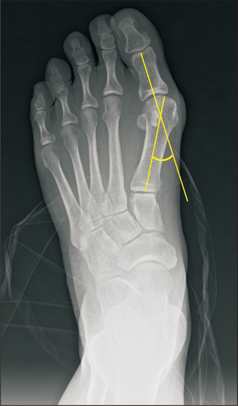
Figure 3 Radiographic measurements of the hallux valgus angle; angle between two yellow line.
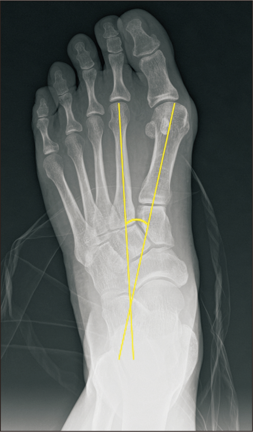
Figure 4 Radiographic measurements of the intermetatarsal angle; angle between two yellow line.

Figure 5 Radiographs showing the sesamoid subluxation grade from normal to severe. (A) Grade 0 shows ≤25% of the lateral sesamoid exposed lateral to the lateral border of 1st metatarsal head, (B) grade 1 shows >25% and ≤50% of lateral sesamoid exposed as same way, (C) grade 2 shows >50% and ≤75% of lateral sesamoid exposed as same way, (D) grade 3 shows >75% of lateral sesamoid exposed as same way.

Figure 6 Distal soft tissue release through the trans-articular approach. With the assistance of C-arm intensifier, release of adductor hallucis tendon and lateral capsulotomy were done.
Reference
-
References
1. Nix S, Smith M, Vicenzino B. 2010; Prevalence of hallux valgus in the general population: a systematic review and meta-analysis. J Foot Ankle Res. 3:21. doi: 10.1186/1757-1146-3-21. DOI: 10.1186/1757-1146-3-21. PMID: 20868524. PMCID: PMC2955707.
Article2. Kakwani M, Kakwani R. 2021; Current concepts review of hallux valgus. J Arthrosc Jt Surg. 8:222–30. doi: 10.1016/j.jajs.2021.04.006. DOI: 10.1016/j.jajs.2021.04.006.
Article3. Saro C, Andrén B, Wildemyr Z, Felländer-Tsai L. 2007; Outcome after distal metatarsal osteotomy for hallux valgus: a prospective randomized controlled trial of two methods. Foot Ankle Int. 28:778–87. doi: 10.3113/FAI.2007.0778. DOI: 10.3113/FAI.2007.0778. PMID: 17666169.
Article4. Thordarson D, Ebramzadeh E, Moorthy M, Lee J, Rudicel S. 2005; Correlation of hallux valgus surgical outcome with AOFAS forefoot score and radiological parameters. Foot Ankle Int. 26:122–7. doi: 10.1177/107110070502600202. Erratum in: Foot Ankle Int. 2005;26:table of contents. DOI: 10.1177/107110070502600202. PMID: 15737253.
Article5. Dreeben S, Mann RA. 1996; Advanced hallux valgus deformity: long-term results utilizing the distal soft tissue procedure and proximal metatarsal osteotomy. Foot Ankle Int. 17:142–4. doi: 10.1177/107110079601700304. DOI: 10.1177/107110079601700304. PMID: 8919617.
Article6. Easley ME, Kiebzak GM, Davis WH, Anderson RB. 1996; Prospective, randomized comparison of proximal crescentic and proximal chevron osteotomies for correction of hallux valgus deformity. Foot Ankle Int. 17:307–16. doi: 10.1177/107110079601700603. DOI: 10.1177/107110079601700603. PMID: 8791076.
Article7. Pochatko DJ, Schlehr FJ, Murphey MD, Hamilton JJ. 1994; Distal chevron osteotomy with lateral release for treatment of hallux valgus deformity. Foot Ankle Int. 15:457–61. doi: 10.1177/107110079401500901. DOI: 10.1177/107110079401500901. PMID: 7820236.
Article8. Resch S, Stenström A, Reynisson K, Jonsson K. 1994; Chevron osteotomy for hallux valgus not improved by additional adductor tenotomy. A prospective, randomized study of 84 patients. Acta Orthop Scand. 65:541–4. doi: 10.3109/17453679409000910. DOI: 10.3109/17453679409000910. PMID: 7801759.
Article9. Silver D. 1923; The operative treatment of hallux valgus. J Bone Joint Surg. 5:225–32.10. Schneider W, Aigner N, Pinggera O, Knahr K. 2004; Chevron osteotomy in hallux valgus. Ten-year results of 112 cases. J Bone Joint Surg Br. 86:1016–20. doi: 10.1302/0301-620x.86b7.15108. DOI: 10.1302/0301-620X.86B7.15108. PMID: 15446530.11. Stamatis ED, Huber MH, Myerson MS. 2004; Transarticular distal soft-tissue release with an arthroscopic blade for hallux valgus correction. Foot Ankle Int. 25:13–8. doi: 10.1177/107110070402500104. DOI: 10.1177/107110070402500104. PMID: 14768959.
Article12. Lee WC, Kim YM. 2007; Correction of hallux valgus using lateral soft-tissue release and proximal Chevron osteotomy through a medial incision. J Bone Joint Surg Am. 89 Suppl 3:82–9. doi: 10.2106/JBJS.G.00483. DOI: 10.2106/JBJS.G.00483. PMID: 17908874.
Article13. Agrawal Y, Desai A, Mehta J. 2011; Lateral sesamoid position in hallux valgus: correlation with the conventional radiological assessment. Foot Ankle Surg. 17:308–11. doi: 10.1016/j.fas.2011.01.001. DOI: 10.1016/j.fas.2011.01.001. PMID: 22017908.
Article14. Chen YJ, Hsu RW, Shih HN, Huang TJ, Hsu KY. 1996; Distal chevron osteotomy with intra-articular lateral soft-tissue release for treatment of moderate to severe hallux valgus deformity. J Formos Med Assoc. 95:776–81.15. Leventen EO. 1990; The Chevron procedure. Orthopedics. 13:973–6. doi: 10.3928/0147-7447-19900901-09. DOI: 10.3928/0147-7447-19900901-09. PMID: 2235745.
Article16. Fraissler L, Konrads C, Hoberg M, Rudert M, Walcher M. 2016; Treatment of hallux valgus deformity. EFORT Open Rev. 1:295–302. doi: 10.1302/2058-5241.1.000005. DOI: 10.1302/2058-5241.1.000005. PMID: 28660074. PMCID: PMC5467633.
Article17. Ko KR, Sung KS. 2017; Corrective osteotomies in hallux valgus. J Korean Foot Ankle Soc. 21:43–9. doi: 10.14193/jkfas.2017.21.2.43. DOI: 10.14193/jkfas.2017.21.2.43.
Article18. Brand JC Jr, Smith RW. 1991; Rupture of the flexor hallucis longus after hallux valgus surgery: case report and comments on technique for adductor release. Foot Ankle. 11:407–10. doi: 10.1177/107110079101100614. DOI: 10.1177/107110079101100614. PMID: 1894238.
Article19. Waldecker U. 2004; Lateral release in hallux valgus surgery: comparison of two approaches. Foot Ankle Surg. 10:195–9. doi: 10.1016/j.fas.2004.09.003. DOI: 10.1016/j.fas.2004.09.003.
Article20. Panchbhavi VK, Rapley J, Trevino SG. 2011; First web space soft tissue release in bunion surgery: functional outcomes of a new technique. Foot Ankle Int. 32:257–61. doi: 10.3113/FAI.2011.0257. Erratum in: Foot Ankle Int. 2011;32:vi. DOI: 10.3113/FAI.2011.0257. PMID: 21477544.
Article21. Park YB, Lee KB, Kim SK, Seon JK, Lee JY. 2013; Comparison of distal soft-tissue procedures combined with a distal chevron osteotomy for moderate to severe hallux valgus: first web-space versus transarticular approach. J Bone Joint Surg Am. 95:e158. doi: 10.2106/JBJS.L.01017. DOI: 10.2106/JBJS.L.01017. PMID: 24196470.
Article22. Li X, Liu D, Wang X. 2021; Correlative study between the sesamoid bones under the head of the first metatarsal and the development of hallux valgus determined with radiographs. Research Square [Preprint]. cited 2023 Sep 14. Available from: https://doi.org/10.21203/rs.3.rs-411401/v1. DOI: 10.21203/rs.3.rs-411401/v1.
Article23. Ahn JY, Lee HS, Chun H, Kim JS, Seo DK, Choi YR, et al. 2013; Comparison of open lateral release and transarticular lateral release in distal chevron metatarsal osteotomy for hallux valgus correction. Int Orthop. 37:1781–7. doi: 10.1007/s00264-013-2023-1. DOI: 10.1007/s00264-013-2023-1. PMID: 23917851. PMCID: PMC3764286.
Article
- Full Text Links
-
- Actions
-
Cited
- CITED
-
- Close
- Share
-
- Similar articles
-
- Comparison of Proximal and Modified Distal Chevron Osteotomy for the Treatment of Moderate to Severe Hallux Valgus Deformity
- Corrective Osteotomies in Hallux Valgus
- Chevron Osteotomy as the Treatment of Moderate to Severe Hallux Valgus Deformity
- A Comparison of Operative Treatment of Hallux Valgus with a Proximal Metatarsal Osteotomy and with a Modified Chevron Osteotomy
- Treatment of Hallux Valgus with Modified McBride Procedure and Proximal Metatarsal Crescentic Osteotomy
