White Matter Structural Connectivity Associated With Visual Field Recovery After Stroke
- Affiliations
-
- 1Asan Institute for Life Sciences, Asan Medical Center, Seoul, Korea
- 2Department of Neurology, Asan Medical Center, University of Ulsan College of Medicine, Seoul, Korea
- 3Nunaps Inc., Seoul, Korea
- KMID: 2551355
- DOI: http://doi.org/10.5853/jos.2023.02222
Figure
-
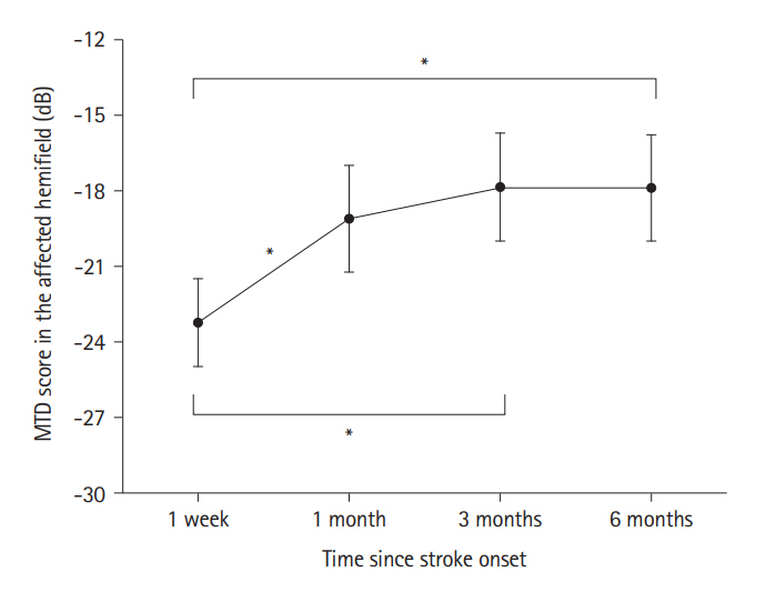
Figure 1. Changes in the MTD scores of the affected hemifield. The MTD scores in the affected hemifield were compared at 1 week, 1, 3, and 6 months after stroke onset using one-way repeated-measures analysis of variance. For post hoc analyses, MTD scores were compared between six pairs of four time points using a Bonferroni-corrected paired t-test with 6 comparisons. The black circles indicate the mean of the MTD scores and the error bars indicate the standard error of the mean. MTD, mean total deviation. *Statistical significance using Bonferroni-corrected paired t-test.
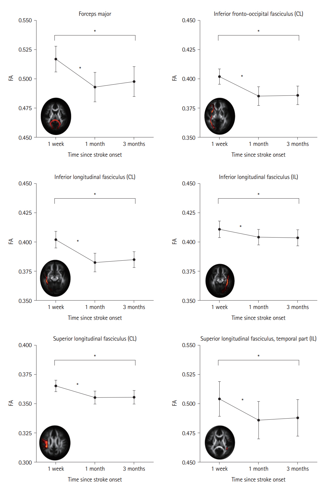
Figure 2. Changes in the FA of the white matter tracts. FA changes in the nine white matter tracts connecting the visual cortex were compared between 1 week, 1 month, and 3 months after stroke onset using one-way repeated-measures analysis of variance. For post hoc analyses, the FA values in the seven tracts with significant changes were compared between three pairs of three time points using the Bonferroni-corrected paired t-tests, with 21 comparisons. The six tracts showed significant temporal changes in the Bonferroni-corrected paired t-tests, as indicated by * symbols. The black circles indicate the mean FA values, and the error bars indicate the standard error of the mean. FA, fractional anisotropy; CL, contralesional; IL, ipsilesional.
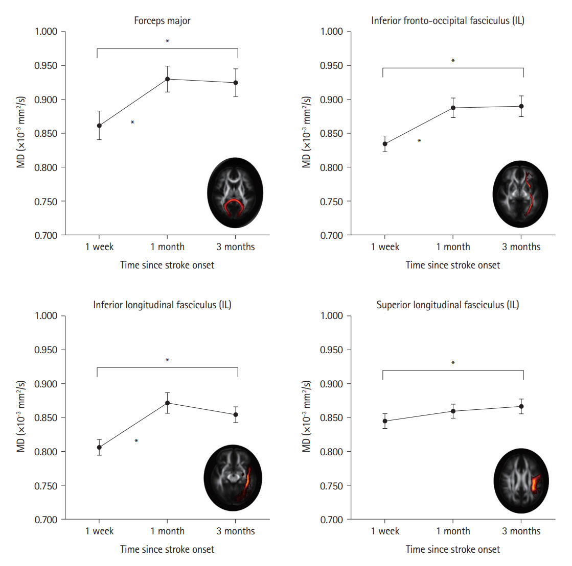
Figure 3. Changes in the MD of the white matter tracts. The MD changes in the nine white matter tracts connecting the visual cortex were compared at 1 week, 1 month, and 3 months after stroke onset using one-way repeated-measures analysis of variance. For post hoc analyses, the MD values in the four tracts with significant changes were compared between three pairs of three time points using the Bonferroni-corrected paired t-tests, with 12 comparisons. The four tracts showed significant temporal changes using Bonferroni-corrected paired t-tests, as indicated by the * symbols. The black circles indicate the mean MD values, and the error bars indicate the standard error of the mean. IL, ipsilesional; MD, mean diffusivity.
Reference
-
References
1. Rowe FJ, Hepworth LR, Howard C, Hanna KL, Cheyne CP, Currie J. High incidence and prevalence of visual problems after acute stroke: an epidemiology study with implications for service delivery. PLoS One. 2019; 14:e0213035.2. Urbanski M, Coubard OA, Bourlon C. Visualizing the blind brain: brain imaging of visual field defects from early recovery to rehabilitation techniques. Front Integr Neurosci. 2014; 8:74.3. Kang DW, Kim D, Chang LH, Kim YH, Takahashi E, Cain MS, et al. Structural and functional connectivity changes beyond visual cortex in a later phase of visual perceptual learning. Sci Rep. 2018; 8:5186.4. Kim YH, Kang DW, Kim D, Kim HJ, Sasaki Y, Watanabe T. Real-time strategy video game experience and visual perceptual learning. J Neurosci. 2015; 35:10485–10492.5. Kim YH, Cho AH, Kim D, Kim SM, Lim HT, Kwon SU, et al. Early functional connectivity predicts recovery from visual field defects after stroke. J Stroke. 2019; 21:207–216.6. Zhang X, Kedar S, Lynn M, Newman N, Biousse V. Homonymous hemianopias: clinical–anatomic correlations in 904 cases. Neurology. 2006; 66:906–910.7. García AO, Brambati SM, Desautels A, Marcotte K. Timing stroke: a review on stroke pathophysiology and its influence over time on diffusion measures. J Neurol Sci. 2022; 441:120377.8. Kern KC, Wright CB, Leigh R. Global changes in diffusion tensor imaging during acute ischemic stroke and post-stroke cognitive performance. J Cereb Blood Flow Metab. 2022; 42:1854–1866.9. Pinter D, Gattringer T, Fandler-Höfler S, Kneihsl M, Eppinger S, Deutschmann H, et al. Early progressive changes in white matter integrity are associated with stroke recovery. Transl Stroke Res. 2020; 11:1264–1272.10. Ortibus E, Verhoeven J, Sunaert S, Casteels I, De Cock P, Lagae L. Integrity of the inferior longitudinal fasciculus and impaired object recognition in children: a diffusion tensor imaging study. Dev Med Child Neurol. 2012; 54:38–43.
- Full Text Links
-
- Actions
-
Cited
- CITED
-
- Close
- Share
-
- Similar articles
-
- Stroke Connectome and Its Implications for Cognitive and Behavioral Sequela of Stroke
- Anatomical Brain Connectivity Map of Korean Children
- Early Functional Connectivity Predicts Recovery from Visual Field Defects after Stroke
- Generation of Mouse Basal Ganglia Diffusion Tractography Using 9.4T MRI
- The Effect of TNF-alpha rs1800629 Polymorphism on White Matter Structures and Memory Function in Patients With Schizophrenia: A Pilot Study
