Prosthetic rehabilitation in a Class III malocclusion patient with increasing occlusal vertical dimension
- Affiliations
-
- 1Department of Prosthodontics and Research Institute of Oral Science, College of Dentistry, Gangneung-Wonju National University, Gangneung, Republic of Korea
- KMID: 2549280
- DOI: http://doi.org/10.14368/jdras.2023.39.3.133
Abstract
- Class III malocclusion with mandibular protrusion can be divided into skeletal and pseudo malocclusion due to tooth displacement. For skeletal malocclusion, favorable treatment results can be obtained by establishing an appropriate vertical and horizontal intermaxillary relationship in order to secure a restoration space and obtain aesthetic and functional results. In this case, complete mouth rehabilitation was performed using an implant and a fixed prosthesis in a patient with mandibular protrusion and anterior teeth wear and reduced occlusal vertical dimension. After cast analysis and digital diagnosis, a provisional restoration with increased vertical dimension was fabricated to secure posterior support and evaluate stable centric occlusion. With the definitive prosthesis reflecting the provisional restoration, favorable function and aesthetics were obtained.
Keyword
Figure
-
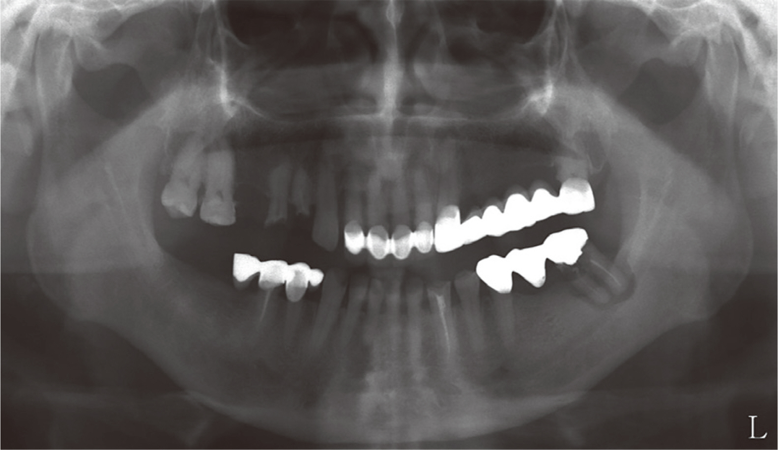
Fig. 1 Initial panoramic radiograph.

Fig. 2 Extraoral photographs. Concave profile and mandible protrusion.

Fig. 3 Pre-operative intraoral photographs. (A) Maxillary occlusal view, (B) Right lateral view, (C) Frontal view, (D) Left lateral view, (E) Mandibular occlusal view.
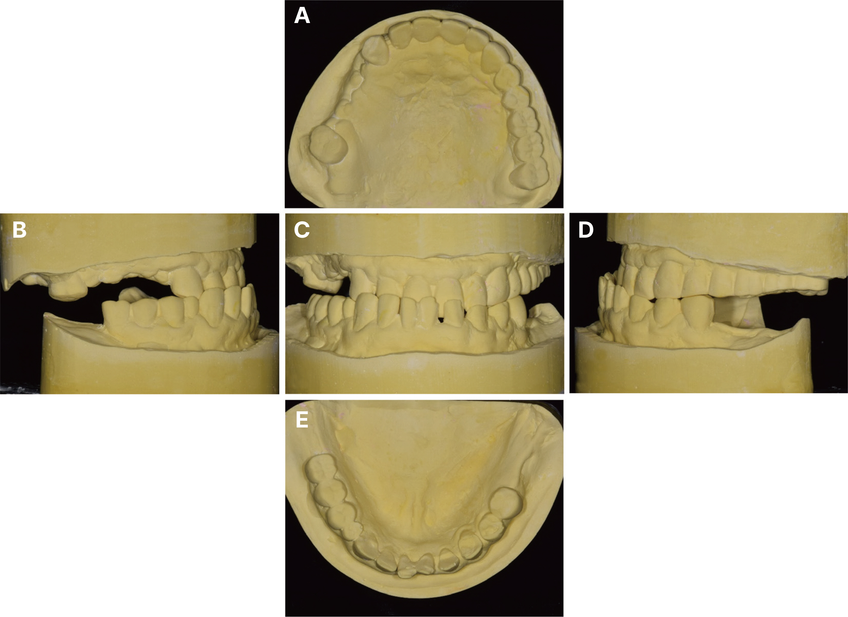
Fig. 4 Diagnostic cast analysis before treatment. (A) Maxillary occlusal view, (B) Right lateral view, (C) Frontal view, (D) Left lateral view, (E) Mandibular occlusal view.
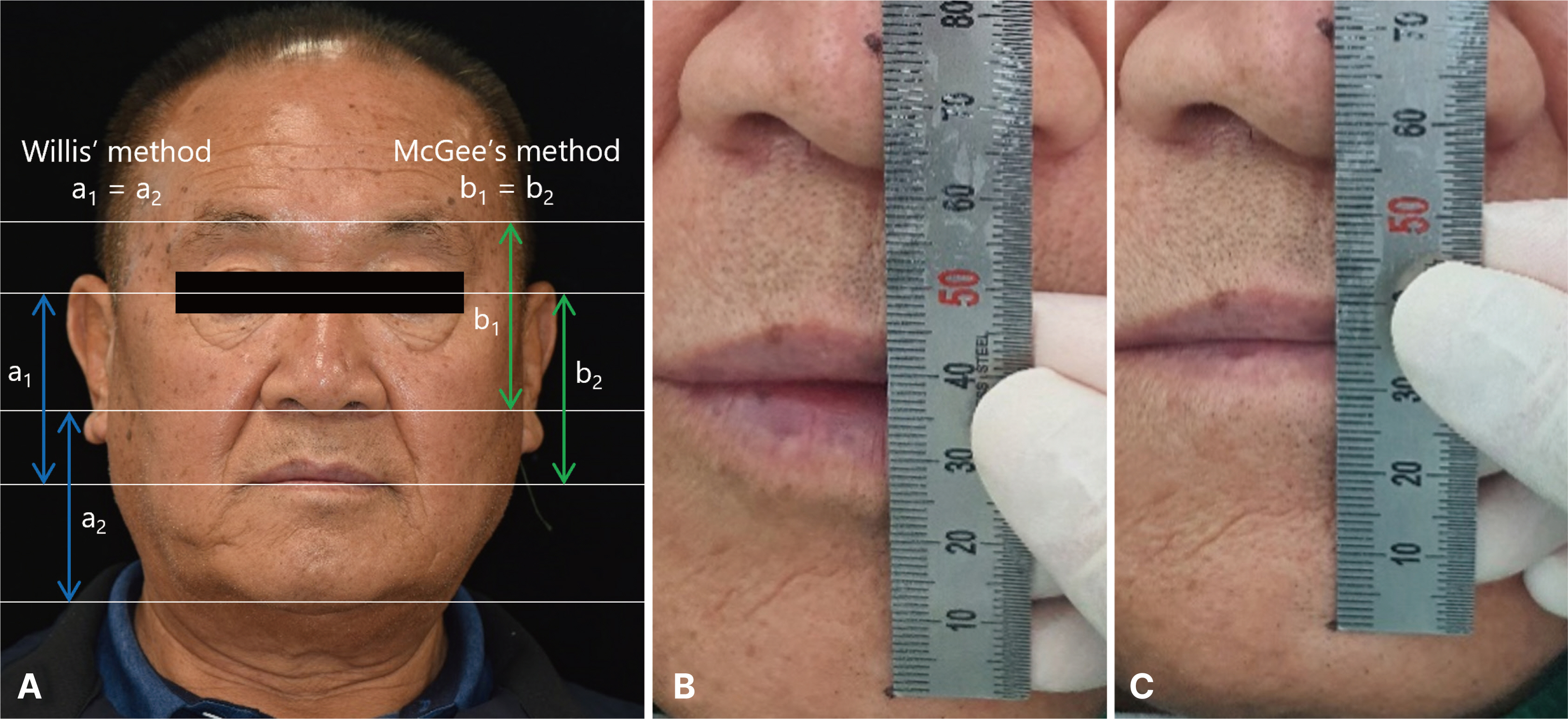
Fig. 5 Vertical dimension evaluation. (A) Facial appearance evaluation, (B) Interocclusal rest distance evaluation (maximum intercuspation), (C) Interocclusal rest distance evaluation (physiologic rest position).
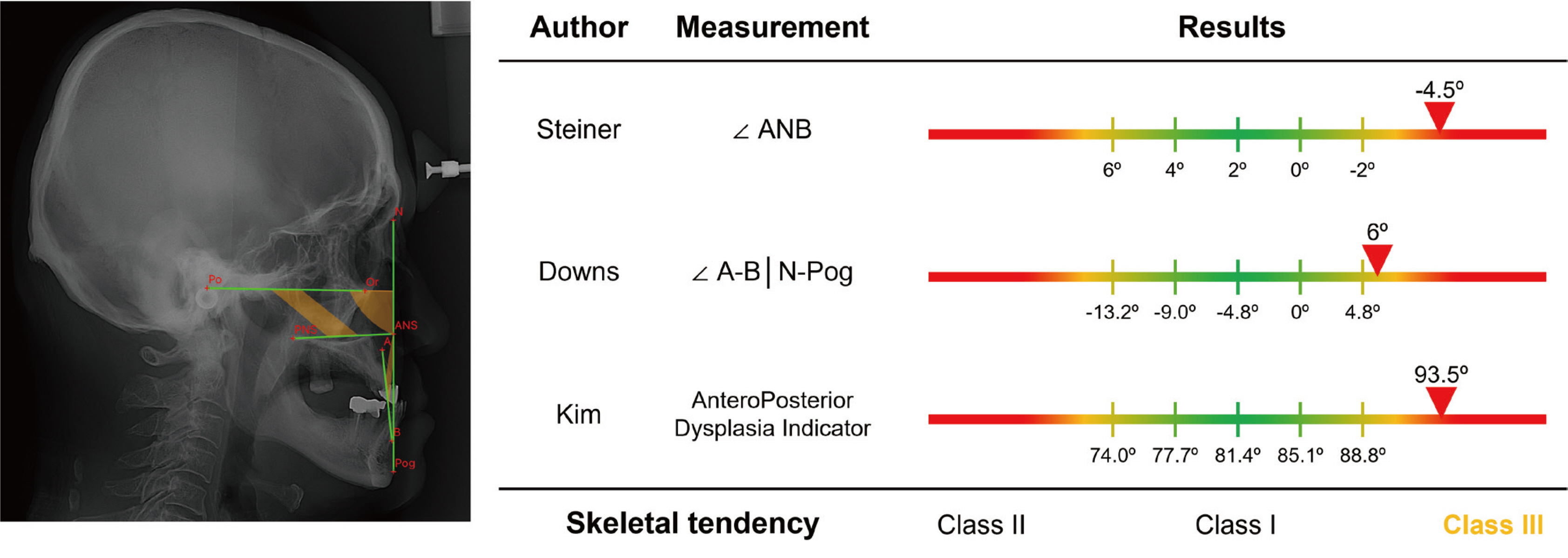
Fig. 6 Cephalometric analysis before treatment.
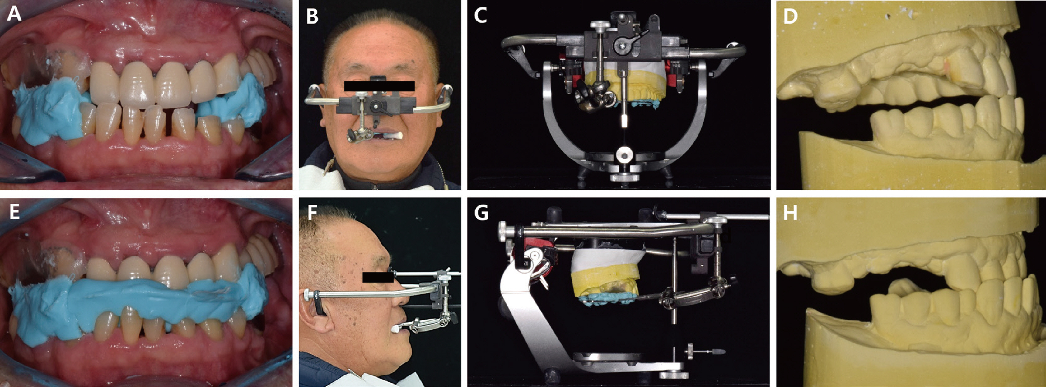
Fig. 7 (A) Interocclusal relationship registration on CR position, (B) Face-bow transfer on CR position, (C, D) Mounting on articulator on CR position, (E) Interocclusal relationship registration on MICP position, (F) Face-bow transfer on MICP position, (G, H) Mounting on articulator on MICP position.

Fig. 8 A digital occlusion analyzer (Arcus Digma II, KaVo, Leutkirch, Germany). (A) Arcus Digma II facebow transfer & individual hinge axis taking, (B, C) Arcus Digma II result; The terminal movement of the arc of closure was vertical.
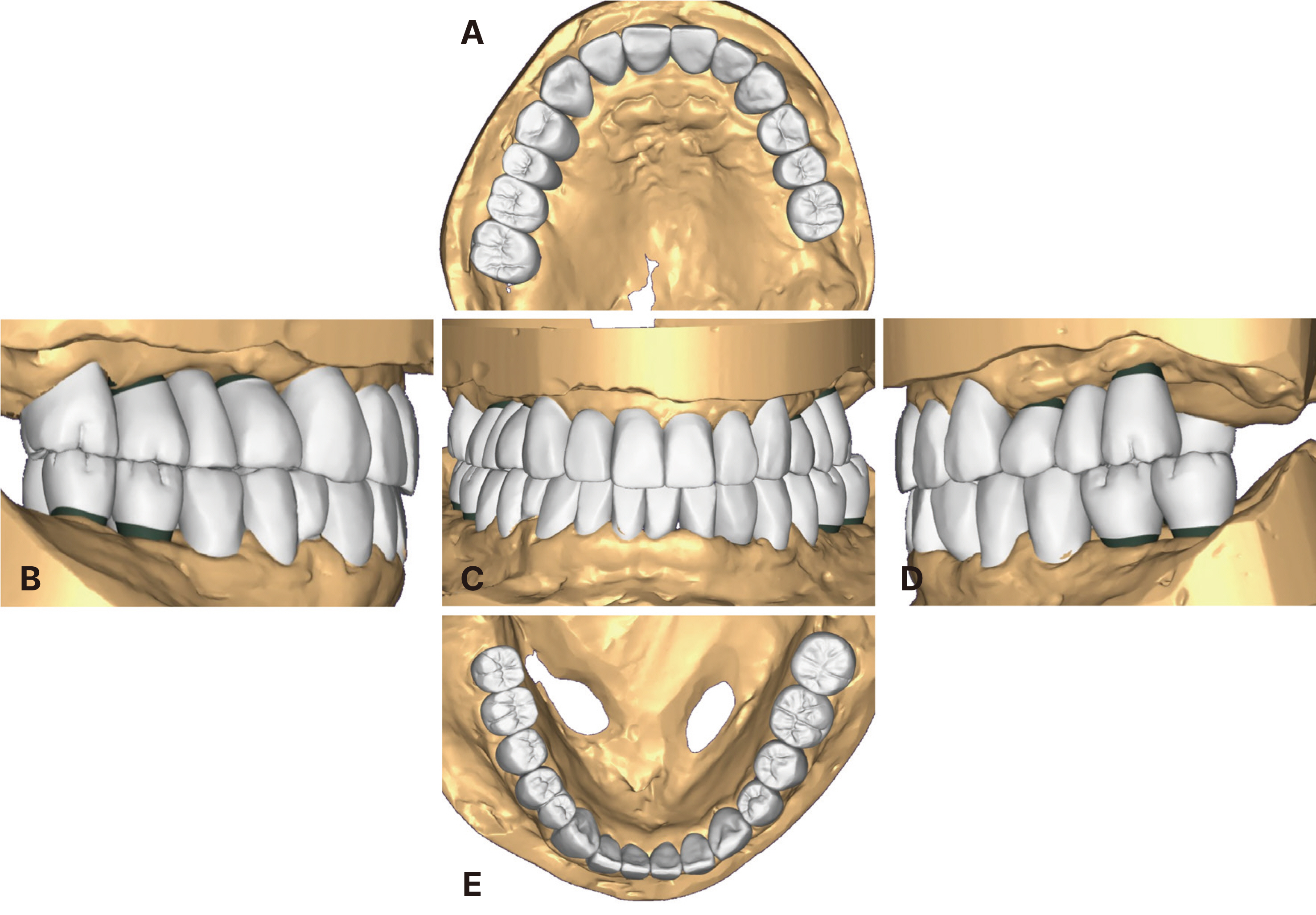
Fig. 9 Provisional restoration design by CAD (computer-assisted design) system. (A) Maxillary occlusal view, (B) Right lateral view, (C) Frontal view, (D) Left lateral view, (E) Mandibular occlusal view.
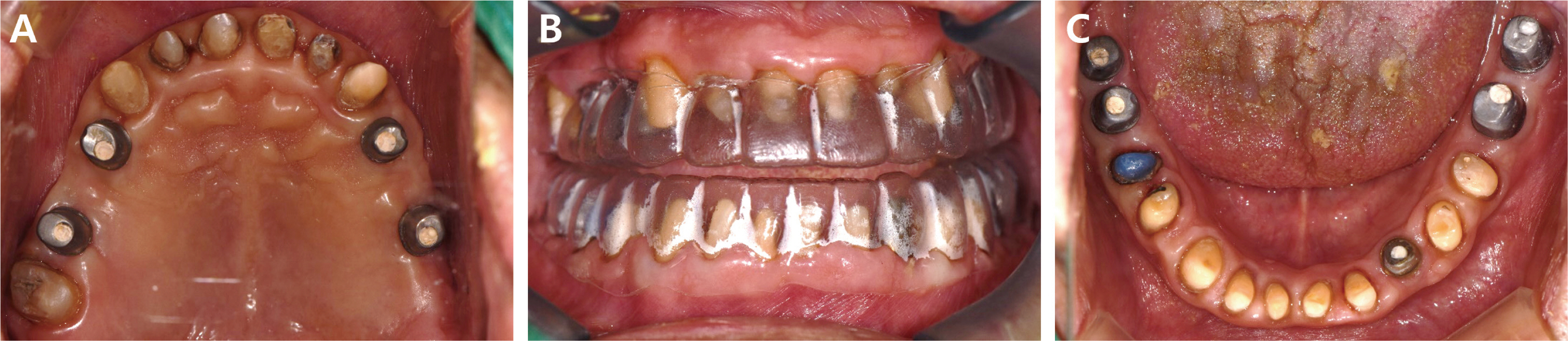
Fig. 10 Teeth preparation procedures. (A) Occlusal view (maxilla), (B) Frontal view of teeth preparation guide, (C) Occlusal view (mandible).
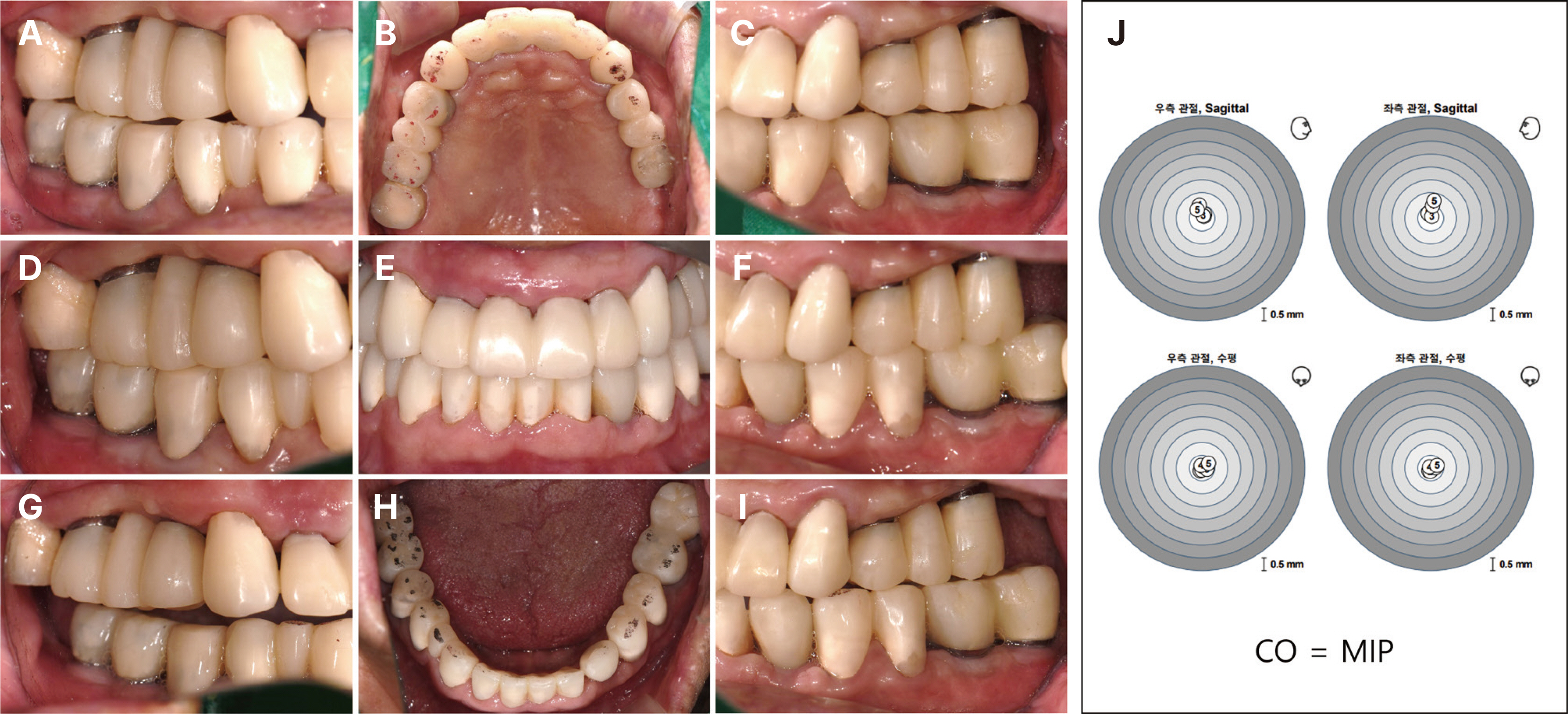
Fig. 11 Intraoral photograph after the placement of provisional restoration. (A) Working side during right lateral excursion, (B) Maxillary occlusal view, (C) Non-working side during right lateral excursion, (D) Left lateral view at centric occlusion, (E) Frontal view at centric occlusion, (F) Right lateral view at centric occlusion, (G) Non-working side during left lateral excursion, (H) Mandibular occlusal view, (I) Working side during left lateral excursion, (J) Arcus Digma II result.
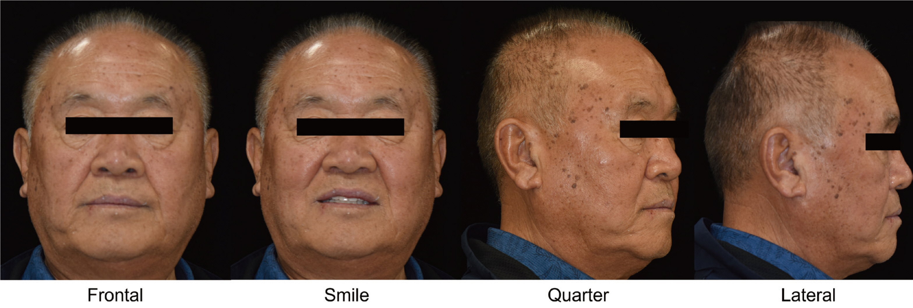
Fig. 12 Extraoral photographs. Straight profile.

Fig. 13 Wax-up pattern for definite prosthesis. (A) Working side during right lateral excursion, (B) Maxillary occlusal view, (C) Non-working side during right lateral excursion, (D) Left lateral view at centric occlusion, (E) Frontal view at centric occlusion, (F) Right lateral view at centric occlusion, (G) Non-working side during left lateral excursion, (H) Mandibular occlusal view, (I) Working side during left lateral excursion, (J) Anterior guide table.
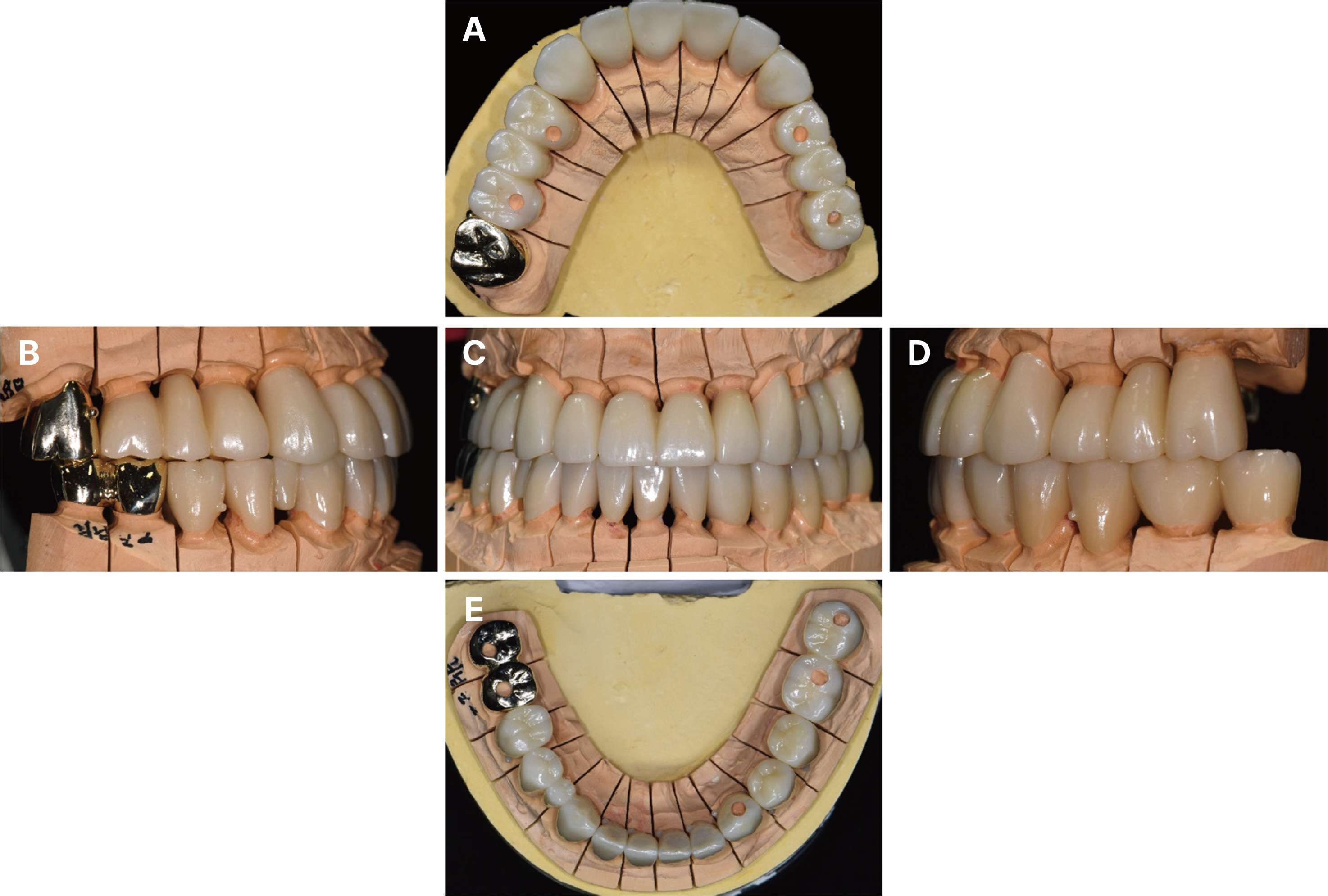
Fig. 14 Definitive prosthesis on master cast. (A) Maxillary occlusal view, (B) Right lateral view, (C) Frontal view, (D) Left lateral view, (E) Mandibular occlusal view.

Fig. 15 Intraoral photograph after the placement of provisional restoration. (A) Working side during right lateral excursion, (B) Maxillary occlusal view, (C) Non-working side during right lateral excursion, (D) Left lateral view at centric occlusion, (E) Frontal view at centric occlusion, (F) Right lateral view at centric occlusion, (G) Non-working side during left lateral excursion, (H) Mandibular occlusal view, (I) Working side during left lateral excursion.
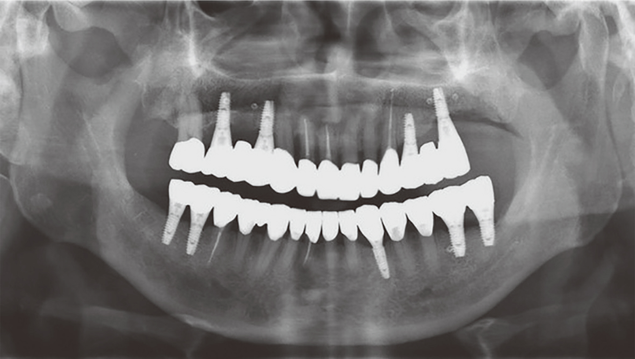
Fig. 16 Posttreatment panoramic radiograph.

Fig. 17 T-scan (T-scan III, Tekscan Inc., Boston, USA) examination on Maximal Intercuspal Position (MICP). (A) 3-D Movie Window. Relative forces are distinguished by column height and or colors. Red color means high force and blue color means low force, (B) 2-D Movie Window. Center of Force (COF) and force percentage per tooth, (C) Force and Timegraph. 3 distinct colored force lines (Red, Green, Grey). Red line means right side force and green line means left side force and grey line means maximum force.
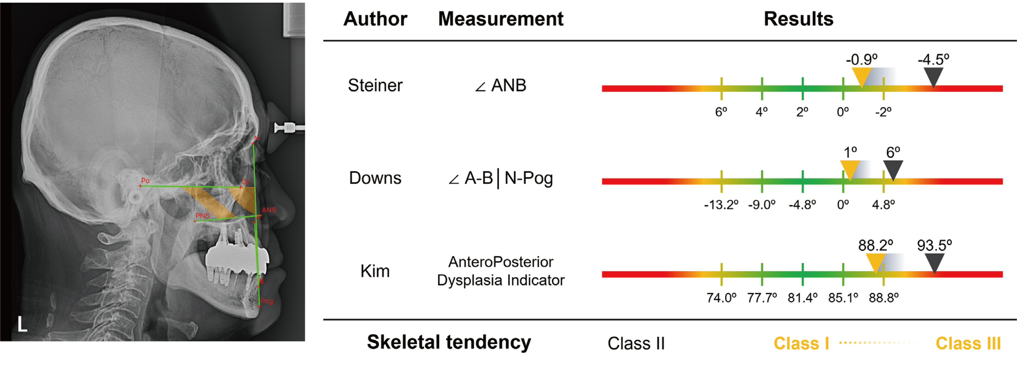
Fig. 18 Cephalometric analysis. The black arrows indicate the values before treatment, and the yellow arrows indicate the values after treatment.
Reference
-
References
1. Dawson PE. 2007. Functional occlusion: from TMJ to smile design. 1st ed. Mosby;St. Louis: p. 429–52. p. 595–602. DOI: 10.4103/0972-4052.32520.2. Lee KY, Kim CY, Jung JH, Kim YL. 2015; Mouth rehabilitation of a patient with severely worn dentition with vertical dimension increase. J Korean Acad Prosthodont. 53:215–21. DOI: 10.4047/jkap.2015.53.3.215.3. Yun AY, Shim HW, An JH. 2014; Full mouth rehabilitation in a patient with loss of vertical dimension caused by severe tooth loss: a case report. J Korean Acad Prosthodont. 52:42–7. DOI: 10.4047/jkap.2014.52.1.42.4. Eichner K. 1990; Renewed examination of the group classification of partially edentulous arches by Eichner and application advices for studies on morbidity statistics. Stomatol DDR. 40:321–5. DOI: 10.1007/978-3-663-13215-8_4.5. Willis FM. 1935; Features of the face involved in full denture prosthesis. Dent Cosmos. 77:851–4.6. Turner KA, Missirlian DM. 1984; Restoration of the extremely worn dentition. J Prosthet Dent. 52:467–74. DOI: 10.1016/0022-3913(84)90326-3. PMID: 6389829.7. Inui M, Fushima K, Sato S. 1999; Facial asymmetry in temporomandibular joint disorders. J Oral Rehabil. 26:402–6. DOI: 10.1046/j.1365-2842.1999.00387.x. PMID: 10373087.8. Ahn SJ, Lee SP, Nahm DS. 2005; Relationship between temporomandibular joint internal derangement and facial asymmetry in women. Am J Orthod Dentofacial Orthop. 128:583–91. DOI: 10.1016/j.ajodo.2004.06.038. PMID: 16286205.9. Zhang YL, Song JL, Xu XC, Zheng LL, Wang QY, Fan YB, Liu Z. 2016; Morphologic Analysis of the Temporomandibular Joint Between Patients With Facial Asymmetry and Asymptomatic Subjects by 2D and 3D Evaluation: A Preliminary Study. Medicine (Baltimore). 95:e3052. DOI: 10.1097/MD.0000000000003052. PMID: 27043669. PMCID: PMC4998530.10. Murphy T. 1958; Mandibular adjustment to functional tooth attrition. Aust Dent J. 3:171–8. DOI: 10.1111/j.1834-7819.1958.tb01832.x.11. Dawson PE. 2007. Functional occlusion: from TMJ to smile design. 1st ed. Mosby;St. Louis: p. 513–24. DOI: 10.4103/0972-4052.32520.12. Dawson PE. 1989. Evaluation, diagnosis, and treatment of occlusal problems. 2nd ed. Mosby;St. Louis:13. Pleasure MA. 1951; Correct vertical dimension and freeway space. J Am Dent Assoc. 43:160–3. DOI: 10.14219/jada.archive.1951.0188. PMID: 14850211.14. Shanahan TE. 2004; Physiologic vertical dimension and centric relation. 1956. J Prosthet Dent. 91:206–9. DOI: 10.1016/j.prosdent.2003.09.002. PMID: 15060486.15. Silverman MM. 2001; The speaking method in measuring vertical dimension. 1952. J Prosthet Dent. 85:427–31. DOI: 10.1067/mpr.2001.116139. PMID: 11357066.16. Abduo J, Lyons K. 2012; Clinical considerations for increasing occlusal vertical dimension: a review. Aust Dent J. 57:2–10. DOI: 10.1111/j.1834-7819.2011.01640.x. PMID: 22369551.17. Millstein P. 2008; Know your indicator. J Mass Dent Soc. 56:30–1.
- Full Text Links
-
- Actions
-
Cited
- CITED
-
- Close
- Share
-
- Similar articles
-
- The evaluation of maximum bite force in the occlusal rehabilitation of patient with Angle Class III malocclusion: a case report
- Rehabilitation of posterior support and vertical dimension in a class 3 malocclusion patient: A case report
- Evaluation methods of occlusal vertical dimension and their clinical applications: A narrative review
- Prosthetic full mouth rehabilitation of patient with mandibular prognathism and asymmetry: a case report
- A study on the vertical dysplasia in the skeletal class iii malocclusion
