ACY-241, a histone deacetylase 6 inhibitor, suppresses the epithelial–mesenchymal transition in lung cancer cells by downregulating hypoxia-inducible factor-1 alpha
- Affiliations
-
- 1College of Pharmacy, Keimyung University, Daegu 42601, Korea
- KMID: 2550291
- DOI: http://doi.org/10.4196/kjpp.2024.28.1.83
Abstract
- Hypoxia-inducible factor-1 alpha (HIF-1α) is a transcription factor activated under hypoxic conditions, and it plays a crucial role in cellular stress regulation. While HIF-1α activity is essential in normal tissues, its presence in the tumor microenvironment represents a significant risk factor as it can induce angiogenesis and confer resistance to anti-cancer drugs, thereby contributing to poor prognoses. Typically, HIF-1α undergoes rapid degradation in normoxic conditions via oxygen-dependent degradation mechanisms. However, certain cancer cells can express HIF-1α even under normoxia. In this study, we observed an inclination toward increased normoxic HIF-1α expression in cancer cell lines exhibiting increased HDAC6 expression, which prompted the hypothesis that HDAC6 may modulate HIF-1α stability in normoxic conditions. To prove this hypothesis, several cancer cells with relatively higher HIF-1α levels under normoxic conditions were treated with ACY-241, a selective HDAC6 inhibitor, and small interfering RNAs for HDAC6 knockdown. Our data revealed a significant reduction in HIF-1α expression upon HDAC6 inhibition. Moreover, the downregulation of HIF-1α under normoxic conditions decreased zinc finger E-box-binding homeobox 1 expression and increased E-cadherin levels in lung cancer H1975 cells, consequently suppressing cell invasion and migration. ACY-241 treatment also demonstrated an inhibitory effect on cell invasion and migration by reducing HIF-1α level. This study confirms that HDAC6 knockdown and ACY-241 treatment effectively decrease HIF-1α expression under normoxia, thereby suppressing the epithelial– mesenchymal transition. These findings highlight the potential of selective HDAC6 inhibition as an innovative therapeutic strategy for lung cancer.
Keyword
Figure
-

Fig. 1 HDAC6 knockdown reduces the protein levels of HIF-1α under normoxic conditions. (A) Western blot analysis of HDAC6 and HIF-1α levels under normoxic conditions in breast cancer and lung cancer cells. (B) Cells were transfected with siHDAC6 for 48 h. HDAC6, HIF-1α, Ac-α-tubulin, and α-tubulin expression levels were measured using western blot analysis. GAPDH was used as the loading control. (C) Cells were transfected with siHDAC6 for 48 h. HIF-1α expression was measured using qRT-PCR. The expression value was normalized to the 18s rRNA level. Statistical analysis was performed using Student’s t-test. Error bars present the mean ± standard deviation (n = 3). NS > 0.05 vs. siCON. HDAC6, histone deacetylase 6; HIF-1α, hypoxia-inducible factor 1 alpha; siHDAC6, small interfering RNA against HDAC6; Ac-α-tubulin, acetylated alpha tubulin; GAPDH, glyceraldehyde 3-phosphate; siCON, negative control siRNA; NS, non-significant.

Fig. 2 ACY-241 decreases the HIF-1α protein level. (A) After treatment with ACY-241 for 48 h, the cell viability was measured using the CCK-8 assay. Statistical analysis was performed using one‐way ANOVA (Tukey's multiple comparisons test). Error bars present the mean ± standard deviation (n = 6). ***p < 0.001 vs. CON. (B) Cells were treated with ACY-241 for 48 h, after which the protein levels of HDAC6, HIF-1α, Ac-α-tubulin, and α-tubulin were measured via western blot analysis. GAPDH was used as the loading control. (C) Cells were treated with ACY-241 for 48 h and with MG-132 4 h before sampling, after which the protein levels of HIF-1α were measured via Western blot analysis. GAPDH was used as the loading control. HDAC6, histone deacetylase 6; HIF-1α, hypoxia-inducible factor 1 alpha; Ac-α-tubulin, acetylated alpha tubulin; GAPDH, glyceraldehyde 3-phosphate; CON, control.
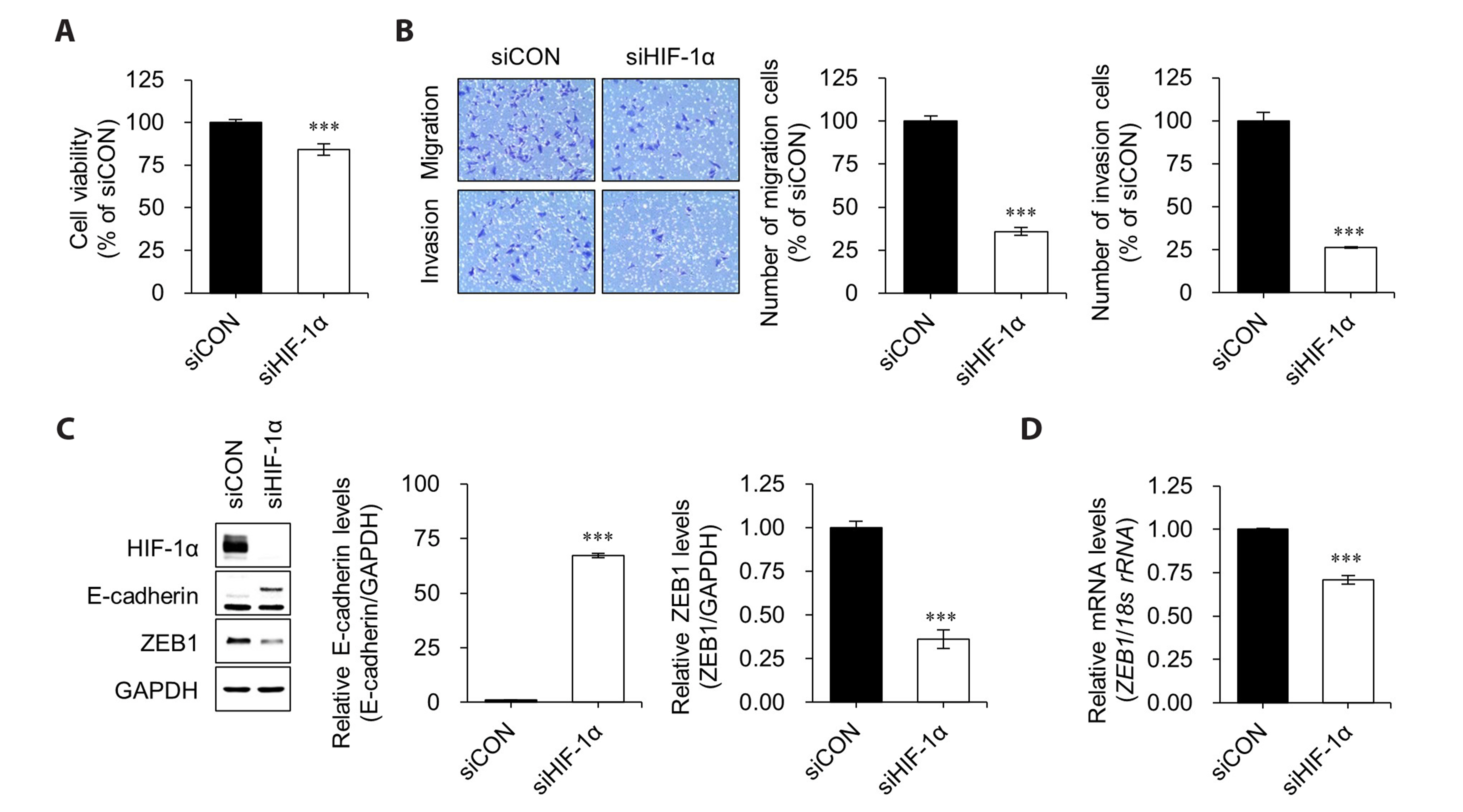
Fig. 3 Knockdown of HIF-1α inhibits invasion and migration of H1975 cells. (A) Cells were transfected with siHIF-1α for 48 h. Cell viability was measured using the CCK-8 assay. Statistical analysis was performed using Student’s t-test. Error bars present the mean ± standard deviation (n = 6). ***p < 0.001 vs. siCON. (B) Transwell assays were performed by incubating H1975 cells for 36 h under normoxic conditions after siHIF-1α treatment. Representative images of cell migration and invasion captured under a microscope. Statistical analysis was performed using one‐way ANOVA. Error bars present the mean ± standard deviation (n = 3). ***p < 0.001 vs. siCON. (C) Cells were transfected with siHIF-1α for 48 h. The HIF-1α, E-cadherin, and ZEB1 expression levels were measured using western blotting. GAPDH was used as a loading control. Quantitation of protein intensity was performed using the ImageJ software. Statistical analysis was performed using Student’s t-test. Error bars present the mean ± standard deviation (n = 3). ***p < 0.001 vs. siCON. (D) qRT-PCR analysis of ZEB1. Cells were transfected with siHIF-1α for 48 h. The expression value was normalized to the 18s rRNA level. Statistical analyses were performed using the Student’s t-test. Error bars present the mean ± standard deviation (n = 3). ***p < 0.001 vs. siCON. HDAC6, histone deacetylase 6; HIF-1α, hypoxia-inducible factor 1 alpha; ZEB1, zinc finger E-box-binding homeobox 1; siHIF-1α, small interfering RNA against HIF-1α; GAPDH, glyceraldehyde 3-phosphate; siCON, negative control siRNA.
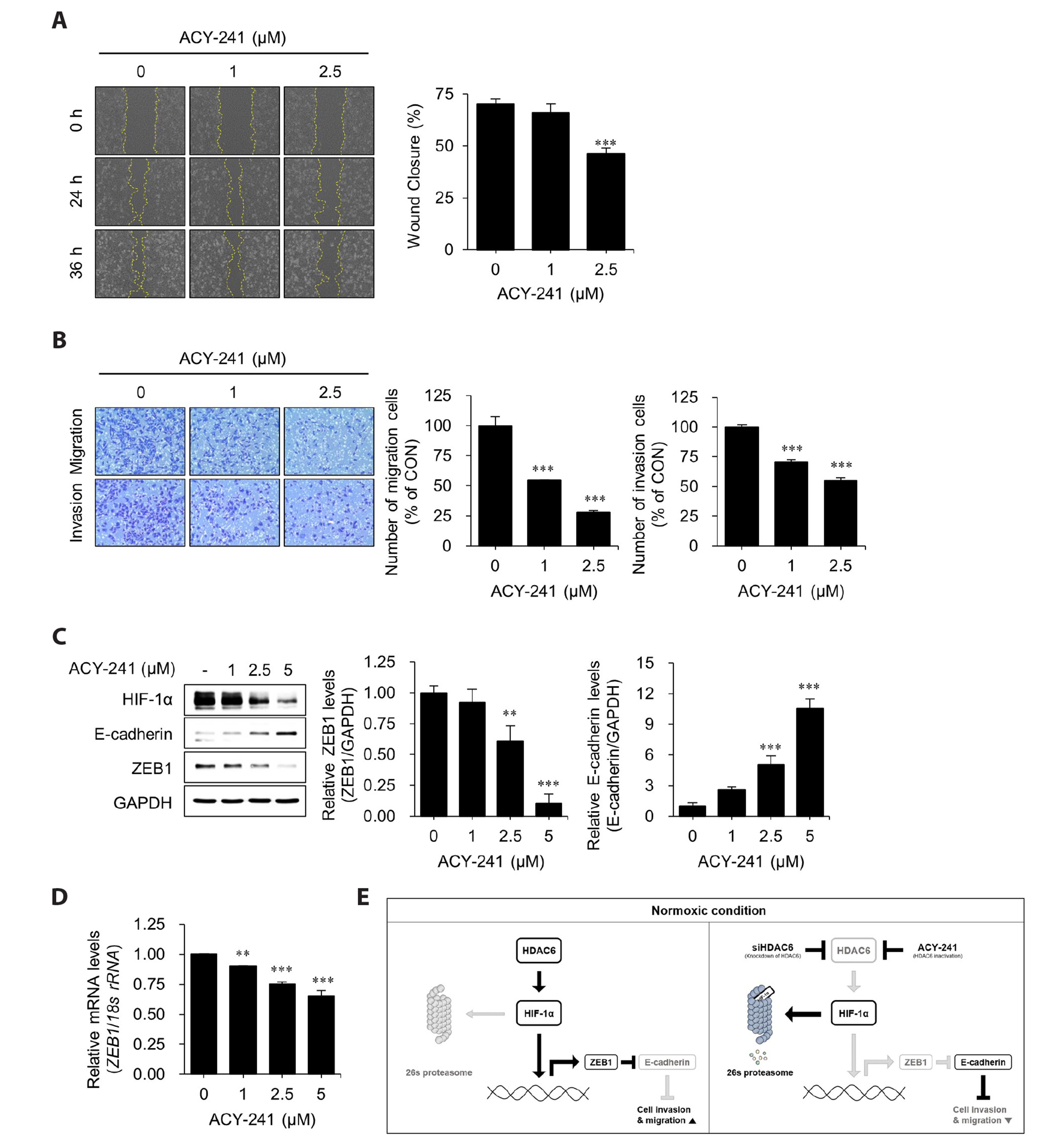
Fig. 4 ACY-241 inhibits migration and invasion in H1975 cell lines. (A) Wound healing assay of H1975 cells treated with different concentrations of ACY-241. Microphotographs of the wounded area were taken at 0, 24, and 36 h to observe cell migration into the wounded area. Statistical analysis was performed using one‐way ANOVA (Tukey's multiple comparisons test). Error bars present the mean ± standard deviation (n = 6). ***p < 0.001 vs. CON. (B) Transwell assays were performed using different concentrations of ACY-241 for 36 h. Representative images of cell migration and invasion were captured under a microscope. Statistical analysis was performed using one‐way ANOVA (Tukey's multiple comparisons test). Error bars present the mean ± standard deviation (n = 3). ***p < 0.001 vs. CON. (C) Cells were treated with ACY-241 for 48 h, after which the protein levels of HDAC6, HIF-1α, E-cadherin, and ZEB1 were monitored via Western blot analysis. GAPDH was used as a loading control. Quantitation of protein intensity was performed using the ImageJ software. Statistical analysis was performed using one‐way ANOVA (Tukey's multiple comparisons test). Error bars present the mean ± standard deviation (n = 3). **p < 0.01; ***p < 0.001 vs. CON. (D) qRT-PCR analysis of ZEB1. Cells were treated with ACY-241 for 48 h. The expression value was normalized to the 18s rRNA level. Statistical analyses were performed using the one‐way ANOVA (Tukey's multiple comparisons test). Error bars present the mean ± standard deviation (n = 3). **p < 0.01; ***p < 0.001 vs. CON. (E) Under normoxic conditions, cancer cells with higher expression of HDAC6 have been shown to increase the stability of HIF-1α protein. The stabilized HIF-1α induces the expression of ZEB1, which decreases the expression of E-cadherin, leading to increased invasion and migration of cancer cells (left). Conversely, reducing HDAC6 through HDAC6 knockdown, or inhibiting HDAC6 activity with ACY-241, decreases the protein level of HIF-1α, which in turn leads to a reduction in epithelial-mesenchymal transition (right). HDAC6, histone deacetylase 6; HIF-1α, hypoxia-inducible factor 1 alpha; ZEB1, zinc finger E-box-binding homeobox 1; GAPDH, glyceraldehyde 3-phosphate ; CON, control.
Reference
-
1. Thai AA, Solomon BJ, Sequist LV, Gainor JF, Heist RS. 2021; Lung cancer. Lancet. 398:535–554. DOI: 10.1016/S0140-6736(21)00312-3. PMID: 34273294.2. Heist RS, Sequist LV, Engelman JA. 2012; Genetic changes in squamous cell lung cancer: a review. J Thorac Oncol. 7:924–933. DOI: 10.1097/JTO.0b013e31824cc334. PMID: 22722794. PMCID: PMC3404741.3. Dong J, Li B, Lin D, Zhou Q, Huang D. 2019; Advances in targeted therapy and immunotherapy for non-small cell lung cancer based on accurate molecular typing. Front Pharmacol. 10:230. DOI: 10.3389/fphar.2019.00230. PMID: 30930778. PMCID: PMC6424010. PMID: a06463d539ec4ce7942b7834c48c2287.4. Berger SL, Kouzarides T, Shiekhattar R, Shilatifard A. 2009; An operational definition of epigenetics. Genes Dev. 23:781–783. DOI: 10.1101/gad.1787609. PMID: 19339683. PMCID: PMC3959995.5. Luo J, Huang Z, Wei W, Sun Y, Gong Y. 2023; Editorial: epigenetic regulation and non-histone post-translational modification in cancer. Front Genet. 14:1176174. DOI: 10.3389/fgene.2023.1176174. PMID: 37091796. PMCID: PMC10116044. PMID: 88d2c1c7ab0e4253b85f93110455a688.6. Delcuve GP, Khan DH, Davie JR. 2012; Roles of histone deacetylases in epigenetic regulation: emerging paradigms from studies with inhibitors. Clin Epigenetics. 4:5. DOI: 10.1186/1868-7083-4-5. PMID: 22414492. PMCID: PMC3320549. PMID: 3837566c859448f1ae5943186a7d0d81.7. Aldana-Masangkay GI, Sakamoto KM. 2011; The role of HDAC6 in cancer. J Biomed Biotechnol. 2011:875824. DOI: 10.1155/2011/875824. PMID: 21076528. PMCID: PMC2975074.8. Wang Z, Tang F, Hu P, Wang Y, Gong J, Sun S, Xie C. 2016; HDAC6 promotes cell proliferation and confers resistance to gefitinib in lung adenocarcinoma. Oncol Rep. 36:589–597. DOI: 10.3892/or.2016.4811. PMID: 27221381.9. Pulya S, Amin SA, Adhikari N, Biswas S, Jha T, Ghosh B. 2021; HDAC6 as privileged target in drug discovery: a perspective. Pharmacol Res. 163:105274. DOI: 10.1016/j.phrs.2020.105274. PMID: 33171304.10. Dong J, Zheng N, Wang X, Tang C, Yan P, Zhou HB, Huang J. 2018; A novel HDAC6 inhibitor exerts an anti-cancer effect by triggering cell cycle arrest and apoptosis in gastric cancer. Eur J Pharmacol. 828:67–79. DOI: 10.1016/j.ejphar.2018.03.026. PMID: 29563065.11. Li J, Yu M, Fu S, Liu D, Tan Y. 2022; Role of selective histone deacetylase 6 inhibitor ACY-1215 in cancer and other human diseases. Front Pharmacol. 13:907981. Erratum in: Front Pharmacol. 2022;13: 1117936. DOI: 10.3389/fphar.2022.907981. PMID: 35652048. PMCID: PMC9149003. PMID: d5a8624566a44df2aec9a2d8779e4720.12. Park SJ, Joo SH, Lee N, Jang WJ, Seo JH, Jeong CH. 2021; ACY-241, an HDAC6 inhibitor, overcomes erlotinib resistance in human pancreatic cancer cells by inducing autophagy. Arch Pharm Res. 44:1062–1075. DOI: 10.1007/s12272-021-01359-x. PMID: 34761352.13. Zhang XH, Kang HQ, Tao YY, Li YH, Zhao JR, Ma LY, Liu HM. Ya-Gao. 2021; Identification of novel 1,3-diaryl-1,2,4-triazole-capped histone deacetylase 6 inhibitors with potential anti-gastric cancer activity. Eur J Med Chem. 218:113392. DOI: 10.1016/j.ejmech.2021.113392. PMID: 33831778.14. Yang MH, Wu KJ. 2008; TWIST activation by hypoxia inducible factor-1 (HIF-1): implications in metastasis and development. Cell Cycle. 7:2090–2096. DOI: 10.4161/cc.7.14.6324. PMID: 18635960.15. Jun JC, Rathore A, Younas H, Gilkes D, Polotsky VY. 2017; Hypoxia-inducible factors and cancer. Curr Sleep Med Rep. 3:1–10. DOI: 10.1007/s40675-017-0062-7. PMID: 28944164. PMCID: PMC5607450.16. Kurtipek E, Koçak N, Esme H, Düzgün N, Akin SE, Ünlü Y, Bekçi TT. 2016; The role of HIF-1 pathway in non-small-cell lung cancer. Eur Respir J. 48:PA2855. DOI: 10.1183/13993003.congress-2016.PA2855.17. Zhu J, Huang Z, Zhang M, Wang W, Liang H, Zeng J, Wu K, Wang X, Hsieh JT, Guo P, Fan J. 2018; HIF-1α promotes ZEB1 expression and EMT in a human bladder cancer lung metastasis animal model. Oncol Lett. 15:3482–3489. Erratum in: Oncol Lett. 2021;22:599. DOI: 10.3892/ol.2021.12860. PMID: 34188701. PMCID: PMC8228379.18. Stroka DM, Burkhardt T, Desbaillets I, Wenger RH, Neil DA, Bauer C, Gassmann M, Candinas D. 2001; HIF-1 is expressed in normoxic tissue and displays an organ-specific regulation under systemic hypoxia. FASEB J. 15:2445–2453. DOI: 10.1096/fj.01-0125com. PMID: 11689469.19. Cao Y, Eble JM, Moon E, Yuan H, Weitzel DH, Landon CD, Nien CY, Hanna G, Rich JN, Provenzale JM, Dewhirst MW. 2013; Tumor cells upregulate normoxic HIF-1α in response to doxorubicin. Cancer Res. 73:6230–6242. DOI: 10.1158/0008-5472.CAN-12-1345. PMID: 23959856. PMCID: PMC3800255.20. Ryu HW, Won HR, Lee DH, Kwon SH. 2017; HDAC6 regulates sensitivity to cell death in response to stress and post-stress recovery. Cell Stress Chaperones. 22:253–261. DOI: 10.1007/s12192-017-0763-3. PMID: 28116619. PMCID: PMC5352599.21. Livak KJ, Schmittgen TD. 2001; Analysis of relative gene expression data using real-time quantitative PCR and the 2(-Delta Delta C(T)) Method. Methods. 25:402–408. DOI: 10.1006/meth.2001.1262. PMID: 11846609.22. Zhang Q, Zhang ZF, Rao JY, Sato JD, Brown J, Messadi DV, Le AD. 2004; Treatment with siRNA and antisense oligonucleotides targeted to HIF-1alpha induced apoptosis in human tongue squamous cell carcinomas. Int J Cancer. 111:849–857. DOI: 10.1002/ijc.20334. PMID: 15300796.23. Masoud GN, Li W. 2015; HIF-1α pathway: role, regulation and intervention for cancer therapy. Acta Pharm Sin B. 5:378–389. DOI: 10.1016/j.apsb.2015.05.007. PMID: 26579469. PMCID: PMC4629436. PMID: 0f5f4982b0944ba585e72b39876cfd33.24. Sánchez-Tilló E, Lázaro A, Torrent R, Cuatrecasas M, Vaquero EC, Castells A, Engel P, Postigo A. 2010; ZEB1 represses E-cadherin and induces an EMT by recruiting the SWI/SNF chromatin-remodeling protein BRG1. Oncogene. 29:3490–3500. DOI: 10.1038/onc.2010.102. PMID: 20418909.25. Haase VH. 2009; The VHL tumor suppressor: master regulator of HIF. Curr Pharm Des. 15:3895–3903. DOI: 10.2174/138161209789649394. PMID: 19671042. PMCID: PMC3622710.26. Li T, Mao C, Wang X, Shi Y, Tao Y. 2020; Epigenetic crosstalk between hypoxia and tumor driven by HIF regulation. J Exp Clin Cancer Res. 39:224. DOI: 10.1186/s13046-020-01733-5. PMID: 33109235. PMCID: PMC7592369. PMID: 3dc64a595d5a44a6be5538d4e2133c8f.27. Liang D, Kong X, Sang N. 2006; Effects of histone deacetylase inhibitors on HIF-1. Cell Cycle. 5:2430–2435. DOI: 10.4161/cc.5.21.3409. PMID: 17102633. PMCID: PMC4505804.28. Yoo YG, Kong G, Lee MO. 2006; Metastasis-associated protein 1 enhances stability of hypoxia-inducible factor-1alpha protein by recruiting histone deacetylase 1. EMBO J. 25:1231–1241. DOI: 10.1038/sj.emboj.7601025. PMID: 16511565. PMCID: PMC1422150.29. Seo HW, Kim EJ, Na H, Lee MO. 2009; Transcriptional activation of hypoxia-inducible factor-1alpha by HDAC4 and HDAC5 involves differential recruitment of p300 and FIH-1. FEBS Lett. 583:55–60. DOI: 10.1016/j.febslet.2008.11.044. PMID: 19071119.30. Kong X, Lin Z, Liang D, Fath D, Sang N, Caro J. 2006; Histone deacetylase inhibitors induce VHL and ubiquitin-independent proteasomal degradation of hypoxia-inducible factor 1alpha. Mol Cell Biol. 26:2019–2028. DOI: 10.1128/MCB.26.6.2019-2028.2006. PMID: 16507982. PMCID: PMC1430285.31. Geng H, Liu Q, Xue C, David LL, Beer TM, Thomas GV, Dai MS, Qian DZ. 2012; HIF1α protein stability is increased by acetylation at lysine 709. J Biol Chem. 287:35496–35505. DOI: 10.1074/jbc.M112.400697. PMID: 22908229. PMCID: PMC3471753.32. Saito S, Zhuang Y, Shan B, Danchuk S, Luo F, Korfei M, Guenther A, Lasky JA. 2017; Tubastatin ameliorates pulmonary fibrosis by targeting the TGFβ-PI3K-Akt pathway. PLoS One. 12:e0186615. DOI: 10.1371/journal.pone.0186615. PMID: 29045477. PMCID: PMC5646855. PMID: e3513edce61f4cd49294030347401e17.33. Schoepflin ZR, Shapiro IM, Risbud MV. 2016; Class I and IIa HDACs mediate HIF-1α stability through PHD2-dependent mechanism, while HDAC6, a Class IIb member, promotes HIF-1α transcriptional activity in nucleus pulposus cells of the intervertebral disc. J Bone Miner Res. 31:1287–1299. DOI: 10.1002/jbmr.2787. PMID: 26765925. PMCID: PMC4891304.34. McLeod AB, Stice JP, Wardell SE, Alley HM, Chang CY, McDonnell DP. 2018; Validation of histone deacetylase 3 as a therapeutic target in castration-resistant prostate cancer. Prostate. 78:266–277. DOI: 10.1002/pros.23467. PMID: 29243324.35. Yang L, Chang Y, Cao P. 2018; CCR7 preservation via histone deacetylase inhibition promotes epithelial-mesenchymal transition of hepatocellular carcinoma cells. Exp Cell Res. 371:231–237. DOI: 10.1016/j.yexcr.2018.08.015. PMID: 30107147.36. Wang J, Xu MQ, Jiang XL, Mei XY, Liu XG. 2018; Histone deacetylase inhibitor SAHA-induced epithelial-mesenchymal transition by upregulating Slug in lung cancer cells. Anticancer Drugs. 29:80–88. DOI: 10.1097/CAD.0000000000000573. PMID: 29176396.37. Wawruszak A, Kalafut J, Okon E, Czapinski J, Halasa M, Przybyszewska A, Miziak P, Okla K, Rivero-Muller A, Stepulak A. 2019; Histone deacetylase inhibitors and phenotypical transformation of cancer cells. Cancers (Basel). 11:148. DOI: 10.3390/cancers11020148. PMID: 30691229. PMCID: PMC6406474. PMID: 542b51f83d824eb3b05c7dac0f82a30c.
- Full Text Links
-
- Actions
-
Cited
- CITED
-
- Close
- Share
-
- Similar articles
-
- Effects of Histone Deacetylase Inhibitor (Valproic Acid) on the Expression of Hypoxia-inducible Factor-1 Alpha in Human Retinal Müller Cells
- Wheatgrass extract inhibits hypoxia-inducible factor-1-mediated epithelial-mesenchymal transition in A549 cells
- The role of hypoxia on the acquisition of epithelial-mesenchymal transition and cancer stemness: a possible link to epigenetic regulation
- Trefoil Factor 1 Suppresses Epithelial-mesenchymal Transition through Inhibition of TGF-beta Signaling in Gastric Cancer Cells
- Hypoxia Induces Epithelial-Mesenchymal Transition in Follicular Thyroid Cancer: Involvement of Regulation of Twist by Hypoxia Inducible Factor-1alpha
