J Korean Med Sci.
2023 Dec;38(50):e421. 10.3346/jkms.2023.38.e421.
Rare Cause of Exertional Angina
- Affiliations
-
- 1Department of Cardiology, Ajou University School of Medicine, Suwon, Korea
- 2Department of Thoracic and Cardiovascular Surgery, Ajou University School of Medicine, Suwon, Korea
- KMID: 2549962
- DOI: http://doi.org/10.3346/jkms.2023.38.e421
Figure
-
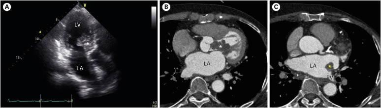
Fig. 1 Images of masses. (A) Transthoracic echocardiography showed a multi-lobulated shaped mass (white asterisk) attached to the anterior leaflet of the mitral valve on the left ventricular side. (B) Cardiac computed tomography showed that the mass (white asterisk) exhibited enhancement on early post-contrast image. (C) Another small mass (yellow asterisk) was observed on the left atrial wall at the opening of pulmonary vein.LV = left ventricle, LA = left atrium, PV = pulmonary vein.
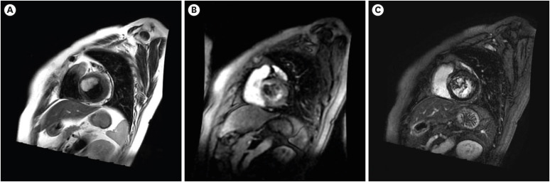
Fig. 2 Cardiac magnetic resonance images, mid-ventricular short axis view. (A) T2-weighted image. (B) Dynamic post-contrast examination. (C) The delayed phase.
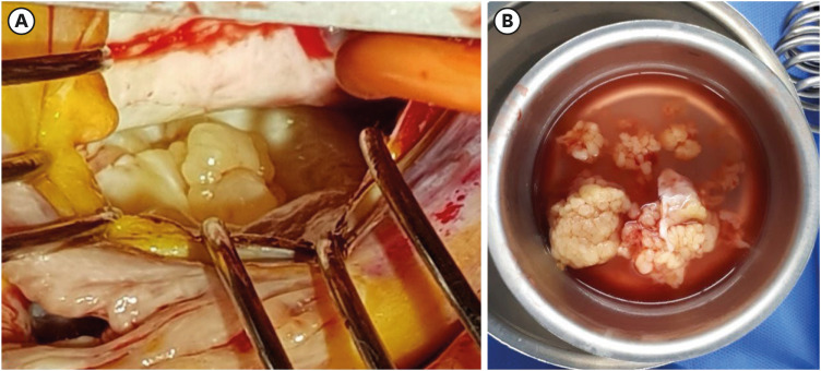
Fig. 3 Intra-op findings. (A) Multi-lobulated masses were noted. (B) Resected masses.
Reference
-
1. Peters PJ, Reinhardt S. The echocardiographic evaluation of intracardiac masses: a review. J Am Soc Echocardiogr. 2006; 19(2):230–240. PMID: 16455432.2. Basso C, Rizzo S, Valente M, Thiene G. Prevalence and pathology of primary cardiac tumours. Cardiovasc Med. 2012; 15(1):18–29.
- Full Text Links
-
- Actions
-
Cited
- CITED
-
- Close
- Share
-
- Similar articles
-
- A case of complete atrioventricular block persisting for 5 days in a patient with variant angina
- Significance of ST-Segment Level, ST-Segment Slope, ST-Segment Index and ST-Segment Integral in Exercise ECG as an Indicator of Myocardial Ischemia
- Complex Coronary Artery Fistula Causing Angina is Resolved Through Coil Embolization
- A Case of Ludwig Angina
- A Case of Cogan's Syndrome With Angina
