Immunoglobulin G4-related hypertrophic pachymeningitis with an isolated scalp mass mimicking a brain tumor: a case report and literature review
- Affiliations
-
- 1Division of Rheumatology, Department of Internal Medicine, Gachon University Gil Medical Center, Incheon, Korea
- 2Department of Neurosurgery, Gachon University Gil Medical Center, Incheon, Korea
- KMID: 2549637
- DOI: http://doi.org/10.4078/jrd.2023.0023
Abstract
- Immunoglobulin G4-related disease (IgG4-RD) is an autoimmune disorder associated with fibroinflammatory conditions that can affect multiple organs. Hallmark histopathological findings of IgG4-RD include lymphocytic infiltration of IgG4-positive plasma cells, storiform fibrosis, and obliterative phlebitis. However, little is known about central nervous system involvement of IgG4-RD. Hypertrophic pachymeningitis (HP) has recently been reported as a manifestation of IgG4-RD, which may have previously been demonstrated in a significant percentage of idiopathic cases. Herein, we report a rare case of a 63-year-old male who presented with a scalp mass that mimicked a brain tumor. He was diagnosed with IgG4-related HP (IgG4-RP) after surgery. This case suggests that awareness of a possibility of IgG4-RP in patients with isolated scalp masses, even in the absence of systemic symptoms, is crucial. A combination of careful history taking, evaluation of serum IgG4-levels and imaging as an initial work-up, followed by tissue biopsy, is important for the differential diagnosis of IgG4-RP, malignancy, and other infectious diseases.
Keyword
Figure
-

Figure 1 Brain magnetic resonance imaging of preoperation shows an enhanced extraaxial mass-like lesion with adjacent subgaleal tissue thickening/enhancement, bony destruction in the right frontal area, dural thickening/enhancement, and leptomeningeal enhancement along the right cerebral convexity. (A) T2-axial, (B) precontrast T1-axial, and (C) postcontrast T1-axial images.

Figure 2 Histopathologic findings of tissue biopsy from scalp lesion. (A) Storiform fibrosis (yellow arrow) with destruction of the skull on H&E stain (×100). (B) Dense fibrosis and infiltration of inflammatory cells including lymphocytes, plasma cells and histiocytes on H&E stain (×200). (C) Increased immunoglobulin G4-positive cells (up to 68/high power field on immunoglobulin G4 immunohistochemistry stain) (×200).
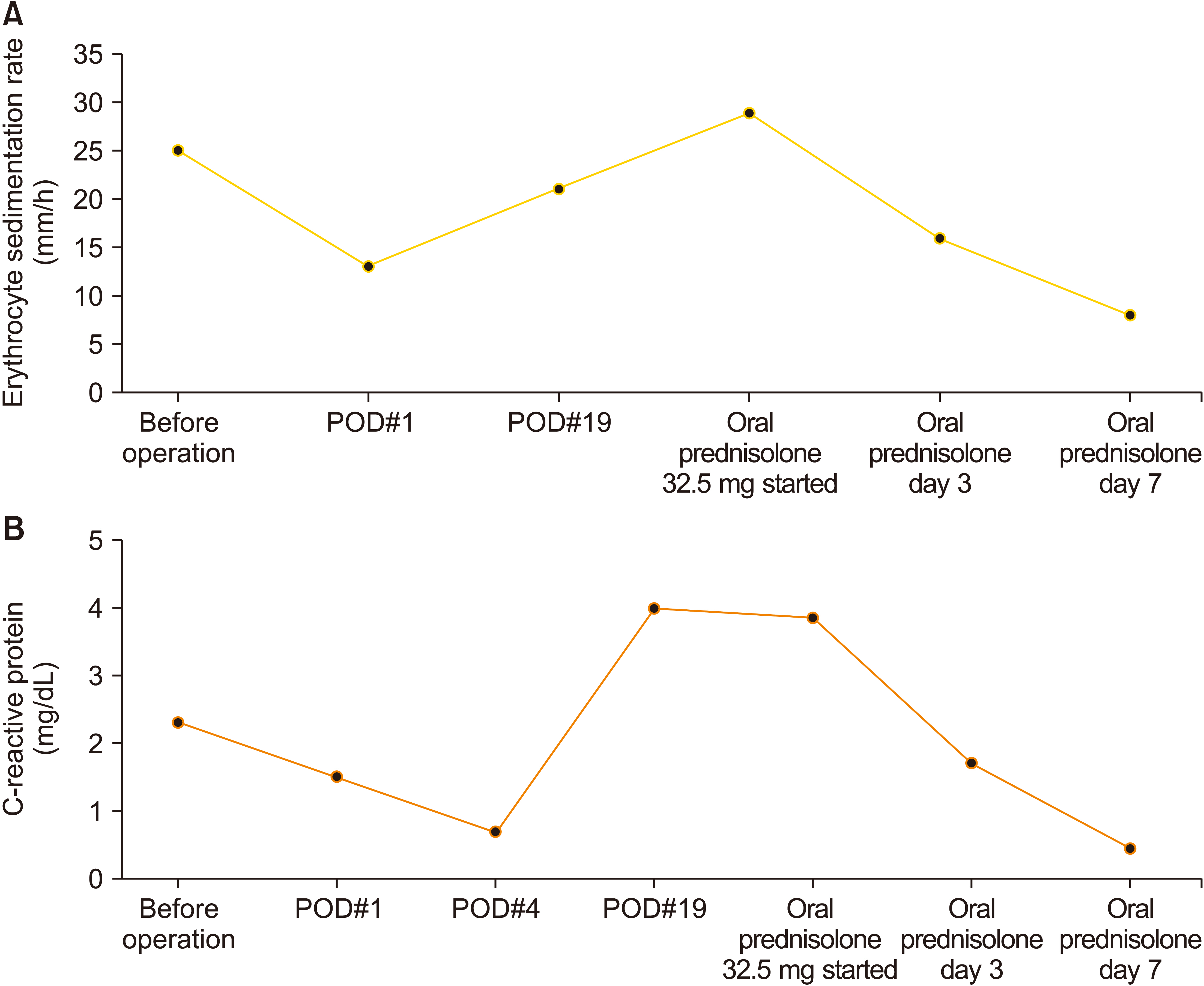
Figure 3 Serial changes of serum acute phase reactants according to time. (A) Erythrocyte sedimentation rate (mm/h); (B) C-reactive protein (mg/dL). POD: postoperation day.
Reference
-
1. Hassan KM, Deb P, Bhatoe HS. 2011; Idiopathic hypertrophic cranial pachymeningitis: three biopsy-proven cases including one case with abdominal pseudotumor and review of the literature. Ann Indian Acad Neurol. 14:189–93. DOI: 10.4103/0972-2327.85891. PMID: 22028532. PMCID: PMC3200042. PMID: f4a28e4185ea463c84aa1c4f9d6510b4.2. Kupersmith MJ, Martin V, Heller G, Shah A, Mitnick HJ. 2004; Idiopathic hypertrophic pachymeningitis. Neurology. 62:686–94. DOI: 10.1212/01.WNL.0000113748.53023.B7. PMID: 15007115.
Article3. Wallace ZS, Naden RP, Chari S, Choi HK, Della-Torre E, Dicaire JF, et al. 2020; The 2019 American College of Rheumatology/European League Against Rheumatism classification criteria for IgG4-related disease. Ann Rheum Dis. 79:77–87. DOI: 10.1136/annrheumdis-2019-216561. PMID: 31796497.
Article4. Deshpande V, Zen Y, Chan JK, Yi EE, Sato Y, Yoshino T, et al. 2012; Consensus statement on the pathology of IgG4-related disease. Mod Pathol. 25:1181–92. DOI: 10.1038/modpathol.2012.72. PMID: 22596100.
Article5. Levraut M, Cohen M, Bresch S, Giordana C, Burel-Vandenbos F, Mondot L, et al. 2019; Immunoglobulin G4-related hypertrophic pachymeningitis: a case-oriented review. Neurol Neuroimmunol Neuroinflamm. 6:e568. DOI: 10.1212/NXI.0000000000000568. PMID: 31355304. PMCID: PMC6624094.
Article6. Dong LL, Sheikh IS, Huang AH, Wu XH, Chen EG, Ying KJ. 2021; Immunoglobulin G4-related disease: case report and literature review. Immunol Res. 69:415–21. DOI: 10.1007/s12026-021-09215-2. PMID: 34374950.
Article7. Lu LX, Della-Torre E, Stone JH, Clark SW. 2014; IgG4-related hypertrophic pachymeningitis: clinical features, diagnostic criteria, and treatment. JAMA Neurol. 71:785–93. DOI: 10.1001/jamaneurol.2014.243. PMID: 24733677.8. Berger JR, Snodgrass S, Glaser J, Post MJ, Norenberg M, Benedetto P. 1989; Multifocal fibrosclerosis with hypertrophic intracranial pachymeningitis. Neurology. 39:1345–9. DOI: 10.1212/WNL.39.10.1345. PMID: 2797457.
Article9. Chan SK, Cheuk W, Chan KT, Chan JK. 2009; IgG4-related sclerosing pachymeningitis: a previously unrecognized form of central nervous system involvement in IgG4-related sclerosing disease. Am J Surg Pathol. 33:1249–52. DOI: 10.1097/PAS.0b013e3181abdfc2. PMID: 19561447.10. Hamano H, Kawa S, Horiuchi A, Unno H, Furuya N, Akamatsu T, et al. 2001; High serum IgG4 concentrations in patients with sclerosing pancreatitis. N Engl J Med. 344:732–8. DOI: 10.1056/NEJM200103083441005. PMID: 11236777.
Article11. Wallace ZS, Carruthers MN, Khosroshahi A, Carruthers R, Shinagare S, Stemmer-Rachamimov A, et al. 2013; IgG4-related disease and hypertrophic pachymeningitis. Medicine (Baltimore). 92:206–16. DOI: 10.1097/MD.0b013e31829cce35. PMID: 23793110. PMCID: PMC4553969.
Article12. Yonekawa T, Murai H, Utsuki S, Matsushita T, Masaki K, Isobe N, et al. 2014; A nationwide survey of hypertrophic pachymeningitis in Japan. J Neurol Neurosurg Psychiatry. 85:732–9. DOI: 10.1136/jnnp-2013-306410. PMID: 24273222.
Article13. Melenotte C, Seguier J, Ebbo M, Kaphan E, Bernit E, Saillier L, et al. 2019; Clinical presentation, treatment and outcome of IgG4-related pachymeningitis: from a national case registry and literature review. Semin Arthritis Rheum. 49:430–7. DOI: 10.1016/j.semarthrit.2019.05.003. PMID: 31155444.
Article14. Saitakis G, Chwalisz BK. 2021; The neurology of IGG4-related disease. J Neurol Sci. 424:117420. DOI: 10.1016/j.jns.2021.117420. PMID: 33845982.
Article15. Masaki Y, Kurose N, Yamamoto M, Takahashi H, Saeki T, Azumi A, et al. 2012; Cutoff values of serum IgG4 and histopathological IgG4+ plasma cells for diagnosis of patients with IgG4-related disease. Int J Rheumatol. 2012:580814. DOI: 10.1155/2012/580814. PMID: 22654917. PMCID: PMC3357966. PMID: 2c5a55b119ac482cbab259cb7a740320.
Article
- Full Text Links
-
- Actions
-
Cited
- CITED
-
- Close
- Share
-
- Similar articles
-
- Immunoglobulin G4-Related Hypertrophic Pachymeningitis with Skull Involvement
- IgG4-Related Hypertrophic Pachymeningitis Mimicking Cerebral Venous Thrombosis
- Immunoglobulin G4-Related Hypertrophic Pachymeningitis Presenting with Multiple Lower Cranial Nerve Palsies
- Immunoglobulin G4-Related Hypertrophic Pachymeningitis Mimicking Chiari Malformation
- IgG4-Related Intracranial Hypertrophic Pachymeningitis : A Case Report and Review of the Literature
