IgG4-Related Intracranial Hypertrophic Pachymeningitis : A Case Report and Review of the Literature
- Affiliations
-
- 1Department of Neurosurgery, National Defense Medical College, Saitama, Japan. s.takeuchi@room.ocn.ne.jp
- KMID: 2018040
- DOI: http://doi.org/10.3340/jkns.2014.55.5.300
Abstract
- Hypertrophic pachymeningitis is an uncommon disorder that causes a localized or diffuse thickening of the dura mater. Recently, the possibility that IgG4-related sclerosing disease may underlie some cases of intracranial hypertrophic pachymeningitis has been suggested. We herein report the tenth case of IgG4-related intracranial hypertrophic pachymeningitis and review the previous literature. A 45-year-old male presented with left-sided focal seizures with generalization. Magnetic resonance imaging (MRI) revealed a diffuse thickening and enhancement of the right convexity dura matter and falx with focal nodularity. The surgically resected specimens exhibited the proliferation of fibroblast-like spindle cells and an infiltration of mononuclear cells, including predominantly plasma cells. The ratio of IgG4-positive plasma cells to the overall IgG-positive cells was 45% in the area containing the highest infiltration of plasma cells. On the basis of the above findings, IgG4-related sclerosing disease arising from the dura mater was suspected. IgG4-related sclerosing disease should be added to the pachymeningitis spectrum.
MeSH Terms
Figure
-
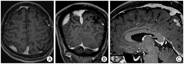
Fig. 1 Preoperative axial (A), coronal (B), and sagittal (C) gadolinium-enhanced magnetic resonance images show irregular thickening and a marked enhancement of the right parieto-occipital dura and falx.
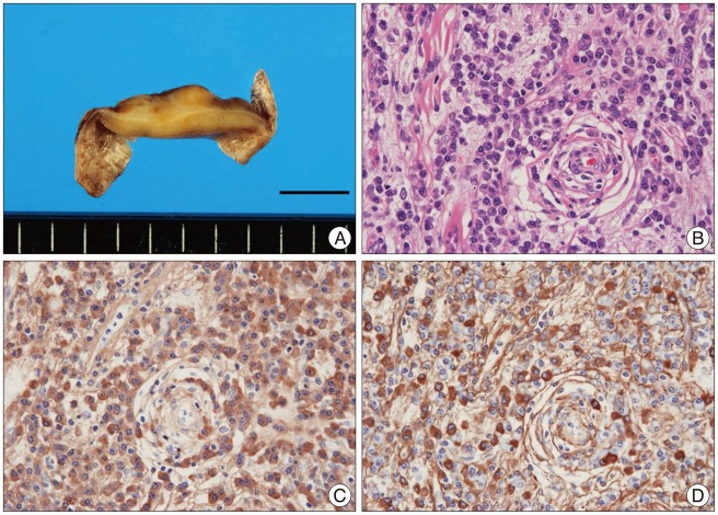
Fig. 2 Pathological findings. A : A macroscopic observation shows a solid yellow to white-colored lesion in the dura mater. B : The photomicrograph shows the solid lesion consisting of the proliferation of fibroblast-like spindle cells and an infiltration of mononuclear cells, including predominantly plasma cells, with abundant collagenous tissue (H&E; original magnification, ×400). C and D : IgG (C) and IgG4 (D) staining reveal that most of the plasma cells infiltrating into the sclerosing lesion are IgG-positive (IgG4-positive plasma cells/high power field, 78), and that the ratio of IgG4-positive plasma cells to the overall IgG-positive cells is 45% in the area containing the highest infiltration of plasma cells (original magnification, ×400).
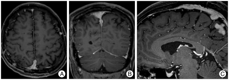
Fig. 3 The twelve-month postoperative axial (A), coronal (B), and sagittal (C) gadolinium-enhanced magnetic resonance images show the disappearance of the mass in the convexity dural region. The mass adjacent to the superior sagittal sinus and falx is left in place.
Reference
-
1. Chan SK, Cheuk W, Chan KT, Chan JK. IgG4-related sclerosing pachymeningitis : a previously unrecognized form of central nervous system involvement in IgG4-related sclerosing disease. Am J Surg Pathol. 2009; 33:1249–1252. PMID: 19561447.2. Choi SH, Lee SH, Khang SK, Jeon SR. IgG4-related sclerosing pachymeningitis causing spinal cord compression. Neurology. 2010; 75:1388–1390. PMID: 20938032.
Article3. Della Torre E, Bozzolo EP, Passerini G, Doglioni C, Sabbadini MG. IgG4-related pachymeningitis : evidence of intrathecal IgG4 on cerebrospinal fluid analysis. Ann Intern Med. 2012; 156:401–403. PMID: 22393144.
Article4. Divatia M, Kim SA, Ro JY. IgG4-related sclerosing disease, an emerging entity : a review of a multi-system disease. Yonsei Med J. 2012; 53:15–34. PMID: 22187229.
Article5. Kim EH, Kim SH, Cho JM, Ahn JY, Chang JH. Imunoglobulin G4-related hypertrophic pachymeningitis involving cerebral parenchyma. J Neurosurg. 2011; 115:1242–1247. PMID: 21854114.
Article6. Kosakai A, Ito D, Yamada S, Ideta S, Ota Y, Suzuki N. A case of definite IgG4-related pachymeningitis. Neurology. 2010; 75:1390–1392. PMID: 20938033.
Article7. Lindstrom KM, Cousar JB, Lopes MB. IgG4-related meningeal disease: clinico-pathological features and proposal for diagnostic criteria. Acta Neuropathol. 2010; 120:765–776. PMID: 20844883.
Article8. Lui PC, Fan YS, Wong SS, Chan AN, Wong G, Chau TK, et al. Inflammatory pseudotumors of the central nervous system. Hum Pathol. 2009; 40:1611–1617. PMID: 19656549.
Article9. Norikane T, Yamamoto Y, Okada M, Maeda Y, Aga F, Kawai N, et al. Hypertrophic cranial pachymeningitis with IgG4-positive plasma cells detected by C-11 methionine PET. Clin Nucl Med. 2012; 37:108–109. PMID: 22157046.
Article
- Full Text Links
-
- Actions
-
Cited
- CITED
-
- Close
- Share
-
- Similar articles
-
- IgG4-Related Hypertrophic Pachymeningitis Mimicking Cerebral Venous Thrombosis
- Immunoglobulin G4-related hypertrophic pachymeningitis with an isolated scalp mass mimicking a brain tumor: a case report and literature review
- Immunoglobulin G4-Related Hypertrophic Pachymeningitis with Skull Involvement
- A Case of IgG4-Related Disease with Pachymeningitis and Periaortitis
- Immunoglobulin G4-Related Hypertrophic Pachymeningitis Presenting with Multiple Lower Cranial Nerve Palsies
