Cerebral fat embolism in sickle cell disease
- Affiliations
-
- 1Service de Médecine Intensive Réanimation, Hôpital Tenon, Assistance Publique – Hôpitaux de Paris, Sorbonne Université, Paris, France
- 2Département de Radiologie, Hôpital Tenon, Assistance Publique – Hôpitaux de Paris, Sorbonne Université, Paris, France
- KMID: 2549492
- DOI: http://doi.org/10.18700/jnc.230024
Figure
-
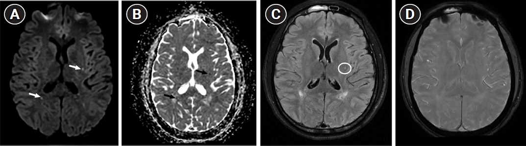
Fig. 1. Initial axial brain magnetic resonance imaging performed 24 hours after admission. (A) Diffusion-weighted imaging (b=1,000 sec/mm2) and (B) the corresponding apparent diffusion coefficient map revealing bilateral scattered punctate foci of restricted diffusion of the supratentorial white matter (arrows). (C) Fluid-attenuated inversion recovery sequence showing hyperintensity of a left putamenal lesion without evidence of collateral flow (circle). (D) Susceptibility-weighted imaging sequence showing no hypointense area in the brain.
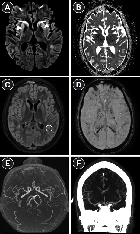
Fig. 2. Second axial brain magnetic resonance imaging performed 26 days after admission. (A) Diffusion-weighted imaging and (B) the corresponding apparent diffusion coefficient map showing bilateral hyperintensity with restricted diffusion of the head of caudate (arrows) and putamen (arrowheads), and left occipital subcortical white matter (asterisks) suggesting additional hypoxic-ischemic encephalopathy. (C) Fluid-attenuated inversion recovery sequence showing hyperintensity of a left occipital white matter lesion (circle) without evidence of collateral flow. (D) The susceptibility-weighted Imaging sequence reveals widespread and consistent pinpoint susceptibilities within the gray matter, gray-white junction, posterior arms of the internal capsules, the splenium of the corpus callosum, basal ganglia, and thalami. This presentation is characteristic of the "walnut kernel microbleed pattern," which strongly indicates cerebral fat embolism. (E, F) The corresponding three-dimensional time-of-flight angiography and computed tomography angiography revealing no abnormalities of intracranial vessels.
Reference
-
1. Dhakal LP, Bourgeois K, Barrett KM, Freeman WD. The "starfield" pattern of cerebral fat embolism from bone marrow necrosis in sickle cell crisis. Neurohospitalist. 2015; 5:74–6.2. Gendreau S, Scholer M, Cecchini J, Habibi A, Razazi K, De Prost N, et al. Cerebral fat embolism in sickle cell disease. Am J Hematol. 2020; 95:E41–5.3. Tsitsikas DA, Vize J, Abukar J. Fat embolism syndrome in sickle cell disease. J Clin Med. 2020; 9:3601.
- Full Text Links
-
- Actions
-
Cited
- CITED
-
- Close
- Share
-
- Similar articles
-
- Gradient-Echo MRI in Defining the Severity of Cerebral Fat Embolism
- Cardiac arrest occurred by cerebral fat embolism
- Cerebral Fat Embolism as a Rare Complication of Postgastrectomy: Case Report
- Cerebral Fat Embolism That Was Initially Negative on DiffusionWeighted Magnetic Resonance Imaging
- Cerebral Fat Embolism after Traumatic Multiple Fracture: A Case Report
