
Gradient-Echo MRI in Defining the Severity of Cerebral Fat Embolism
- Affiliations
-
- 1Department of Neurology, Yeungnam University School of Medicine, Daegu, Korea. junlee@ynu.ac.kr
- KMID: 2287670
- DOI: http://doi.org/10.3988/jcn.2008.4.4.164
Abstract
- BACKGROUND
A few studies have found that abnormal findings on diffusion-weighted magnetic resonance imaging (MRI) are useful for diagnosing cerebral fat embolism in the acute stage. CASE REPORT: We applied serial MRI to a case of cerebral fat embolism with cognitive impairment lasting for 2 months. Although marked resolution of the previous abnormal findings was demonstrated, T2*-weighted gradient-echo MRI revealed multiple tiny lesions. CONCLUSIONS:We suggest that T2*-weighted gradient-echo MRI is useful in defining the clinical severity of patients with cerebral fat embolism.
Figure
-
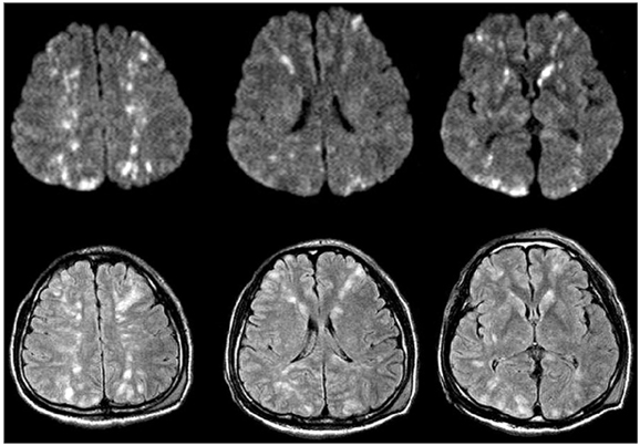
Fig. 1 Multiple scattered high signals were evident on DWI and FLAIR images obtained 10 hours after the onset of drowsiness. DWI: diffusion-weighted MRI, FLAIR: fluid-attenuated inversion recovery.
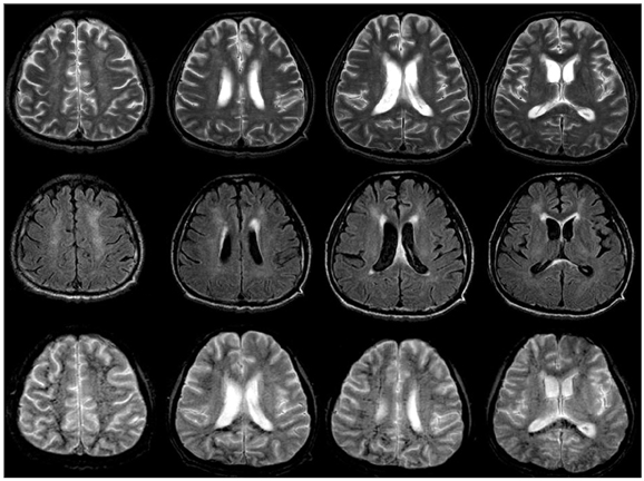
Fig. 2 T2WI, FLAIR, and T2*-weighted gradient-echo MRI was performed on day 50. Most of the lesions had disappeared, but ill-defined high-signal lesions remained in the periventricular and subcortical white matter and in the cortex. T2*-weighted gradient-echo MRI showed numerous punctate low-signal lesions in the centrum semiovale, subcortical white matter, periventricular white matter, and corpus callosum. T2WI: T2-weighted imaging, FLAIR: fluid-attenuated inversion recovery.
Cited by 1 articles
-
Cerebral Fat Embolism after Intramedullary Nailing for Femur and Tibia Fractures: A Case Report
Ki-Hoon Kim, Aleum Lee, Sun-Chul Hwang
Korean J Neurotrauma. 2013;9(2):157-162. doi: 10.13004/kjnt.2013.9.2.157.
Reference
-
1. Marshall GB, Heale VR, Herx L, Abdeen A, Mrkonjic L, Powell J, et al. Magnetic resonance diffusion weighted imaging in cerebral fat embolism. Can J Neurol Sci. 2004. 31:417–421.2. Parizel PM, Demey HE, Veeckmans G, Verstreken F, Cras P, Jorens PG, et al. Early diagnosis of cerebral fat embolism syndrome by diffusion-weighted MRI (starfield pattern). Stroke. 2001. 32:2942–2944.
Article3. Ryu CW, Lee DH, Kim TK, Kim SJ, Kim HS, Lee JH, et al. Cerebral fat embolism: diffusion-weighted magnetic resonance imaging findings. Acta Radiol. 2005. 46:528–533.
Article4. Eguia P, Medina A, Garcia-Monco JC, Martin V, Monton FI. The value of diffusion-weighted MRI in the diagnosis of cerebral fat embolism. J Neuroimaging. 2007. 17:78–80.
Article5. Kamenar E, Burger PC. Cerebral fat embolism: a neuropathological study of a microembolic state. Stroke. 1980. 11:477–484.
Article6. Kim HJ, Lee CH, Kim HG, Lee SD, Son SM, Kim YW, et al. Reversible MR changes in the cat brain after cerebral fat embolism induced by triolein emulsion. AJNR Am J Neuroradiol. 2004. 25:958–963.7. Kamano M, Honda Y, Kitaguchi M, Kazuki K. Cerebral fat embolism after a nondisplaced tibial fracture: case report. Clin Orthop Relat Res. 2001. 206–209.8. Butteriss DJ, Mahad D, Soh C, Walls T, Weir D, Birchall D. Reversible cytotoxic cerebral edema in cerebral fat embolism. AJNR Am J Neuroradiol. 2006. 27:620–623.9. Simon AD, Ulmer JL, Strottmann JM. Contrast-enhanced MR imaging of cerebral fat embolism: case report and review of the literature. AJNR Am J Neuroradiol. 2003. 24:97–101.10. Takahashi M, Suzuki R, Osakabe Y, Asai JI, Miyo T, Nagashima G, et al. Magnetic resonance imaging findings in cerebral fat embolism: correlation with clinical manifestations. J Trauma. 1999. 46:324–327.11. Kim JE, Lee BR, Chun JE, Lee SJ, Lee BH, Yu IK, et al. Cognitive dysfunction in 16 patients with carotid stenosis: detailed neuropsychological findings. J Clin Neurol. 2007. 3:9–17.
Article12. Desmond DW. Cognition and white matter lesions. Cerebrovasc Dis. 2002. 13:Suppl 2. 53–57.
Article13. Parizel PM, Van Goethem JW, Ozsarlak O, Maes M, Phillips CD. New developments in the neuroradiological diagnosis of craniocerebral trauma. Eur Radiol. 2005. 15:569–581.
Article14. Kim BJ, Lee SH. Silent microbleeds and hemorrhagic conversion of an embolic infarction. J Clin Neurol. 2007. 3:147–149.
Article
- Full Text Links
-
- Actions
-
Cited
- CITED
-
- Close
- Share
-
- Similar articles
-
- Cerebral Fat Embolism as a Rare Complication of Postgastrectomy: Case Report
- Cerebral Fat Embolism That Was Initially Negative on DiffusionWeighted Magnetic Resonance Imaging
- Cardiac arrest occurred by cerebral fat embolism
- Magnetic Resonance Findings in Two Episodes of Repeated Cerebral Fat Embolisms in a Patient with Autologous Fat Injection into the Face
- MRI Findings of Cerebral Fat Embolism


