Positional deviation between CBCT-based digital facebow transfer and analog facebow transfer: case series
- Affiliations
-
- 1Department of Prosthodontics, School of Dentistry, Kyungpook National University, Daegu, Republic of Korea
- 2Advanced Dental Device Development Institute, Kyungpook National University, Daegu, Republic of Korea
- 3Department of Dental Science, Graduate School, Kyungpook National University, Daegu, Republic of Korea
- KMID: 2549284
- DOI: http://doi.org/10.14368/jdras.2023.39.3.176
Abstract
- Facebow transfer is essential for accurately mounting a dental cast onto a semi-adjustable articulator. The precision of traditional analog facebow transfer is influenced by both the accuracy of the equipment used and the skill level of the operator. Considering that substantial positional deviations can adversely affect the quality of a fabricated dental prosthesis; it is critical to assess the positional accuracy of casts mounted using analog facebow transfer. This case report evaluates the linear and angular deviations of
the occlusal plane for maxillary casts mounted through both analog facebow transfer and cone-beam computed tomography-based
methods
. The findings indicate that analog facebow transfer produced a linear deviation ranging from 3 to 16 mm and an angular deviation of the occlusal plane between 5 to 7 degrees. This case report confirms that, across two patients, analog facebow transfer can result in varying degrees of positional deviation, thereby potentially leading to inaccuracies in the fabrication of dental pros-theses. These results suggest that, in clinical practice, the use of analog facebow transfer may yield significant deviations during the process of mounting maxillary casts.
Figure
-

Fig. 1 Intraoral photograph and panoramic radiographic image. (A) Frontal view, (B) Maxillary occlusal view, (C) Mandibular occlusal view, (D) Right lateral view, (E) Left lateral view, (F) panoramic radiographic image.

Fig. 2 Facebow transfer procedure.

Fig. 3 Working casts mounted on semi-adjustable articulator. (A) Frontal view, (B) Right lateral view, (C) Left lateral view.
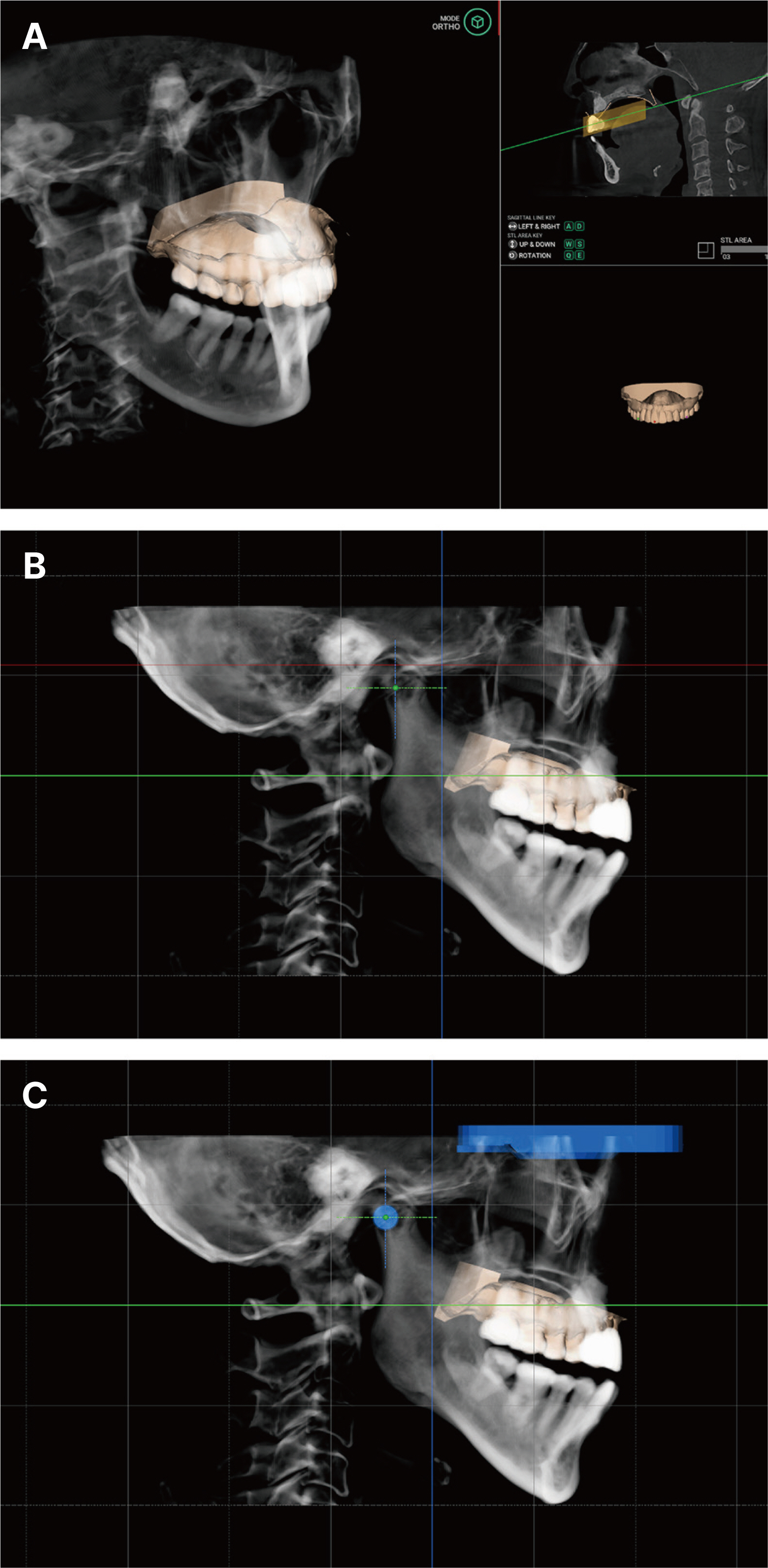
Fig. 4 CBCT-based digital facebow transfer process via software. (A) Aligning a virtual maxillary cast based on CBCT data, (B) Frankfort horizontal plane (red line) and condylar position (green cross) settings, (C) Semiadjustable articulator settings (blue objects indicate the positions of the articulator and condyle).

Fig. 5 Position deviation analysis process via analysis software. (A) Alignment process of virtual maxillary cast (violet color) acquired via conventional facebow based on semi-adjustable articulator data (blue color) acquired CBCT-based digital facebow, (B) Distance deviation in left central incisor and left and right first molars, (C) Measurement of angle deviation of occlusal planes.

Fig. 6 Intraoral photograph and panoramic radiographic image. (A) Frontal view, (B) Maxillary occlusal view, (C) Mandibular occlusal view, (D) Right lateral view, (E) Left lateral view, (F) panoramic radiographic image.
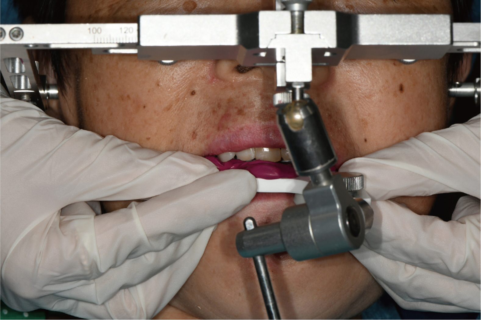
Fig. 7 Facebow transfer procedure.

Fig. 8 Working casts mounted on semi-adjustable articulator. (A) Frontal view, (B) Right lateral view, (C) Left lateral view.
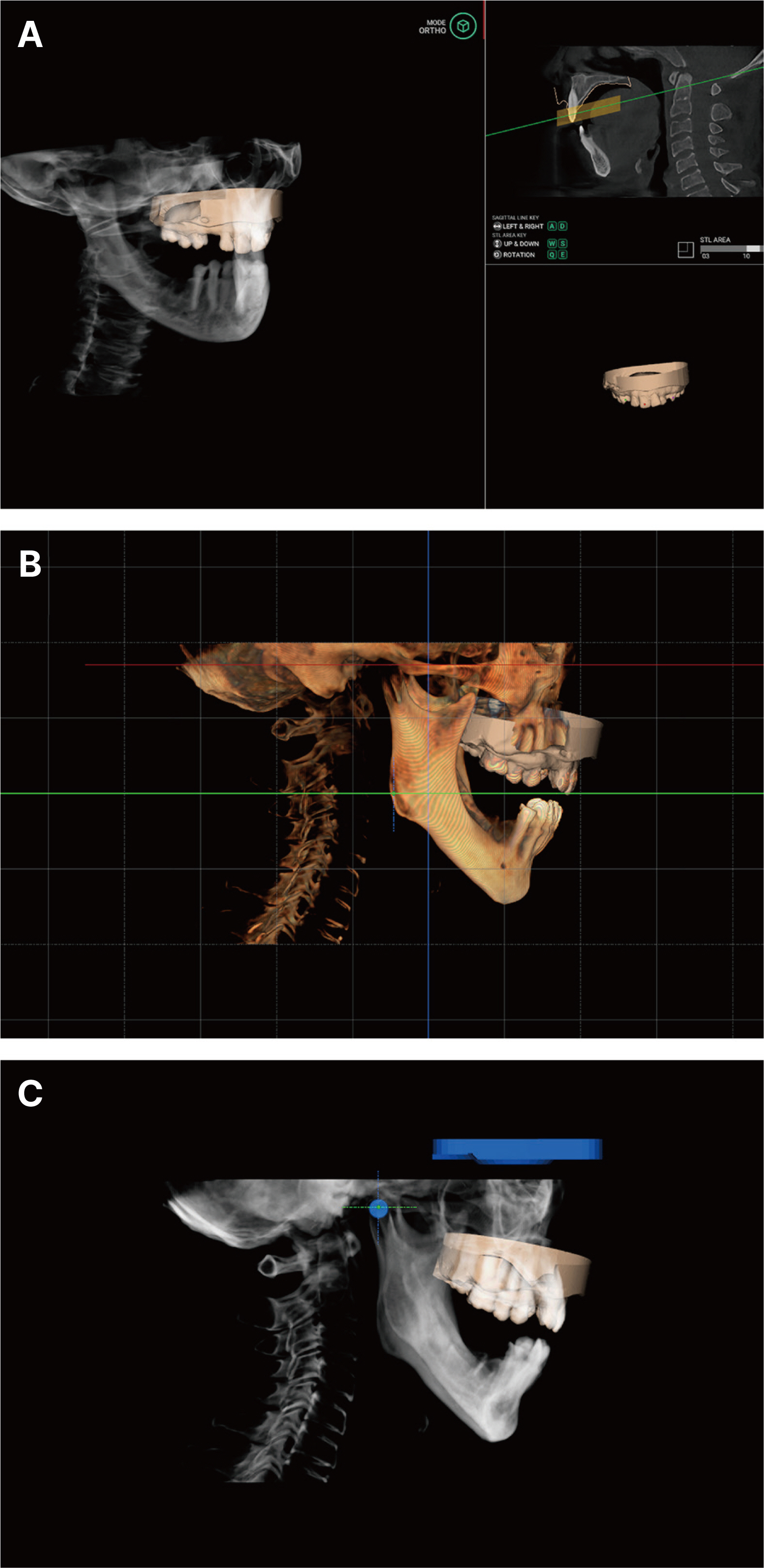
Fig. 9 CBCT-based digital facebow transfer process via software. (A) Aligning a virtual maxillary cast based on CBCT data, (B) Frankfort horizontal plane (red line) settings, (C) Semi-adjustable articulator settings (blue objects indicate the positions of the articulator and condyle).

Fig. 10 Position deviation analysis process via analysis software. (A) Alignment process of virtual maxillary cast (cyan color) acquired via conventional facebow based on semi-adjustable articulator data ellow color) acquired CBCT-based digital facebow, (B) Distance deviation in left central incisor and left and right first molars, (C) Measurement of angle deviation of occlusal planes.
Reference
-
References
1. Son K, Lee JM, Lee KB. 2022; Marginal and internal fit and intaglio surface trueness of temporary crowns fabricated with stereolithography, digital light processing, and milling technology. Int J Prosthodont. 35:697–701. DOI: 10.11607/ijp.7764. PMID: 36511795.2. Lee H, Son K, Lee DH, Kim SY, Lee KB. 2022; Comparison of wear of interim crowns in accordance with the build angle of digital light processing 3D printing: A preliminary in vivo study. Bioengineering. 9:417. DOI: 10.3390/bioengineering9090417. PMID: 36134963. PMCID: PMC9495768.3. Son K, Kim GR, Kim WG, Kang W, Lee DH, Kim SY, Lee JM, Kim YG, Kim JW, Lee ST, Jin MU, Kim HJ, Lee J, Kim JR, Lee KB. 2023; Requirements for Dental CAD Software: A Survey of Korean Dental Personnel. Appl Sci. 13:2803. DOI: 10.3390/app13052803.4. Yang S, Dong B, Zhang Q, Li J, Yuan Q, Yue L. 2022; An indirect digital technique to transfer 3D printed casts to a mechanical articulator with individual sagittal condylar inclination settings using CBCT and intraoral scans. J Prosthodont. 31:822–7. DOI: 10.1111/jopr.13570. PMID: 35864801.5. Revilla-León M, Zeitler JM, Barmak AB, Kois JC. 2022; Jun. 27. Accuracy of the 3-dimensional virtual patient representation obtained by using 4 different techniques: an in vitro study. J Prosthet Dent. S0022-3913(22)00342-0. doi: 10.1016/j.prosdent.2022.05.016. Online ahead of print. DOI: 10.1016/j.prosdent.2022.05.016.6. Li J, Sommer C, Wang HL, Lepidi L, Joda T, Mendonca G. 2021; Creating a virtual patient for completely edentulous computer-aided implant surgery: A dental technique. J Prosthet Dent. 125:564–8. DOI: 10.1016/j.prosdent.2020.02.026. PMID: 32376032.7. Mai HN, Win TT, Tong MS, Lee CH, Lee KB, Kim SY, Lee HW, Lee DH. 2023; Three-dimensional morphometric analysis of facial units in virtual smiling facial images with different smile expressions. J Adv Prosthodont. 15:1–10. DOI: 10.4047/jap.2023.15.1.1. PMID: 36908751. PMCID: PMC9992697.8. Urdalleta DPC, Olmedo RR, Fábrega FRC, Picand JLB. 2022; Individual variation of the distance between nasion and Frankfort horizontal plane - an error factor of facebow in semi-adjustable articulators. J Prosthet Dent. 128:604.e1–5. DOI: 10.1016/j.prosdent.2022.07.007. PMID: 36309468.9. Bowley JF, Michaels GC, Lai TW, Lin PP. 1992; Reliability of a facebow transfer procedure. J Prosthet Dent. 67:491–8. DOI: 10.1016/0022-3913(92)90079-P. PMID: 1507132.10. Thompson GA, Nick C, Francisco P, Lux L, Wiens JP. 2022; Comparison of two arbitrary cast transfer systems with a kinematic facebow for mounting a maxillary cast on a semiadjustable articulator. J Prosthet Dent. 128:597–603. DOI: 10.1016/j.prosdent.2020.12.023. PMID: 33736862.11. Pitchford JH. 1991; A reevaluation of the axis-orbital plane and the use of orbitale in a facebow transfer record. J Prosthet Dent. 66:349–55. DOI: 10.1016/0022-3913(91)90262-U. PMID: 1800732.12. Zambrana N, Sesma N, Fomenko I, Dakir EI, Pieralli S. 2022; Mar. 16. Jaw tracking integration to the virtual patient: A 4D dynamic approach. J Prosthet Dent. S0022-3913(22)00110-X. doi: 10.1016/j.prosdent.2022.02.011. Online ahead of print. DOI: 10.1016/j.prosdent.2022.02.011.13. Li J, Sommer C, Wang HL, Lepidi L, Joda T, Mendonca G. 2021; Creating a virtual patient for completely edentulous computer-aided implant surgery: A dental technique. J Prosthet Dent. 125:564–8. DOI: 10.1016/j.prosdent.2020.02.026. PMID: 32376032.14. Li J, Chen Z, Decker AM, Wang HL, Joda T, Mendonca G, Lepidi L. 2022; Trueness and precision of economical smartphone-based virtual facebow records. J Prosthodont. 31:22–9. DOI: 10.1111/jopr.13366. PMID: 33876857. PMCID: PMC8526632.15. Inoue N, Scialabba R, Lee JD, Lee SJ. 2022; Apr. 2. A comparison of virtually mounted dental casts from traditional facebow records, average values, and 3D facial scans. J Prosthet Dent. S0022-3913(22)00146-9. doi: 10.1016/j.prosdent.2022.03.001. Online ahead of print. DOI: 10.1016/j.prosdent.2022.03.001. PMID: 35382941.16. Amezua X, Iturrate M, Garikano X, Solaberrieta E. 2022; Analysis of the influence of the facial scanning method on the transfer accuracy of a maxillary digital scan to a 3D face scan for a virtual facebow technique: An in vitro study. J Prosthet Dent. 128:1024–31. DOI: 10.1016/j.prosdent.2021.02.007. PMID: 33722381.17. Palaskar JN, Joshi N, Gullapalli P, Shah P. 2020; Comparative evaluation of sagittal inclination of the occlusal plane with Frankfort horizontal plane in facebow transfers to semiadjustable and fully adjustable articulators. J Prosthet Dent. 123:299–304. DOI: 10.1016/j.prosdent.2018.12.024. PMID: 31227235.18. Maveli TC, Suprono MS, Kattadiyil MT, Goodacre CJ, Bahjri K. 2015; In vitro comparison of the maxillary occlusal plane orientation obtained with five facebow systems. J Prosthet Dent. 114:566–73. DOI: 10.1016/j.prosdent.2015.02.030. PMID: 26139043.
- Full Text Links
-
- Actions
-
Cited
- CITED
-
- Close
- Share
-
- Similar articles
-
- Fabrication of CAD-CAM complete denture using existing provisional denture and digital facebow transfer
- Posterior rehabilitation considering mandibular movement with digital facebow transfer and virtual articulator: A case report
- In vitro comparison of the accuracy of an occlusal plane transfer method between facebow and POP bow systems in asymmetric ear position
- Full-mouth rehabilitation of a patient with severe wear using digital facebow transfer and virtual articulator
- Full-mouth rehabilitation of severely attrited dentition with missing posterior teeth: a case report using digital workflow with jaw motion tracking
