Various hand tumors: over 20 years of clinical experience in Korea
- Affiliations
-
- 1Department of Plastic and Reconstructive Surgery, Keimyung University Dongsan Hospital, Keimyung University School of Medicine, Daegu, Korea
- 2Department of Plastic and Reconstructive Surgery, Saeson Hospital, Daejeon, Korea
- KMID: 2548596
- DOI: http://doi.org/10.12790/ahm.23.0026
Abstract
- Purpose
Although the hand only constitutes 2% of the body, it is a site where a wide variety of tumors occur, and the results of surgery substantially impact its function. Therefore, an accurate diagnosis and appropriate selection of individual treatment strategies are very important. This study reviewed the diagnostic characteristics and clinical features of surgically treated hand tumors at our facility.
Methods
We retrospectively reviewed 65 patients who underwent surgery for suspected hand tumors at our institution between 2003 and 2020. Data, including patient demographics, diagnosis, pathology, type of surgery, comorbidities, and tumor characteristics (e.g., location and tumor size), were collected from medical records.
Results
In total, 59 (90.7%) and six patients (9.3%) were diagnosed with benign and malignant tumors, respectively. Ganglion cysts were the most common tumors (n=11), followed by vascular anomalies (n=10), epidermoid cysts (n=7), and glomus tumors (n=4). There were four cases of bone tumors (6.2%), including one chondroma, one chondrosarcoma, and two giant cell tumors. In 53 cases with an imaging workup, the pathological diagnosis was concordant in 44 cases. Of the nine cases with inconsistent results, eight were tumors located in the finger; this association was significant (p=0.048).
Conclusion
In this study, ganglion cyst and squamous cell carcinoma were the most common benign and malignant tumors, respectively, and four cases of bone tumors were found. Radiologic misdiagnosis was more frequent in finger tumors than in other tumors. Thus, hand tumors, especially those suspected to be malignant, should be assessed using a multidisciplinary approach.
Figure
-

Fig. 1. A 55-year-old man with a suspected glomus tumor underwent surgery; after the first operation, it was diagnosed as a fibroma. (A, B) Gross views of the finger lesion. (C) Postoperative biopsy. (D) The lesion was diagnosed as a suspected glomus tumor by a radiologist following preoperative ultrasonography. (E) The tumor recurred after 1 year. Following reexcision, it was diagnosed as clear cell sarcoma. (F) The defect was covered by a cross-finger flap. (G) After 3 weeks, flap detachment was performed. (H) After 6 months, the lesion had healed well, without complications.
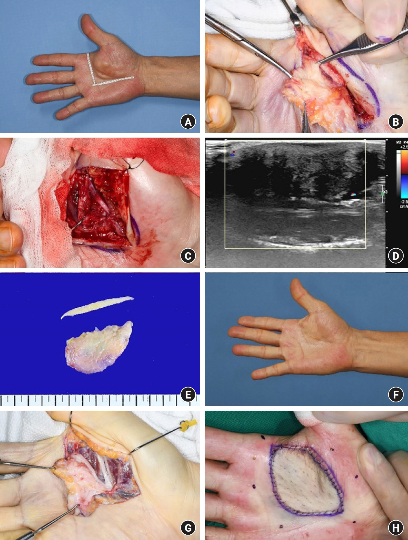
Fig. 2. A 62-year-old man, suspected to have a fibroma, underwent surgery and was diagnosed with a granular cell tumor (benign). (A, B) Gross views of the palm lesion. (C) Fibrotic tissue was found, involving the common distal nerve to the index finger. Neurorrhaphy was performed following partial excision of the tumor. (D) The lesion was diagnosed as suspected fibromatosis by a radiologist following preoperative ultrasonography. (E) Postoperative biopsy. (F) After 1 year, a remnant mass was observed at the same site. (G) A secondary excision was performed. (H) The defect was covered by a full-thickness skin graft.
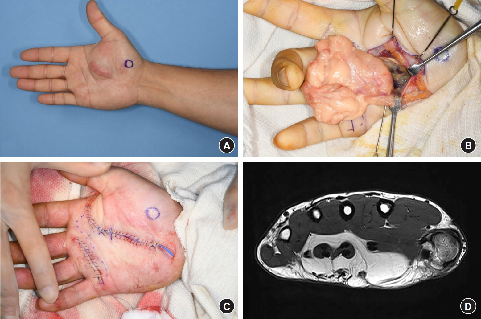
Fig. 3. A 43-year-old man suspected to have a lipoid tumor underwent surgery and was diagnosed as having a benign lipoma. (A) A gross view of the palm lesion. (B) Mass excision was done. (C) Postoperative view. (D) The lesion was diagnosed as an atypical lipoid tumor or well-differentiated liposarcoma by a radiologist following preoperative magnetic resonance imaging.
Reference
-
References
1. Pertea M, Grosu OM, Filip A, et al. Rare benign tumors and tumor-like lesions of the hand without skin damage-clinical, imagistic and histopathological diagnosis, retrospective study. Diagnostics (Basel). 2023; 13:1204.
Article2. Rodriguez FJ, Folpe AL, Giannini C, Perry A. Pathology of peripheral nerve sheath tumors: diagnostic overview and update on selected diagnostic problems. Acta Neuropathol. 2012; 123:295–319.
Article3. Garcia J, Bianchi S. Diagnostic imaging of tumors of the hand and wrist. Eur Radiol. 2001; 11:1470–82.
Article4. Nepal P, Songmen S, Alam SI, Gandhi D, Ghimire N, Ojili V. Common soft tissue tumors involving the hand with histopathological correlation. J Clin Imaging Sci. 2019; 9:15.
Article5. Fujibuchi T, Imai H, Miyawaki J, Kidani T, Kiyomatsu H, Miura H. Hand tumors: a review of 186 patients at a single institute. J Orthop Surg (Hong Kong). 2021; 29:2309499021993994.
Article6. Amer K, Otero K, Smith B, Datiashvili R. Hand tumors: an individual surgeon’s retrospective review. Eplasty. 2022; 22:e1.7. AbuMoussa S, Roshan MP, Souza FF, et al. Soft tissue masses of the hand: a review of clinical presentation and imaging features. Curr Oncol. 2023; 30:2032–48.
Article8. Thornburg LE. Ganglions of the hand and wrist. J Am Acad Orthop Surg. 1999; 7:231–8.
Article9. Mavrogenis AF, Panagopoulos GN, Angelini A, et al. Tumors of the hand. Eur J Orthop Surg Traumatol. 2017; 27:747–62.
Article10. Datta NK, Das KP, Aish PK, et al. Management of the hand tumors. Mymensingh Med J. 2023; 32:135–43.11. Henderson M, Neumeister MW, Bueno RA Jr. Hand tumors: II. Benign and malignant bone tumors of the hand. Plast Reconstr Surg. 2014; 133:814e–821e.12. Henderson MM, Neumeister MW, Bueno RA Jr. Hand tumors: I. skin and soft-tissue tumors of the hand. Plast Reconstr Surg. 2014; 133:154e–164e.13. Lin PP, Guzel VB, Pisters PW, et al. Surgical management of soft tissue sarcomas of the hand and foot. Cancer. 2002; 95:852–61.
Article14. Bray PW, Bell RS, Bowen CV, Davis A, O’Sullivan B. Limb salvage surgery and adjuvant radiotherapy for soft tissue sarcomas of the forearm and hand. J Hand Surg Am. 1997; 22:495–503.
Article15. Pradhan A, Cheung YC, Grimer RJ, et al. Soft-tissue sarcomas of the hand: oncological outcome and prognostic factors. J Bone Joint Surg Br. 2008; 90:209–14.
