Resin infiltrant protects deproteinized dentin against erosive and abrasive wear
- Affiliations
-
- 1Department of Dentistry, School of Dentistry, University of Fortaleza, Fortaleza, CE, Brazil
- 2Department of Restorative Dentistry, School of Pharmacy, Dentistry and Nursing, Federal University of Ceará, Fortaleza, CE, Brazil
- 3Dental Biomedical Sciences Ph.D. Program, University of Maryland School of Dentistry, Baltimore, MD, USA
- 4Division of Operative Dentistry, Department of General Dentistry, University of Maryland School of Dentistry, Baltimore, MD, USA
- KMID: 2548133
- DOI: http://doi.org/10.5395/rde.2022.47.e29
Abstract
Objectives
This study aimed to investigate the anti-erosive/abrasive effect of resin infiltration of previous deproteinized dentin.
Materials and Methods
Dentin slabs were randomly assigned to 3 groups (n = 15): Control (no deproteinization; no resin infiltrant applied), RI (no deproteinization; resin infiltrant applied), and DRI (deproteinization; resin infiltrant applied). After undergoing the assigned treatment, all slabs were subjected to an in vitro cycling model for 5 days. The specimens were immersed in citric acid (0.05 M, pH = 3.75; 60 seconds; 3 times/day) and brushed (150 strokes). Between the challenges, the specimens were exposed to a remineralizing solution (60 minutes). The morphological alterations were analyzed by mechanical profilometry (µm) and scanning electron microscopy (SEM). Data were submitted to one-way analysis of variance (ANOVA) and Tukey tests (p < 0.05).
Results
Control and RI groups presented mineral wear and did not significantly differ from each other (p = 0.063). DRI maintained a protective layer preserving the dentin (p < 0.001). After erosive/abrasive cycles, it was observed that in group RI, only 25% of the slabs partially evidenced the presence of the infiltrating, while, in the DRI group, 80% of the slabs presented the treated surface entirely covered by a resin-component layer protecting the dentin surface as observed in SEM images.
Conclusions
The removal of the organic content allows the resin infiltrant to efficiently protect the dentin surface against erosive/abrasive lesions.
Figure
-
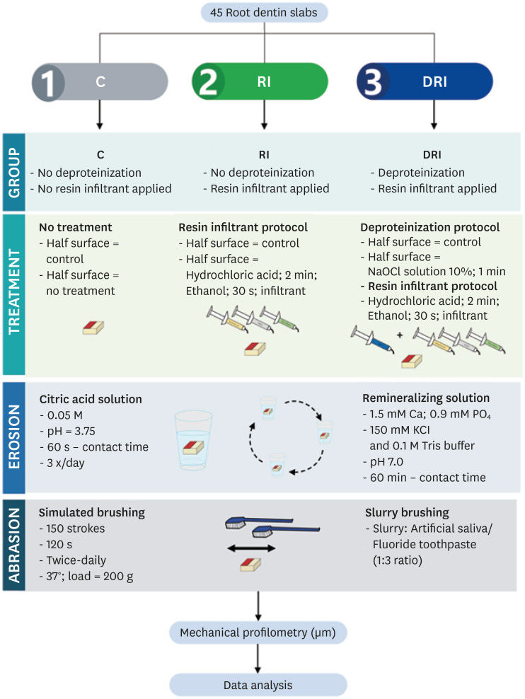
Figure 1 Experimental design.C, no deproteinization performed and no resin infiltrant applied (control); RI, no deproteinization performed and resin infiltrant applied; DRI, deproteinization performed and resin infiltrant applied.
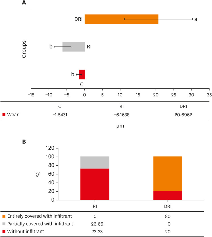
Figure 2 Wear and presence of infiltrant data on the dentin surface. (A) Mean and standard deviation of the wear/protective barrier obtained in the experimental groups. (B) Distribution of the presence of the infiltrant on the surface from groups that received the resinous infiltration.C, no deproteinization performed and no resin infiltrant applied (control); RI, no deproteinization performed and resin infiltrant applied; DRI, deproteinization performed and resin infiltrant applied.
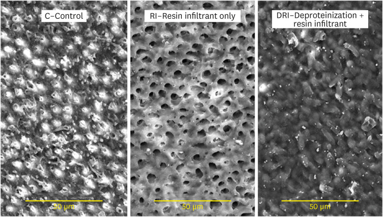
Figure 3 Scanning electron microscopy images of the dentin surface after different surface treatments. (×2,000).
Reference
-
1. Bartlett DW, Lussi A, West NX, Bouchard P, Sanz M, Bourgeois D. Prevalence of tooth wear on buccal and lingual surfaces and possible risk factors in young European adults. J Dent. 2013; 41:1007–1013. PMID: 24004965.
Article2. Grippo JO, Simring M, Coleman TA. Abfraction, abrasion, biocorrosion, and the enigma of noncarious cervical lesions: a 20-year perspective. J Esthet Restor Dent. 2012; 24:10–23. PMID: 22296690.
Article3. Lussi A, Carvalho TS. Erosive tooth wear: a multifactorial condition of growing concern and increasing knowledge. Monogr Oral Sci. 2014; 25:1–15. PMID: 24993253.4. Ganss C. Definition of erosion and links to tooth wear. Monogr Oral Sci. 2006; 20:9–16. PMID: 16687881.
Article5. Ganss C, Lussi A. Diagnosis of erosive tooth wear. Monogr Oral Sci. 2006; 20:32–43. PMID: 16687883.
Article6. O’Toole S, Bartlett D. The relationship between dentine hypersensitivity, dietary acid intake and erosive tooth wear. J Dent. 2017; 67:84–87. PMID: 29017845.
Article7. Peumans M, Politano G, Van Meerbeek B. Treatment of noncarious cervical lesions: when, why, and how. Int J Esthet Dent. 2020; 15:16–42. PMID: 31994534.8. de Moraes MDR, Carneiro JR, Passos VF, Santiago SL. Effect of green tea as a protective measure against dental erosion in coronary dentine. Braz Oral Res. 2016; 30:1–6.
Article9. Machado AC, Bezerra SJC, João-Souza SH, Caetano TM, Russo LC, Carvalho TS, Scaramucci T. Using fluoride mouthrinses before or after toothbrushing: effect on erosive tooth wear. Arch Oral Biol. 2019; 108:104520. PMID: 31445424.
Article10. Passos VF, de Vasconcellos AA, Pequeno JH, Rodrigues LK, Santiago SL. Effect of commercial fluoride dentifrices against hydrochloric acid in an erosion-abrasion model. Clin Oral Investig. 2015; 19:71–76.
Article11. Passos VF, Melo MAS, Lima JPM, Marçal FF, Costa CAGA, Rodrigues LKA, Santiago SL. Active compounds and derivatives of camellia sinensis responding to erosive attacks on dentin. Braz Oral Res. 2018; 32:e40. PMID: 29846385.
Article12. Passos VF, Rodrigues LKA, Santiago SL. The effect of magnesium hydroxide-containing dentifrice using an extrinsic and intrinsic erosion cycling model. Arch Oral Biol. 2018; 86:46–50. PMID: 29156417.
Article13. Wegehaupt FJ, Tauböck TT, Sener B, Attin T. Long-term protective effect of surface sealants against erosive wear by intrinsic and extrinsic acids. J Dent. 2012; 40:416–422. PMID: 22326565.
Article14. Wegehaupt FJ, Tauböck TT, Attin T. Durability of the anti-erosive effect of surfaces sealants under erosive abrasive conditions. Acta Odontol Scand. 2013; 71:1188–1194. PMID: 23294118.
Article15. Zhao X, Pan J, Zhang S, Malmstrom HS, Ren YF. Effectiveness of resin-based materials against erosive and abrasive enamel wear. Clin Oral Investig. 2017; 21:463–468.
Article16. Paris S, Bitter K, Krois J, Meyer-Lueckel H. Seven-year-efficacy of proximal caries infiltration - randomized clinical trial. J Dent. 2020; 93:103277. PMID: 31931026.
Article17. Oliveira GC, Boteon AP, Ionta FQ, Moretto MJ, Honório HM, Wang L, Rios D. In vitro effects of resin infiltration on enamel erosion inhibition. Oper Dent. 2015; 40:492–502. PMID: 25587972.18. Rios D, Oliveira GC, Zampieri CR, Jordão MC, Dionisio EJ, Buzalaf M, Wang L, Honório HM. Resin-based materials protect against erosion/abrasion-a prolonged in situ study. Oper Dent. 2019; 44:302–311. PMID: 30629465.
Article19. Skucha-Nowak M, Machorowska-Pieniążek A, Tanasiewicz M. Assessing the penetrating abilities of experimental preparation with dental infiltrant features using optical microscope: preliminary study. Adv Clin Exp Med. 2016; 25:961–969. PMID: 28028962.
Article20. Alshaikh KH, Hamama HHH, Mahmoud SH. Effect of smear layer deproteinization on bonding of self-etch adhesives to dentin: a systematic review and meta-analysis. Restor Dent Endod. 2018; 43:e14. PMID: 29765895.
Article21. Thanatvarakorn O, Prasansuttiporn T, Thittaweerat S, Foxton RM, Ichinose S, Tagami J, Hosaka K, Nakajima M. Smear layer-deproteinizing improves bonding of one-step self-etch adhesives to dentin. Dent Mater. 2018; 34:434–441. PMID: 29233540.
Article22. Augusto MG, Torres C, Pucci CR, Schlueter N, Borges AB. Bond stability of a universal adhesive system to eroded/abraded dentin after deproteinization. Oper Dent. 2018; 43:291–300. PMID: 29676982.23. Esteves SR, Huhtala MF, Gomes AP, Ye Q, Spencer P, De Paiva Gonçalves SE. Longitudinal effect of surface treatments modified by NaOCl-induced deproteinization and Nd:YAG laser on dentin permeability. Photomed Laser Surg. 2016; 34:68–75. PMID: 26799835.
Article24. Reddy A, Norris DF, Momeni SS, Waldo B, Ruby JD. The pH of beverages in the United States. J Am Dent Assoc. 2016; 147:255–263. PMID: 26653863.25. Abdelmegid FY. Effect of deproteinization before and after acid etching on the surface roughness of immature permanent enamel. Niger J Clin Pract. 2018; 21:591–596. PMID: 29735859.
Article26. Soares LE, Soares AL, De Oliveira R, Nahórny S. The effects of acid erosion and remineralization on enamel and three different dental materials: FT-Raman spectroscopy and scanning electron microscopy analysis. Microsc Res Tech. 2016; 79:646–656. PMID: 27145291.
Article27. Arnold WH, Gaengler P. Light- and electronmicroscopic study of infiltration of resin into initial caries lesions--a new methodological approach. J Microsc. 2012; 245:26–33. PMID: 21919904.
Article28. Yetkiner E, Wegehaupt FJ, Attin R, Wiegand A, Attin T. Stability of two resin combinations used as sealants against toothbrush abrasion and acid challenge in vitro . Acta Odontol Scand. 2014; 72:825–830. PMID: 24850503.
Article29. Schlueter N, Hara A, Shellis RP, Ganss C. Methods for the measurement and characterization of erosion in enamel and dentine. Caries Res. 2011; 45(Supplement 1):13–23.
Article
- Full Text Links
-
- Actions
-
Cited
- CITED
-
- Close
- Share
-
- Similar articles
-
- Compatibility of self-etching dentin adhesives with resin luting cements
- The effects of desensitizing agents, bonding resin and tooth brushing on dentin permeability, in vitro
- Diagnostic Utilization of Laser Fluorescence for Resin Infiltration in Primary Teeth
- COMPARISON OF WEAR RESISTANCE AMONG RESIN DENTURE TEETH OPPOSING VARIOUS RESTORATIVE MATERIALS
- The effect of repeated bonding on the shear bond strength of different resin cements to enamel and dentin
