Chitosan-induced biomodification on demineralized dentin to improve the adhesive interface
- Affiliations
-
- 1Department of Restorative Dentistry, School of Dentistry of Ribeirão Preto, University of São Paulo, Ribeirão Preto, SP, Brazil
- KMID: 2548132
- DOI: http://doi.org/10.5395/rde.2022.47.e28
Abstract
Objectives
Metalloproteinase-inhibiting agents, such as chitosan, can prevent collagen degradation in demineralized dental substrates, thereby improving the adhesive interface. This study evaluated the bond strength (BS) and chemical and morphological characterization of the adhesive interface after applying chitosan solution to demineralized dentin.
Materials and Methods
The 80 third molars were selected. Forty teeth underwent caries induction using the pH cycling method. The teeth were divided according to the treatment: distilled water (control) and 2.5% chitosan solution. The surfaces were restored using adhesive and composite resins. Half of the specimens in each group were aged, and the other half underwent immediate analyses. The teeth were sectioned and underwent the microtensile bond strength test (µTBS), and chemical and morphological analyses using energy-dispersive spectroscopy and scanning electron microscopy, respectively. Data analysis was performed using 3-way analysis of variance.
Results
For µTBS, sound dentin was superior to demineralized dentin (p < 0.001), chitosantreated specimens had higher bond strength than the untreated ones (p < 0.001), and those that underwent immediate analysis had higher values than the aged specimens (p = 0.019). No significant differences were observed in the chemical or morphological compositions.
Conclusions
Chitosan treatment improved bond strength both immediately and after aging, even in demineralized dentin.
Keyword
Figure
-
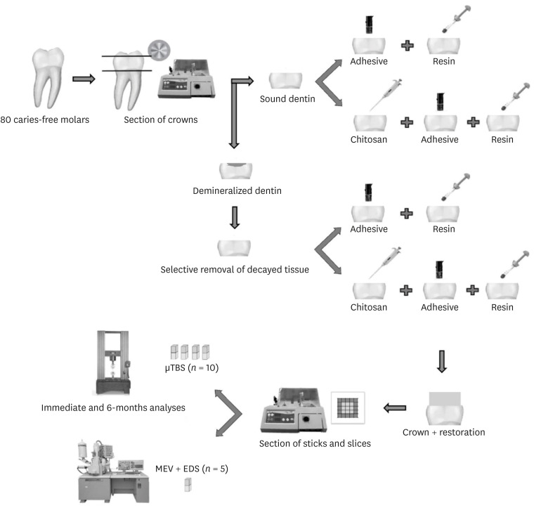
Figure 1 Schematic diagram of the experimental workflow.
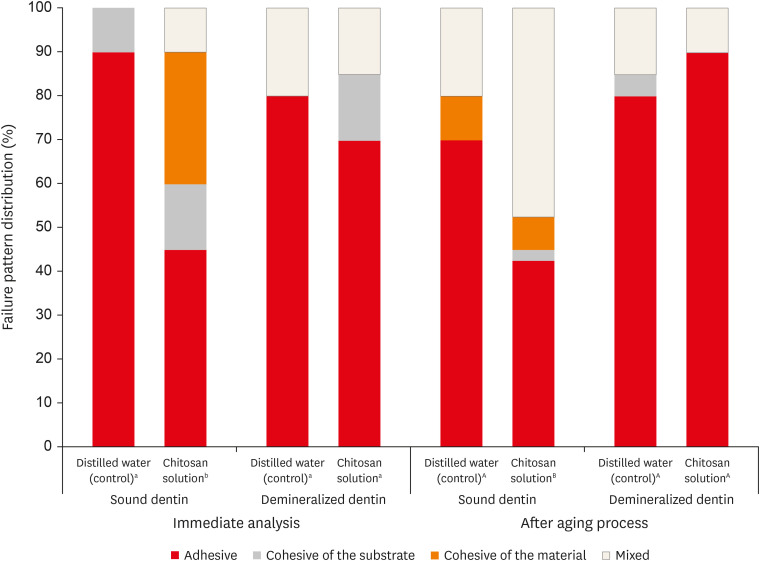
Figure 2 Frequency distribution of the failure patterns (%) after microtensile bond strength (µTBS) test. Different letters indicate significant difference among groups. Capital letters correspond to immediate analysis and lowercase letters to after aging process (χ2 test, p < 0.05).
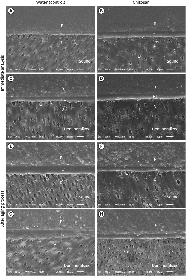
Figure 3 Photomicrographs (×1,000) of the adhesive interface - Immediately after the adhesive procedure: (A) Sound dentin with water (control); (B) Sound dentin with chitosan; (C) Demineralized dentin with water and (D) Demineralized dentin with chitosan. After aging of the adhesive interface: (E) Sound dentin with water (control); F) Sound dentin with chitosan; (G) Demineralized dentin with water; and (H) Demineralized dentin with chitosan. R, resin; HL, hybrid layer; D, dentin; T, resin tags.
Reference
-
1. Brostek AM, Walsh LJ. Minimal intervention dentistry in general practice. Oral Health Dent Manag. 2014; 13:285–294. PMID: 24984635.2. Comert S, Oz AA. Clinical effect of a fluoride-releasing and rechargeable primer in reducing white spot lesions during orthodontic treatment. Am J Orthod Dentofacial Orthop. 2020; 157:67–72. PMID: 31901283.
Article3. Perdigão J. Dentin bonding-variables related to the clinical situation and the substrate treatment. Dent Mater. 2010; 26:e24–e37. PMID: 20005565.
Article4. Chaussain-Miller C, Fioretti F, Goldberg M, Menashi S. The role of matrix metalloproteinases (MMPs) in human caries. J Dent Res. 2006; 85:22–32. PMID: 16373676.5. Kato MT, Leite AL, Hannas AR, Calabria MP, Magalhães AC, Pereira JC, Buzalaf MA. Impact of protease inhibitors on dentin matrix degradation by collagenase. J Dent Res. 2012; 91:1119–1123. PMID: 23023765.
Article6. Park KM, Lee HJ, Koo KT, Ben Amara H, Leesungbok R, Noh K, Lee SC, Lee SW. Oral soft tissue regeneration using nano controlled system inducing sequential release of trichloroacetic acid and epidermal growth factor. Tissue Eng Regen Med. 2020; 17:91–103. PMID: 31970697.
Article7. Mazzi-Chaves JF, Martins CV, Souza-Gabriel AE, Brito-Jùnior M, Cruz-Filho AM, Steier L, Sousa-Neto MD. Effect of a chitosan final rinse on the bond strength of root canal fillings. Gen Dent. 2019; 67:54–57. PMID: 31454324.8. Machado AH, Garcia IM, Motta AS, Leitune VC, Collares FM. Triclosan-loaded chitosan as antibacterial agent for adhesive resin. J Dent. 2019; 83:33–39. PMID: 30794843.
Article9. Rodrigues MR. Synthesis and investigation of chitosan derivatives formed by reaction with acyl chlorides. J Carbohydr Chem. 2005; 24:41–54.
Article10. Kong M, Chen XG, Xing K, Park HJ. Antimicrobial properties of chitosan and mode of action: a state of the art review. Int J Food Microbiol. 2010; 144:51–63. PMID: 20951455.
Article11. Chronopoulou L, Nocca G, Castagnola M, Paludetti G, Ortaggi G, Sciubba F, Bevilacqua M, Lupi A, Gambarini G, Palocci C. Chitosan based nanoparticles functionalized with peptidomimetic derivatives for oral drug delivery. N Biotechnol. 2016; 33:23–31. PMID: 26257139.
Article12. Curylofo-Zotti FA, Tanta GS, Zucoloto ML, Souza-Gabriel AE, Corona SA. Selective removal of carious lesion with Er:YAG laser followed by dentin biomodification with chitosan. Lasers Med Sci. 2017; 32:1595–1603. PMID: 28762194.
Article13. Baena E, Cunha SR, Maravić T, Comba A, Paganelli F, Alessandri-Bonetti G, Ceballos L, Tay FR, Breschi L, Mazzoni A. Effect of chitosan as a cross-linker on matrix metalloproteinase activity and bond stability with different adhesive systems. Mar Drugs. 2020; 18:18.
Article14. Persadmehr A, Torneck CD, Cvitkovitch DG, Pinto V, Talior I, Kazembe M, Shrestha S, McCulloch CA, Kishen A. Bioactive chitosan nanoparticles and photodynamic therapy inhibit collagen degradation in vitro . J Endod. 2014; 40:703–709. PMID: 24767568.
Article15. Anjana J, Mohandas A, Seethalakshmy S, Suresh MK, Menon R, Biswas R, Jayakumar R. Bi-layered nanocomposite bandages for controlling microbial infections and overproduction of matrix metalloproteinase activity. Int J Biol Macromol. 2018; 110:124–132. PMID: 29233714.
Article16. Curylofo-Zotti FA, Scheffel DL, Macedo AP, Souza-Gabriel AE, Hebling J, Corona SA. Effect of Er:YAG laser irradiation and chitosan biomodification on the stability of resin/demineralized bovine dentin bond. J Mech Behav Biomed Mater. 2019; 91:220–228. PMID: 30597375.17. Pini NI, Lima DA, Luka B, Ganss C, Schlueter N. Viscosity of chitosan impacts the efficacy of F/Sn containing toothpastes against erosive/abrasive wear in enamel. J Dent. 2020; 92:103247. PMID: 31743693.
Article18. Fawzy AS, Nitisusanta LI, Iqbal K, Daood U, Beng LT, Neo J. Chitosan/Riboflavin-modified demineralized dentin as a potential substrate for bonding. J Mech Behav Biomed Mater. 2013; 17:278–289. PMID: 23127636.
Article19. Hashimoto M, Tay FR, Svizero NR, de Gee AJ, Feilzer AJ, Sano H, Kaga M, Pashley DH. The effects of common errors on sealing ability of total-etch adhesives. Dent Mater. 2006; 22:560–568. PMID: 16289724.
Article20. Marquezan PK, Alves LS, Dalla Nora A, Maltz M, do Amaral Zenkner JE. Radiographic pattern of underlying dentin lesions (ICDAS 4) in permanent teeth. Clin Oral Investig. 2019; 23:3879–3883.
Article21. Daood U, Iqbal K, Nitisusanta LI, Fawzy AS. Effect of chitosan/riboflavin modification on resin/dentin interface: spectroscopic and microscopic investigations. J Biomed Mater Res A. 2013; 101:1846–1856. PMID: 23184366.
Article22. Profeta AC, Mannocci F, Foxton RM, Thompson I, Watson TF, Sauro S. Bioactive effects of a calcium/sodium phosphosilicate on the resin-dentine interface: a microtensile bond strength, scanning electron microscopy, and confocal microscopy study. Eur J Oral Sci. 2012; 120:353–362. PMID: 22813227.
Article23. Borsatto MC, Martinelli MG, Contente MM, Mellara TS, Pécora JD, Galo R. Bond durability of Er:YAG laser-prepared primary tooth enamel. Braz Dent J. 2013; 24:330–334. PMID: 24173250.
Article24. Ganss C, Klimek J, Brune V, Schürmann A. Effects of two fluoridation measures on erosion progression in human enamel and dentine in situ . Caries Res. 2004; 38:561–566. PMID: 15528912.
Article25. Castellan CS, Bedran-Russo AK, Antunes A, Pereira PN. Effect of dentin biomodification using naturally derived collagen cross-linkers: one-year bond strength study. Int J Dent. 2013; 2013:918010. PMID: 24069032.
Article26. Ururahy MS, Curylofo-Zotti FA, Galo R, Nogueira LF, Ramos AP, Corona SA. Wettability and surface morphology of eroded dentin treated with chitosan. Arch Oral Biol. 2017; 75:68–73. PMID: 28061390.
Article27. Gu LS, Cai X, Guo JM, Pashley DH, Breschi L, Xu HH, Wang XY, Tay FR, Niu LN. Chitosan-based extrafibrillar demineralization for dentin bonding. J Dent Res. 2019; 98:186–193. PMID: 30326766.
Article28. Costa AR, Garcia-Godoy F, Correr-Sobrinho L, Naves LZ, Raposo LH, Carvalho FG, Sinhoreti MA, Puppin-Rontani RM. Influence of different dentin substrate (caries-affected, caries-infected, sound) on long-term μTBS. Braz Dent J. 2017; 28:16–23. PMID: 28301013.
Article29. Yoshiyama M, Tay FR, Torii Y, Nishitani Y, Doi J, Itou K, Ciucchi B, Pashley DH. Resin adhesion to carious dentin. Am J Dent. 2003; 16:47–52. PMID: 12744413.30. Stenhagen IS, Rukke HV, Dragland IS, Kopperud HM. Effect of methacrylated chitosan incorporated in experimental composite and adhesive on mechanical properties and biofilm formation. Eur J Oral Sci. 2019; 127:81–88. PMID: 30412313.
Article31. Diolosà M, Donati I, Turco G, Cadenaro M, Di Lenarda R, Breschi L, Paoletti S. Use of methacrylate-modified chitosan to increase the durability of dentine bonding systems. Biomacromolecules. 2014; 15:4606–4613. PMID: 25347288.
Article32. Perdigão J. New developments in dental adhesion. Dent Clin North Am. 2007; 51:333–357. PMID: 17532916.
Article
- Full Text Links
-
- Actions
-
Cited
- CITED
-
- Close
- Share
-
- Similar articles
-
- Bone Induction by Demineralized Dentin Matrix in Nude Mouse Muscles
- Bonding efficacy of cured or uncured dentin adhesives in indirect resin
- Microleakage and characteristics of resin-tooth tissues interface of a selfetch and an etch-and-rinse adhesive systems
- Pomegranate extract on eroded dentin: antioxidant action, bond strength and morphology of the adhesive interface after aging
- Quantitative comparison of permeability in the adhesive interface of four adhesive systems
