Effects of calcium silicate cements on neuronal conductivity
- Affiliations
-
- 1Department of Endodontics, Faculty of Dentistry, Hacettepe University, Ankara, Turkey
- 2Department of Biology, Faculty of Science, Hacettepe University, Ankara, Turkey
- 3Department of Biology, Faculty of Science and Letters, Aksaray University, Aksaray, Turkey
- 4Adult Restorative Dentistry and Endodontics, Oman Dental College, Muscat, Oman
- 5Private Practice, Fort Lauderdale, FL, USA
- KMID: 2548122
- DOI: http://doi.org/10.5395/rde.2022.47.e18
Abstract
Objectives
This study evaluated alterations in neuronal conductivity related to calcium silicate cements (CSCs) by investigating compound action potentials (cAPs) in rat sciatic nerves.
Materials and Methods
Sciatic nerves were placed in a Tyrode bath and cAPs were recorded before, during, and after the application of test materials for 60-minute control, application, and recovery measurements, respectively. Freshly prepared ProRoot MTA, MTA Angelus, Biodentine, Endosequence RRM-Putty, BioAggregate, and RetroMTA were directly applied onto the nerves. Biopac LabPro version 3.7 was used to record and analyze cAPs. The data were statistically analyzed.
Results
None of the CSCs totally blocked cAPs. RetroMTA, Biodentine, and MTA Angelus caused no significant alteration in cAPs (p > 0.05). Significantly lower cAPs were observed in recovery measurements for BioAggregate than in the control condition (p < 0.05). ProRoot MTA significantly but transiently reduced cAPs in the application period compared to the control period (p < 0.05). Endosequence RRM-Putty significantly reduced cAPs.
Conclusions
Various CSCs may alter cAPs to some extent, but none of the CSCs irreversibly blocked them. The usage of fast-setting CSCs during apexification or regeneration of immature teeth seems safer than slow-setting CSCs due to their more favorable neuronal effects.
Keyword
Figure
-
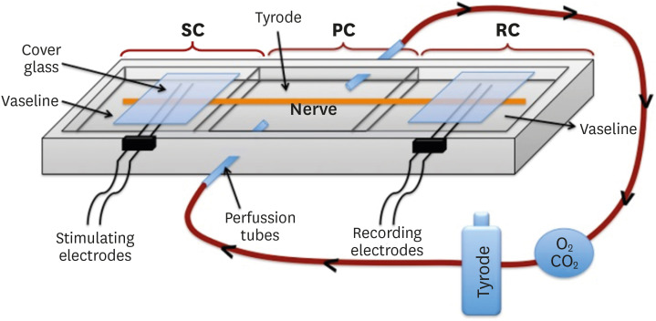
Figure 1 Diagrammatic representation of the experimental set-up. A 3-chambered bath was used, consisting of a stimulating chamber (SC), a perfusion chamber (PC), and a recording chamber (RC). The sciatic nerve is placed through the bath, contacts stimulating electrodes, and passes through the perfusion chamber and reaches the recording electrodes. The Tyrode solution, O2, and CO2 flow in the direction of the arrows and perfuse the nerve.
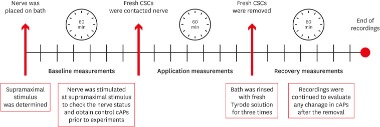
Figure 2 Diagrammatic timetable of the experimental process, showing 3 measurement intervals: baseline, application, and recovery measurements.cAP, compound action potential; CSC, calcium silicate cement.
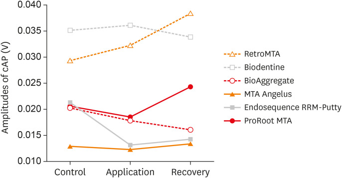
Figure 3 Changes in compound action potential (cAP) amplitudes of the tested calcium silicate cements versus time.
Reference
-
1. Dawood AE, Parashos P, Wong RHK, Reynolds EC, Manton DJ. Calcium silicate-based cements: composition, properties, and clinical applications. J Investig Clin Dent. 2017; 8:e12195.2. Torabinejad M, Hong CU, McDonald F, Pitt Ford TR. Physical and chemical properties of a new root-end filling material. J Endod. 1995; 21:349–353. PMID: 7499973.
Article3. Parirokh M, Torabinejad M. Mineral trioxide aggregate: a comprehensive literature review--part I: chemical, physical, and antibacterial properties. J Endod. 2010; 36:16–27. PMID: 20003930.
Article4. Song JS, Mante FK, Romanow WJ, Kim S. Chemical analysis of powder and set forms of Portland cement, gray ProRoot MTA, white ProRoot MTA, and gray MTA-Angelus. Oral Surg Oral Med Oral Pathol Oral Radiol Endod. 2006; 102:809–815. PMID: 17138186.5. Malkondu Ö, Karapinar Kazandağ M, Kazazoğlu E. A review on biodentine, a contemporary dentine replacement and repair material. BioMed Res Int. 2014; 2014:160951. PMID: 25025034.
Article6. Roberts HW, Toth JM, Berzins DW, Charlton DG. Mineral trioxide aggregate material use in endodontic treatment: a review of the literature. Dent Mater. 2008; 24:149–164. PMID: 17586038.
Article7. Walsh RM, Woodmansey KF, Glickman GN, He J. Evaluation of compressive strength of hydraulic silicate-based root-end filling materials. J Endod. 2014; 40:969–972. PMID: 24935545.
Article8. Kang SH, Shin YS, Lee HS, Kim SO, Shin Y, Jung IY, Song JS. Color changes of teeth after treatment with various mineral trioxide aggregate-based materials: an ex vivo study. J Endod. 2015; 41:737–741. PMID: 25732402.
Article9. Torabinejad M, Pitt Ford TR. Root end filling materials: a review. Endod Dent Traumatol. 1996; 12:161–178. PMID: 9028180.
Article10. Gomes Cornélio AL, Salles LP, Campos da Paz M, Cirelli JA, Guerreiro-Tanomaru JM, Tanomaru Filho M. Cytotoxicity of Portland cement with different radiopacifying agents: a cell death study. J Endod. 2011; 37:203–210. PMID: 21238803.
Article11. Chang SW, Oh TS, Lee W, Cheung GS, Kim HC. Long-term observation of the mineral trioxide aggregate extrusion into the periapical lesion: a case series. Int J Oral Sci. 2013; 5:54–57. PMID: 23558344.
Article12. Asgary S, Fayazi S. Endodontic surgery of a symptomatic overfilled MTA apical plug: a histological and clinical case report. Iran Endod J. 2017; 12:376–380. PMID: 28808469.13. Comin Chiaramonti L, Cavalleri G. Effect of unintentionally extruded mineral trioxide aggregate in treatment of root perforation with periradicular lesion: a case report. Minerva Stomatol. 2011; 60:217–222. PMID: 21471944.14. Islam I, Chng HK, Yap AU. Comparison of the physical and mechanical properties of MTA and portland cement. J Endod. 2006; 32:193–197. PMID: 16500224.
Article15. Asgary S, Eghbal MJ, Mehrdad L, Kheirieh S, Nosrat A. Surgical management of a failed internal root resorption treatment: a histological and clinical report. Restor Dent Endod. 2014; 39:137–142. PMID: 24790928.
Article16. Tuğ Kılkış B, Er K, Taşdemir T, Yildirim M, Taskesen F, Tümkaya L, Kalkan Y, Serper A. Neurotoxicity of various root canal sealers on rat sciatic nerve: an electrophysiologic and histopathologic study. Clin Oral Investig. 2015; 19:2091–2100.
Article17. Ahlgren FK, Johannessen AC, Hellem S. Displaced calcium hydroxide paste causing inferior alveolar nerve paraesthesia: report of a case. Oral Surg Oral Med Oral Pathol Oral Radiol Endod. 2003; 96:734–737. PMID: 14676765.
Article18. Pogrel MA. Damage to the inferior alveolar nerve as the result of root canal therapy. J Am Dent Assoc. 2007; 138:65–69. PMID: 17197403.
Article19. He S, Teagle HFB, Buchman CA. The electrically evoked compound action potential: from laboratory to clinic. Front Neurosci. 2017; 11:339. PMID: 28690494.
Article20. Onur MA, Cehreli ZC, Tasman F, Gümrükçuoğlu A. Neurotoxic effects of fifth-generation dentin adhesives on rat sciatic nerve. J Endod. 2001; 27:676–678. PMID: 11716079.21. Damas BA, Wheater MA, Bringas JS, Hoen MM. Cytotoxicity comparison of mineral trioxide aggregates and EndoSequence bioceramic root repair materials. J Endod. 2011; 37:372–375. PMID: 21329824.
Article22. Asrari M, Lobner D. In vitro neurotoxic evaluation of root-end-filling materials. J Endod. 2003; 29:743–746. PMID: 14651282.23. Abbasipour F, Rastqar A, Bakhtiar H, Khalilkhani H, Aeinehchi M, Janahmadi M. The nociceptive and anti-nociceptive effects of white mineral trioxide aggregate. Int Endod J. 2009; 42:794–801. PMID: 19549151.
Article24. González-Martín M, Torres-Lagares D, Gutiérrez-Pérez JL, Segura-Egea JJ. Inferior alveolar nerve paresthesia after overfilling of endodontic sealer into the mandibular canal. J Endod. 2010; 36:1419–1421. PMID: 20647109.
Article25. Sarkar NK, Caicedo R, Ritwik P, Moiseyeva R, Kawashima I. Physicochemical basis of the biologic properties of mineral trioxide aggregate. J Endod. 2005; 31:97–100. PMID: 15671817.
Article26. Sinkar RC, Patil SS, Jogad NP, Gade VJ. Comparison of sealing ability of ProRoot MTA, RetroMTA, and Biodentine as furcation repair materials: an ultraviolet spectrophotometric analysis. J Conserv Dent. 2015; 18:445–448. PMID: 26752836.
Article27. Duarte MA, De Oliveira Demarchi AC, Yamashita JC, Kuga MC, De Campos Fraga S. Arsenic release provided by MTA and Portland cement. Oral Surg Oral Med Oral Pathol Oral Radiol Endod. 2005; 99:648–650. PMID: 15829892.
Article28. Monteiro Bramante C, Demarchi AC, de Moraes IG, Bernadineli N, Garcia RB, Spångberg LS, Duarte MA. Presence of arsenic in different types of MTA and white and gray Portland cement. Oral Surg Oral Med Oral Pathol Oral Radiol Endod. 2008; 106:909–913. PMID: 18835535.
Article29. Hughes MF. Arsenic toxicity and potential mechanisms of action. Toxicol Lett. 2002; 133:1–16. PMID: 12076506.
Article30. International Standardization Organization. Dentistry - water based cements part 1: powder/liquid acid-base cements. Geneva: International Standardization Organization;2003. p. 1–22.
- Full Text Links
-
- Actions
-
Cited
- CITED
-
- Close
- Share
-
- Similar articles
-
- Chemical characteristics of mineral trioxide aggregate and its hydration reaction
- Conservative approach of a symptomatic carious immature permanent tooth using a tricalcium silicate cement (Biodentine): a case report
- Calcium silicate-based root canal sealers: a literature review
- The Ocular Electrical Conductivity by Vitreous Substitutes in rabbits
- Biomineralization of three calcium silicate-based cements after implantation in rat subcutaneous tissue
