Interface between calcium silicate cement and adhesive systems according to adhesive families and cement maturation
- Pradelle-Plasse N
 1,2,3
1,2,3 - Mocquot C1,2,3
- Semennikova K1,2,3
- Colon P1,2,3
- Grosgogeat B3,4,5
- Affiliations
-
- 1Department of Conservative Dentistry - Endodontics, Faculty of Dental Surgery, University of Paris, Paris, France
- 2Rothschild Hospital, Assistance Publique Hôpitaux de Paris, Paris, France
- 3Multimaterials and Interfaces Laboratory (UMR 5615), Biomaterials Team, Villeurbanne, France
- 4Department of Biomaterials, Faculty of Dental Surgery, University of Lyon 1, Lyon, France
- 5Hospices Civils de Lyon, Lyon, France
- KMID: 2548053
- DOI: http://doi.org/10.5395/rde.2021.46.e3
Abstract
Objectives
This study aimed to evaluate the interface between a calcium silicate cement (CSC), Biodentine and dental adhesives in terms of sealing ability.
Materials and Methods
Microleakage test: 160 standardized class II cavities were prepared on 80 extracted human molars. The cavities were filled with Biodentine and then divided into 2 experimental groups according to the time of restoration: composite resin obturation 15 minutes after Biodentine handling (D0); restoration after 7 days (D7). Each group was then divided into 8 subgroups (n = 5) according to the adhesive system used: etch-and-rinse adhesive (Prime & Bond); self-etch adhesive 2 steps (Optibond XTR and Clearfil SE Bond); self-etch adhesive 1 step (Xeno III, G-aenial Bond, and Clearfil Tri-S Bond); and universal used as etch-and-rinse or self-etch (ScotchBond Universal ER or SE). After thermocycling, the teeth were immersed in a silver nitrate solution, stained, longitudinally sectioned, and the Biodentine/adhesive percolation was quantified. Scanning electron microscopic observations: Biodentine/adhesive interfaces were observed.
Results
A tendency towards less microleakage was observed when Biodentine was etched (2.47%) and when restorations were done without delay (D0: 4.31%, D7: 6.78%), but this was not significant. The adhesives containing 10-methacryloyloxydecyl dihydrogen phosphate monomer showed the most stable results at both times studied. All Biodentine/adhesive interfaces were homogeneous and regular.
Conclusions
The good sealing of the CSC/adhesive interface is not a function of the system adhesive family used or the cement maturation before restoration. Biodentine can be used as a dentine substitute.
Keyword
Figure
-
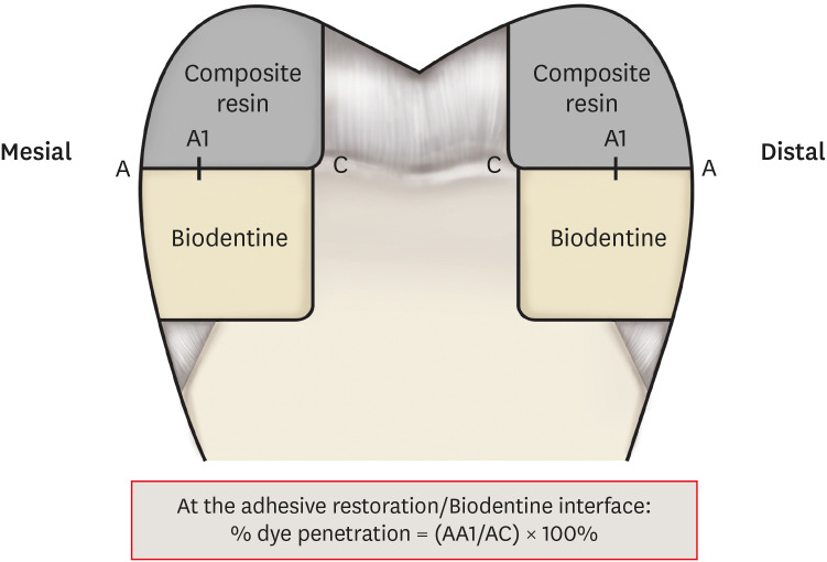
Figure 1 Cavity preparation and measurements of the dye penetration length.

Figure 2 Tooth section: on the left, the arrow shows silver nitrate penetration along the interface; on the right no penetration.
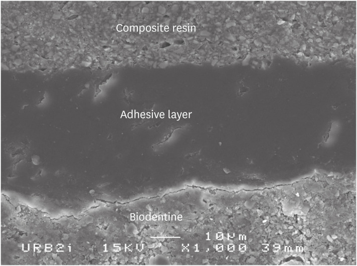
Figure 3 Scanning electron microscopic (SEM) observation of Biodentine/2 steps self-etch system (Clearfil SE Bond) interface (×1,000).

Figure 4 Scanning electron microscopic (SEM) observation of Biodentine/one step self-etch system (Clearfil Tri-S Bond) interface (×1,000).

Figure 5 Scanning electron microscopic (SEM) observation of Biodentine/universal system used as one step self-etch system (Scotchbond Universal) interface (×1,000).
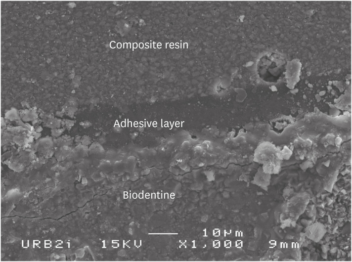
Figure 6 Scanning electron microscopic (SEM) observation of Biodentine/universal system used as etch-and-rinse system (Scotchbond Universal) interface (×1,000).
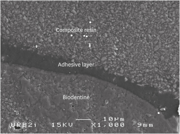
Figure 7 Scanning electron microscopic (SEM) observation of Biodentine/etch-and-rinse system (Prime & Bond) interface (×1,000).
Reference
-
1. Bachoo IK, Seymour D, Brunton P. A biocompatible and bioactive replacement for dentine: Is this a reality? The properties and uses of a novel calcium-based cement. Br Dent J. 2013; 214:E5. PMID: 23348482.
Article2. Torabinejad M, Hong CU, McDonald F, Pitt Ford TR. Physical and chemical properties of a new root-end filling material. J Endod. 1995; 21:349–353. PMID: 7499973.
Article3. Schwartz RS, Mauger M, Clement DJ, Walker WA 3rd. Mineral trioxide aggregate: a new material for endodontics. J Am Dent Assoc. 1999; 130:967–975. PMID: 10422400.
Article4. Torabinejad M, Parirokh M. Mineral trioxide aggregate: a comprehensive literature review--part II: leakage and biocompatibility investigations. J Endod. 2010; 36:190–202. PMID: 20113774.
Article5. Sarkar NK, Caicedo R, Ritwik P, Moiseyeva R, Kawashima I. Physicochemical basis of the biologic properties of mineral trioxide aggregate. J Endod. 2005; 31:97–100. PMID: 15671817.
Article6. Bozeman TB, Lemon RR, Eleazer PD. Elemental analysis of crystal precipitate from gray and white MTA. J Endod. 2006; 32:425–428. PMID: 16631841.
Article7. Watson TF, Atmeh AR, Sajini S, Cook RJ, Festy F. Present and future of glass-ionomers and calcium-silicate cements as bioactive materials in dentistry: biophotonics-based interfacial analyses in health and disease. Dent Mater. 2014; 30:50–61. PMID: 24113131.
Article8. Atmeh AR, Chong EZ, Richard G, Festy F, Watson TF. Dentin-cement interfacial interaction: calcium silicates and polyalkenoates. J Dent Res. 2012; 91:454–459. PMID: 22436906.9. Pradelle-Plasse N, Tran X, Colon P. Biocompatibility or cytotoxic effects of dental composites. Oxfordshire: Coxmoor Publishing Company;2009.10. Villat C, Tran XV, Pradelle-Plasse N, Ponthiaux P, Wenger F, Grosgogeat B, Colon P. Impedance methodology: a new way to characterize the setting reaction of dental cements. Dent Mater. 2010; 26:1127–1132. PMID: 20728209.
Article11. Koubi G, Colon P, Franquin JC, Hartmann A, Richard G, Faure MO, Lambert G. Clinical evaluation of the performance and safety of a new dentine substitute, Biodentine, in the restoration of posterior teeth - a prospective study. Clin Oral Investig. 2013; 17:243–249.
Article12. Bergenholtz G, Cox CF, Loesche WJ, Syed SA. Bacterial leakage around dental restorations: its effect on the dental pulp. J Oral Pathol. 1982; 11:439–450. PMID: 6819352.
Article13. Browne RM, Tobias RS. Microbial microleakage and pulpal inflammation: a review. Endod Dent Traumatol. 1986; 2:177–183. PMID: 3539583.
Article14. Camilleri J. Investigation of Biodentine as dentine replacement material. J Dent. 2013; 41:600–610. PMID: 23685034.
Article15. Meraji N, Camilleri J. Bonding over dentin replacement materials. J Endod. 2017; 43:1343–1349. PMID: 28662878.
Article16. Hashem DF, Foxton R, Manoharan A, Watson TF, Banerjee A. The physical characteristics of resin composite-calcium silicate interface as part of a layered/laminate adhesive restoration. Dent Mater. 2014; 30:343–349. PMID: 24418628.
Article17. Cengiz E, Ulusoy N. Microshear bond strength of tri-calcium silicate-based cements to different restorative materials. J Adhes Dent. 2016; 18:231–237. PMID: 27045140.18. Çolak H, Tokay U, Uzgur R, Uzgur Z, Ercan E, Hamidi MM. The effect of different adhesives and setting times on bond strength between Biodentine and composite. J Appl Biomater Funct Mater. 2016; 14:e217–e222. PMID: 27149941.
Article19. Odabaş ME, Bani M, Tirali RE. Shear bond strengths of different adhesive systems to Biodentine. ScientificWorldJournal. 2013; 2013:626103. PMID: 24222742.
Article20. Abraham SB, Gaintantzopoulou MD, Eliades G. Cavity adaptation of water-based restoratives placed as liners under a resin composite. Int J Dent. 2017; 2017:5957107. PMID: 28465685.
Article21. Aggarwal V, Singla M, Yadav S, Yadav H, Ragini . Marginal adaptation evaluation of Biodentine and MTA plus in ‘open sandwich’ class II restorations. J Esthet Restor Dent. 2015; 27:167–175. PMID: 25771941.
Article22. Raskin A, Eschrich G, Dejou J, About I. In vitro microleakage of Biodentine as a dentin substitute compared to Fuji II LC in cervical lining restorations. J Adhes Dent. 2012; 14:535–542. PMID: 22724110.23. Besnault C, Attal JP. Simulated oral environment and microleakage of class II resin-based composite and sandwich restorations. Am J Dent. 2003; 16:186–190. PMID: 12967073.24. Gale MS, Darvell BW. Thermal cycling procedures for laboratory testing of dental restorations. J Dent. 1999; 27:89–99. PMID: 10071465.
Article25. Cervino G, Fiorillo L, Spagnuolo G, Bramanti E, Laino L, Lauritano F, Cicciù M. Interface between MTA and dental bonding agents: scanning electron microscope evaluation. J Int Soc Prev Community Dent. 2017; 7:64–68. PMID: 28316952.
Article26. Garrault S, Behr T, Nonat A. Formation of the C-S-H layer during early hydration of tricalcium silicate grains with different sizes. J Phys Chem B. 2006; 110:270–275. PMID: 16471532.
Article27. Rengo C, Goracci C, Juloski J, Chieffi N, Giovannetti A, Vichi A, Ferrari M. Influence of phosphoric acid etching on microleakage of a self-etch adhesive and a self-adhering composite. Aust Dent J. 2012; 57:220–226. PMID: 22624765.
Article28. Kayahan MB, Nekoofar MH, Kazandağ M, Canpolat C, Malkondu O, Kaptan F, Dummer PM. Effect of acid-etching procedure on selected physical properties of mineral trioxide aggregate. Int Endod J. 2009; 42:1004–1014. PMID: 19732179.
Article29. Atabek D, Sillelioğlu H, Olmez A. Bond strength of adhesive systems to mineral trioxide aggregate with different time intervals. J Endod. 2012; 38:1288–1292. PMID: 22892753.
Article30. Silva e Souza MH Jr, Carneiro KG, Lobato MF, Silva e Souza PA, de Góes MF. Adhesive systems: important aspects related to their composition and clinical use. J Appl Oral Sci. 2010; 18:207–214. PMID: 20856995.
Article31. Karaman E, Yazici AR, Aksoy B, Karabulut E, Ozgunaltay G, Dayangac B. Effect of operator variability on microleakage with different adhesive systems. Eur J Dent. 2013; 7:S060–S065. PMID: 24966730.
Article32. Palma PJ, Marques JA, Falacho RI, Vinagre A, Santos JM, Ramos JC. Does delayed restoration improve shear bond strength of different restorative protocols to calcium silicate-based cements? Materials (Basel). 2018; 11:2216.
Article33. Sultana N, Nawal RR, Chaudhry S, Sivakumar M, Talwar S. Effect of acid etching on the micro-shear bond strength of resin composite-calcium silicate interface evaluated over different time intervals of bond aging. J Conserv Dent. 2018; 21:194–197. PMID: 29674824.
Article34. Yoshioka M, Yoshida Y, Inoue S, Lambrechts P, Vanherle G, Nomura Y, Okazaki M, Shintani H, Van Meerbeek B. Adhesion/decalcification mechanisms of acid interactions with human hard tissues. J Biomed Mater Res. 2002; 59:56–62. PMID: 11745537.
Article35. Yoshida Y, Nagakane K, Fukuda R, Nakayama Y, Okazaki M, Shintani H, Inoue S, Tagawa Y, Suzuki K, De Munck J, Van Meerbeek B. Comparative study on adhesive performance of functional monomers. J Dent Res. 2004; 83:454–458. PMID: 15153451.
Article36. Carrilho E, Cardoso M, Marques Ferreira M, Marto CM, Paula A, Coelho AS. 10-MDP based dental adhesives: adhesive interface characterization and adhesive stability-a systematic review. Materials (Basel). 2019; 12:790.
Article37. Yoshihara K, Yoshida Y, Nagaoka N, Hayakawa S, Okihara T, De Munck J, Maruo Y, Nishigawa G, Minagi S, Osaka A, Van Meerbeek B. Adhesive interfacial interaction affected by different carbon-chain monomers. Dent Mater. 2013; 29:888–897. PMID: 23768795.
Article38. Chatel E, Pradelle-Plasse N, Colon P, Grosgogeat B. Degradation mechanisms of modern self–etching adhesives. Clin Oral Investig. 2013; 17:1035.39. Koubi S, Elmerini H, Koubi G, Tassery H, Camps J. Quantitative evaluation by glucose diffusion of microleakage in aged calcium silicate-based open-sandwich restorations. Inter J Dent. 2012; 2012:105863.
Article40. Aksel H, Küçükkaya Eren S, Askerbeyli Õrs S, Karaismailoğlu E. Surface and vertical dimensional changes of mineral trioxide aggregate and Biodentine in different environmental conditions. J Appl Oral Sci. 2018; 27:e20180093. PMID: 30540071.
Article41. Agrafioti A, Tzimpoulas N, Chatzitheodoridis E, Kontakiotis EG. Comparative evaluation of sealing ability and microstructure of MTA and Biodentine after exposure to different environments. Clin Oral Investig. 2016; 20:1535–1540.
Article
- Full Text Links
-
- Actions
-
Cited
- CITED
-
- Close
- Share
-
- Similar articles
-
- Two Cases of Cement Burn
- The effect of different adhesive system applications on push-out bond strengths of glass fiber posts
- Effect of adhesive luting on the fracture resistance of zirconia compared to that of composite resin and lithium disilicate glass ceramic
- Effect of applying adhesive after enamel etching on the shear bond strength of orthodontic brackets using light curing resin cements
- The shear bond strength of two adhesives bonded to composite resin and glass ionomer cement restorations
