Value of Thallium-201 SPECT and SPECT/CT Brain Imaging in Differentiating Malignant From Nonmalignant Lesions: A Comparative Case-Series Study With Pathologic and/or Clinical Correlation
- Affiliations
-
- 1Institute for Neurosciences, St. Luke’s Medical Center-Quezon City and Global City, Philippines
- 2Department of Neurosciences, College of Medicine and Philippine General Hospital, University of the Philippines Manila, Manila, Philippines
- 3Department of Nuclear Medicine, St. Luke’ Medical Center-Global City, Taguig, Philippines
- KMID: 2547412
- DOI: http://doi.org/10.14791/btrt.2023.0022
Abstract
- Background
Thallium-201 single-photon emission computed tomography/computed tomography (SPECT/CT) brain scan is an imaging modality which can be done to differentiate between malignant and nonmalignant lesions among patients with nonconclusive findings on conventional neuroimaging. This study describes the results of thallium-201 SPECT/CT brain imaging and relate it to histopathologic and/or clinical findings and evaluate the value of thallium-201 SPECT/CT brain imaging in differentiating malignant from nonmalignant lesions.
Methods
This is a retrospective case series study of 10 patients with cerebral lesions who un-derwent thallium-201 SPECT/CT brain imaging in a hospital in the Philippines from 2010 to 2021.
Results
A total of 10 patients underwent thallium-201 SPECT/CT brain scan. Six had nega- tive results while 4 had positive results. All of the patients who had positive results were found to have malignancy, whether recurrent or newly diagnosed. All of the patients with negative scan were found to have either an infectious and inflammatory disease and responded to treatment albeit in different degrees. Two of the 10 patients underwent biopsy whose results were consistent with the thallium-201 SPECT/CT brain scan results.
Conclusion
Thallium-201 brain scan combined with SPECT and SPECT/CT has been demon- strated to be useful in distinguishing malignant from nonmalignant lesions and is more cost-effective versus other imaging techniques. The findings in this study support the role of thallium scintigraphy in the diagnosis of patients with brain lesions most significantly when there is a need to differentiate between a malignant and benign condition.
Keyword
Figure
-

Fig. 1 Thallium-201 brain scan findings and histopathologic image of Patient 3. A and B: A negative thallium brain scintigraphy planar (A) and SPECT/CT images (B) showing no increased tracer accumulation. C: Cerebellar tissues with gliosis (H&E, ×10). SPECT/CT, single-photon emission computed tomography/computed tomography.
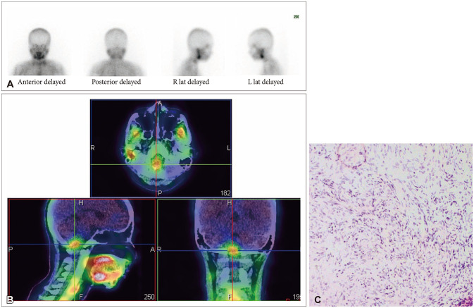
Fig. 2 Thallium-201 brain scan findings and histopathologic image of Patient 9. A: Initial planar images after 90 minutes of thallium-201 intravenous injection showing no focus of increased tracer uptake. B: SPECT/CT imaging with a focus of thallium-201 uptake in the cervicomedullary junction and right brachium pontis. C: Cellular portion of brain tissue populated by bland round and spindle cells (H&E ×100), immunohistochemistry results revealed positive GFAP and P16, focally positive SOX10, IDH1 negative, and retained nuclear staining on ATRX favoring the diagnosis of pilocytic astrocytoma. SPECT/CT, single-photon emission computed tomography/computed tomography.
Reference
-
1. Norden AD, Pope WB, Chang SM. Current concepts in brain tumor imaging. Am Soc Clin Oncol Educ Book. 2012; 32:119–124.
Article2. Smego RA Jr, Orlovic D, Wadula J. An algorithmic approach to intracranial mass lesions in HIV/AIDS. Int J STD AIDS. 2006; 17:271–276. PMID: 16595052.
Article3. Di Bonaventura R, Montano N, Giordano M, Gessi M, Gaudino S, Izzo A, et al. Reassessing the role of brain tumor biopsy in the era of advanced surgical, molecular, and imaging techniques—a single-center experience with long-term follow-up. J Pers Med. 2021; 11:909. PMID: 34575685.
Article4. Lorberboym M, Estok L, Machac J, Germano I, Sacher M. Thallium-201 brain spect and CT/MRI imaging for differential diagnosis of cerebral toxoplasmosis and primary CNS lymphoma. Clin Nucl Med. 1996; 21:353.
Article5. Lorberboym M, Estok L, Machac J, Germano I, Sacher M, Feldman R, et al. Rapid differential diagnosis of cerebral toxoplasmosis and primary central nervous system lymphoma by thallium-201 SPECT. J Nucl Med. 1996; 37:1150–1154. PMID: 8965186.6. Hussain FS, Hussain NS. Clinical utility of thallium-201 single photon emission computed tomography and cerebrospinal fluid Epstein-Barr virus detection using polymerase chain reaction in the diagnosis of AIDS-related primary central nervous system lymphoma. Cureus. 2016; 8:e606. PMID: 27330874.
Article7. Yang M, Sun J, Bai HX, Tao Y, Tang X, States LJ, et al. Diagnostic accuracy of SPECT, PET, and MRS for primary central nervous system lymphoma in HIV patients: a systematic review and meta-analysis. Medicine (Baltimore). 2017; 96:e6676. PMID: 28489744.8. Kaplan WD, Takvorian T, Morris JH, Rumbaugh CL, Connolly BT, Atkins HL. Thallium-201 brain tumor imaging: a comparative study with pathologic correlation. J Nucl Med. 1987; 28:47–52. PMID: 3467030.9. Lorberboym M, Baram J, Feibel M, Hercbergs A, Lieberman L. A prospective evaluation of thallium-201 single photon emission computerized tomography for brain tumor burden. Int J Radiat Oncol Biol Phys. 1995; 32:249–254. PMID: 7721624.
Article10. Kim KT, Black KL, Marciano D, Mazziotta JC, Guze BH, Grafton S, et al. Thallium-201 SPECT imaging of brain tumors: methods and results. J Nucl Med. 1990; 31:965–969. PMID: 2161453.11. Atkins HL, Budinger TF, Lebowitz E, Ansari AN, Greene MW, Fairchild RG, et al. Thallium-201 for medical use. Part 3: human distribution and physical imaging properties. J Nucl Med. 1977; 18:133–140. PMID: 833658.12. Bénard F, Romsa J, Hustinx R. Imaging gliomas with positron emission tomography and single-photon emission computed tomography. Semin Nucl Med. 2003; 33:148–162. PMID: 12756647.
Article13. Kline JL, Noto RB, Glantz M. Single-photon emission CT in the evaluation of recurrent brain tumor in patients treated with gamma knife radiosurgery or conventional radiation therapy. AJNR Am J Neuroradiol. 1996; 17:1681–1686. PMID: 8896622.14. Tie J, Gunawardana DH, Rosenthal MA. Differentiation of tumor recurrence from radiation necrosis in high-grade gliomas using 201Tl-SPECT. J Clin Neurosci. 2008; 15:1327–1334. PMID: 18845440.
Article15. Valotassiou V, Leondi A, Angelidis G, Psimadas D, Georgoulias P. SPECT and PET imaging of meningiomas. ScientificWorldJournal. 2012; 2012:412580. PMID: 22623896.
Article16. Matsunaga S, Shuto T, Takase H, Ohtake M, Tomura N, Tanaka T, et al. Semiquantitative analysis using thallium-201 SPECT for differential diagnosis between tumor recurrence and radiation necrosis after gamma knife surgery for malignant brain tumors. Int J Radiat Oncol Biol Phys. 2013; 85:47–52. PMID: 22541963.
Article17. Smith J, English J, Gilmore D. Evaluation of PET/CT and thallium-201 imaging for brain tumors vs. necrosis. J Nucl Med. 2010; 51(supplement 2):2120.18. Chochrek C, Stokes M, Gilmore D. Differentiating recurrent brain tumors from radiation necrosis with the use of thallium brain imaging. J Nucl Med. 2009; 50(supplement 2):2207.19. Verma N, Cowperthwaite MC, Burnett MG, Markey MK. Differentiating tumor recurrence from treatment necrosis: a review of neuro-oncologic imaging strategies. Neuro Oncol. 2013; 15:515–534. PMID: 23325863.
Article20. Dierckx RA, Martin JJ, Dobbeleir A, Crols R, Neetens I, De Deyn PP. Sensitivity and specificity of thallium-201 single-photon emission tomography in the functional detection and differential diagnosis of brain tumours. Eur J Nucl Med. 1994; 21:621–633. PMID: 7957348.
Article21. Munir S, Khan SA, Hanif H, Khan M. Diagnostic accuracy of magnetic resonance imaging in detection of intra-axial gliomas. Pak J Med Sci. 2021; 37:125–130. PMID: 33437263.
- Full Text Links
-
- Actions
-
Cited
- CITED
-
- Close
- Share
-
- Similar articles
-
- The Significance of Thallium-201 SPECT in the Diagnosis of Brain Tumors: Clinical Analysis
- Accumulation of Thallium-201 in Hemorrhagic Cerebral Infarction
- Thallium-201 SPECT Imaging of Brain Tumors
- Preoperative Evaluation of Brain Lesion with 201Tl Brain SPECT: Is It Useful to Differentiate Benign and Malignant Lesions?
- Thallium-201 SPECT in Differential Diagnosis of Malignancy from Benign Pathology in Patients with a Solitary Pulmonary Lesion
