Clinical and anatomical importance of foramen magnum and craniocervical junction structures in the perspective of surgical approaches
- Affiliations
-
- 1Department of Anatomy, School of Medicine, Ankara Medipol University, Ankara, Türkiye
- KMID: 2546462
- DOI: http://doi.org/10.5115/acb.23.006
Abstract
- This study was conducted to investigate the clinical and anatomical importance of the relevant region from the perspective of surgical approaches by determining the morphometric analysis of the craniocervical junction and foramen magnum (FM) region and determining their distances from important anatomical points. This research was carried out with 59 skulls found at the Anatomy Laboratories of Erciyes and Ankara Medipol University. Metric measurements of FM and condyle, FM shape, condyle-fossa relationship, and pharyngeal tubercle (PT) were made in mm-based dry bone samples of unknown age and sex. The distance between the anterior notches and the FM was 87.01±4.35, the distance between the anterior notches and the PT was 77.70±4.24, the distance between the PT-sphenooccipital junction was 13.23±2.42, and the FM index was 81.86±7.47. The anteroposterior and transverse lengths of FM were determined as 33.80±2.99 and 27.72±2.30, respectively. The morphometric and morphological data available regarding the craniocervical junction showed significant differences between populations. Comprehensive knowledge of this topic will provide a better approach to treat Arnold Chiari Malformation, FM meningiomas, and other posterior cranial fossa lesions. Therefore, we believe that FM and craniocervical junction morphology will be a guide not only for anatomists, but also for radiologists, neurosurgeons, ENT surgeons, and orthopedists.
Figure
-
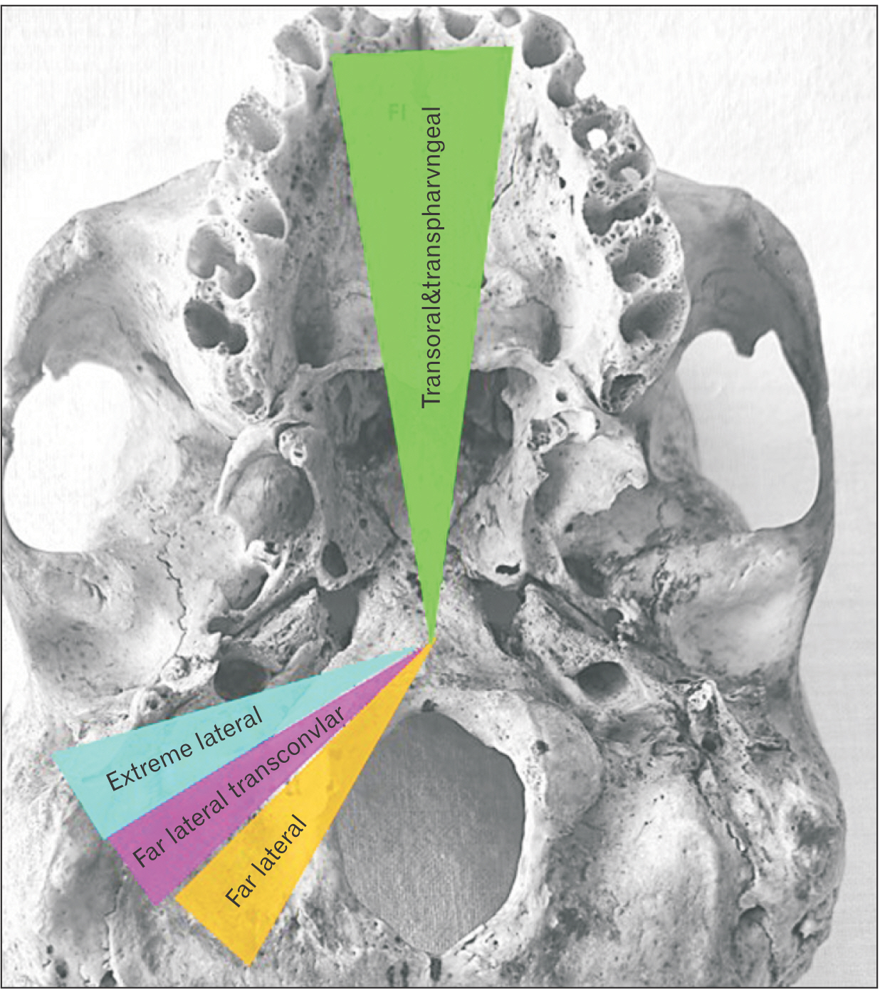
Fig. 1 Posterolateral, transoral and transpharygeal approaches. Adapted from Luzzi et al. (Turk Neurosurg 2019;29:875-86) [7].

Fig. 2 Morphological measurements of foramen magnum (FM) and occipital condyle (OC). (A) 1: FM anteroposterior length, 2: FM transverse length, 3: OC length, 4: OC width. (B) f1: the distance from the anterior midpoint of the FM to the most protruding point of the medial edge of the OC, f2: posterior of the FM distance from its midpoint to the most protruding point of the medial edge of the OC, f3: from the anterior midpoint of the FM to the midpoint of the medial edge of the OC, f4: from the posterior midpoint of the foramen magnum to the midpoint of the medial edge of the occipital condyle.
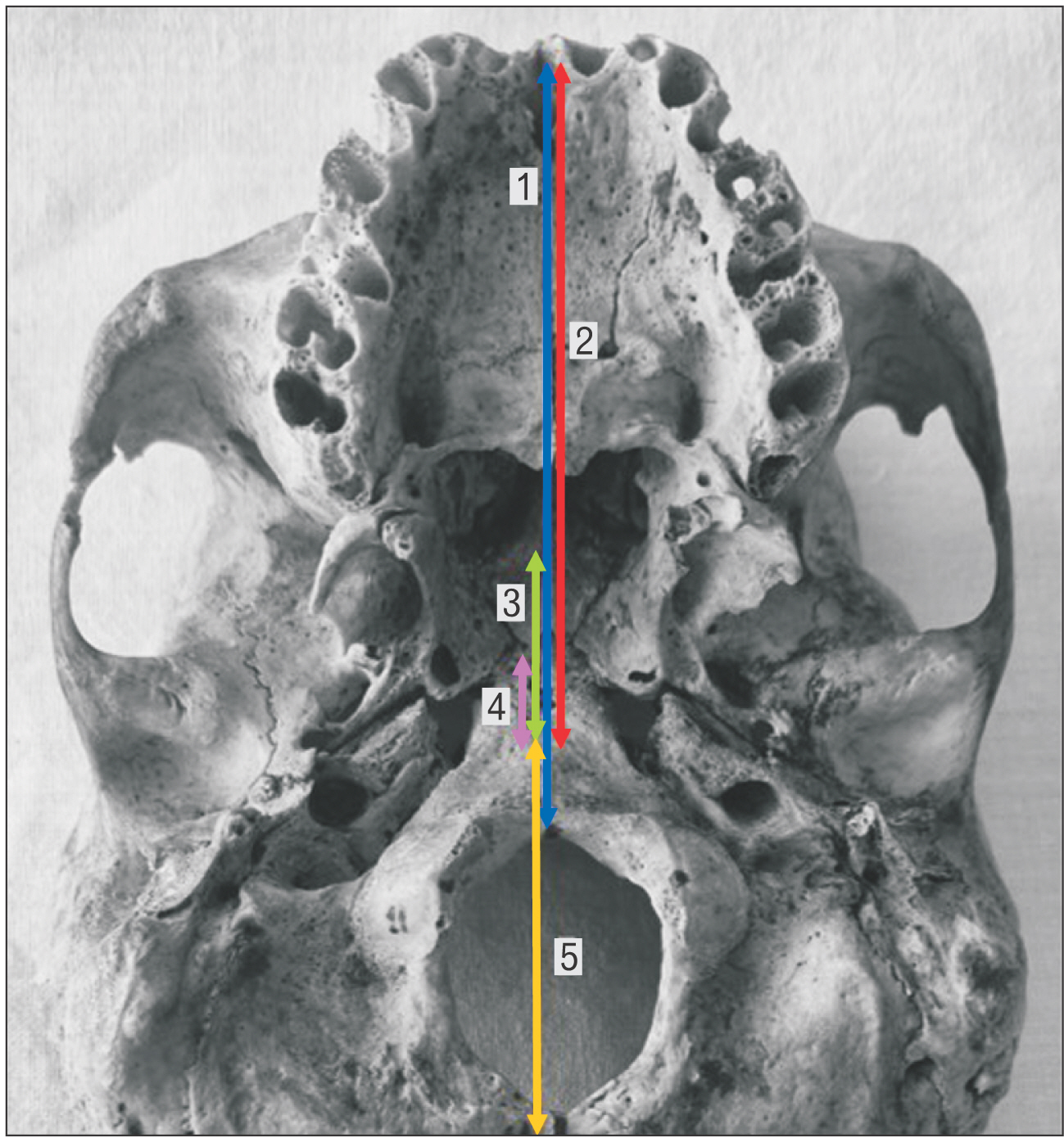
Fig. 3 Relationship of PT with key points. 1: distance between anterior notchs-medial of FM, 2: distance between anterior notchs-TP, 3: distance between PT-sphenooccipital junction, 4: distance between PT-ala of vomer, 5: distance between PT-basion. PT, pharyngeal tubercle; FM, foramen magnum.
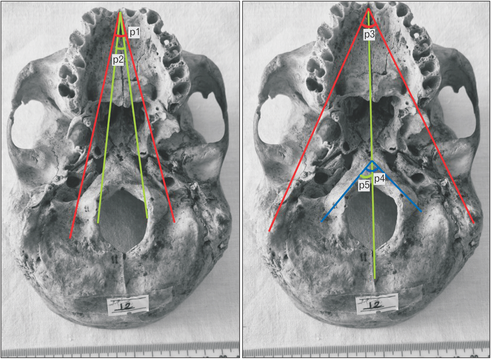
Fig. 4 Angles associated with FM. p1: angle between anterior notchs and lateral of occipital condyle (AN-OC/L), p2: angle between anterior notchs and medial of occipital condyle (AN-OC/M), p3: anterior notchs angle between mastoid process (AN-MP), p4: sagittal intercondylar angle (SIA), p5: sagittal condylar angle (SCA).
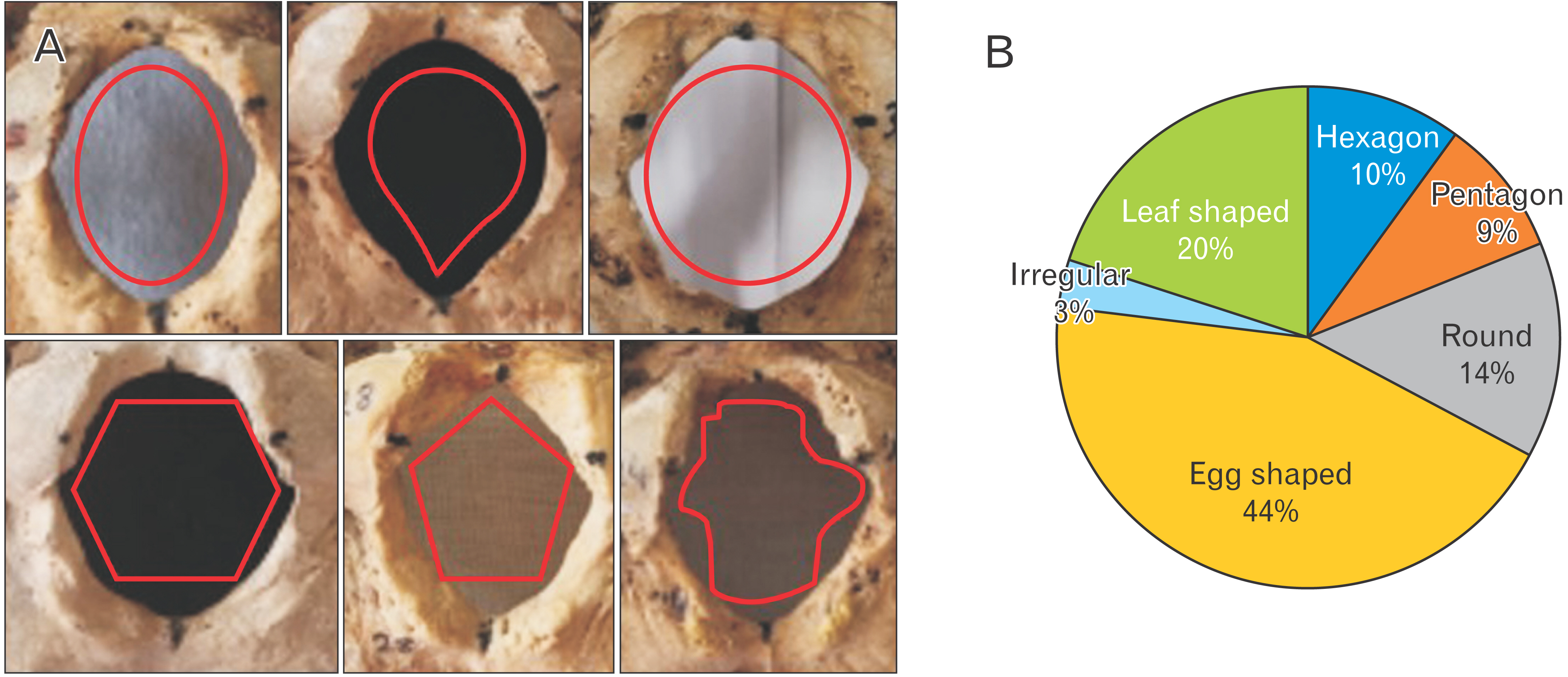
Fig. 5 Foramen magnum (FM) shapes. (A) FM shapes, (B) percentages of FM shapes.
Reference
-
References
1. Dasegowda G, Padmalatha K, Priyanka BP, Mirmire S. 2020; Morphology and morphometric analysis of foramen magnum in adult human skulls. Int J Anat Res. 8:7771–6. DOI: 10.16965/ijar.2020.212.
Article2. Natsis K, Piagkou M, Skotsimara G, Piagkos G, Skandalakis P. 2013; A morphometric anatomical and comparative study of the foramen magnum region in a Greek population. Surg Radiol Anat. 35:925–34. DOI: 10.1007/s00276-013-1119-z. PMID: 23620089.
Article3. Nishikawa M, Sakamoto H, Hakuba A, Nakanishi N, Inoue Y. 1997; Pathogenesis of Chiari malformation: a morphometric study of the posterior cranial fossa. J Neurosurg. 86:40–7. DOI: 10.3171/jns.1997.86.1.0040. PMID: 8988080.
Article4. Tubbs RS, Elton S, Grabb P, Dockery SE, Bartolucci AA, Oakes WJ. 2001; Analysis of the posterior fossa in children with the Chiari 0 malformation. Neurosurgery. 48:1050–4. discussion 1054–5. DOI: 10.1227/00006123-200105000-00016. PMID: 11334271.
Article5. Radinsky L. 1967; Relative brain size: a new measure. Science. 155:836–8. DOI: 10.1126/science.155.3764.836. PMID: 4959814.
Article6. Tai AX, Herur-Raman A, Jean WC. 2020; The benefits of progressive occipital condylectomy in enhancing the far lateral approach to the foramen magnum. World Neurosurg. 134:e144–52. DOI: 10.1016/j.wneu.2019.09.152. PMID: 31605848.
Article7. Luzzi S, Maestro MD, Elia A, Vincitorio F, Perna GD, Zenga F, Garbossa D, Elbabaa SK, Galzio R. 2019; Morphometric and radiomorphometric study of the correlation between the foramen magnum region and the anterior and posterolateral approaches to ventral intradural lesions. Turk Neurosurg. 29:875–86. DOI: 10.5137/1019-5149.JTN.26052-19.2. PMID: 31452176.
Article8. Vidya HK, Nagashree MV. 2018; Morphometric evaluation of foramen magnum. Medpulse Int J Anat. 8:1–4. DOI: 10.26611/1001811.9. Zhong S, Ren J, Zhang Y, Li W, Li H, Song S, Zhao G, Cheng Y. 2018; Anatomic study of craniocervical junction and its surrounding structures in endoscopic transoral-transpharyngeal approach. J Craniofac Surg. 29:1973–7. DOI: 10.1097/SCS.0000000000004648. PMID: 29863562.
Article10. Ceri NG, Sahmelikoglu AG, Cantas F, Sakalli G. 2021; The anatomic study of extracranial structures related to tuberculum pharyngeum. Eur J Anat. 25:241–6.11. Subramanian S, Vivek J. 2021; Transoral transpharyngeal approach to craniovertebral junction: indications, management and outcomes in a survey of 7 cases. Univ J Surg Surg Spec. 7:5.12. Muthukumar N, Swaminathan R, Venkatesh G, Bhanumathy SP. 2005; A morphometric analysis of the foramen magnum region as it relates to the transcondylar approach. Acta Neurochir (Wien). 147:889–95. DOI: 10.1007/s00701-005-0555-x. PMID: 15924208.
Article13. Sampada PK, Poornima B, Mallikarjun M, Sakri SB. 2017; Morphometric and morphological study of foramen magnum in dried human skull bones. Int J Anat Res. 5:3682–6. DOI: 10.16965/ijar.2017.139.
Article14. Singh D, Patnaik P, Gupta N. 2019; Morphology and morphometric analysis of the foramen magnum in dried adult skulls in North Indian region. Int J Health Sci Res. 9:36–42.15. Degno S, Abrha M, Asmare Y, Muche A. 2019; Anatomical variation in morphometry and morphology of the foramen magnum and occipital condyle in dried adult skulls. J Craniofac Surg. 30:256–9. DOI: 10.1097/SCS.0000000000004925. PMID: 30480625.
Article16. Gocmen Mas N, Cirpan S, Aksu F, Yonguc Demirci GN, Lafci Fahrioglu S, Durmaz O, Karabekir S. 2018; Comparison of three methods used for estimating area of foramen magnum. J Craniofac Surg. 29:792–5. DOI: 10.1097/SCS.0000000000004250. PMID: 29419586.
Article17. Nagwani M, Srivastava G, Rani A. 2020; To study the correlation between foramen magnum index and cranial index in Indian population. Innov J Med Health Sci. 10:780–3. DOI: 10.32388/kcd97y.18. Zdilla MJ, Russell ML, Bliss KN, Mangus KR, Koons AW. 2017; The size and shape of the foramen magnum in man. J Craniovertebr Junction Spine. 8:205–21. DOI: 10.4103/jcvjs.JCVJS_62_17. PMID: 29021672. PMCID: PMC5634107. PMID: 07f785a13a064502a4f376a336797978.
Article19. Bhatnagar S, Iwanaga J, Decater T, Loukas M, Tubbs RS. 2020; Foramen magnum variant with elongation of the anterior notch. Cureus. 12:e8506. DOI: 10.7759/cureus.8506. PMID: 32656022. PMCID: PMC7346306.
Article20. Ukoha U, Egwu OA, Okafor IJ, Anyabolu AE, Ndukwe GU, Okpala I. 2011; Sexual dimorphism in the foramen magnum of Nigerian adult. Int J Biol Med Res. 2:878–81.21. Chethan P, Prakash KG, Murlimanju BV, Prashanth KU, Prabhu LV, Saralaya VV, Krishnamurthy A, Somesh MS, Kumar CG. 2012; Morphological analysis and morphometry of the foramen magnum: an anatomical investigation. Turk Neurosurg. 22:416–9. DOI: 10.5137/1019-5149.JTN.4297-11.1. PMID: 22843456.
Article22. Aljarrah K, Packirisamy V, Al Anazi N, Nayak SB. 2022; Morphometric analysis of foramen magnum and occipital condyle using CT images for sex determination in a Saudi Arabian population. Morphologie. 106:260–70. DOI: 10.1016/j.morpho.2021.07.006. PMID: 34391659.
Article23. Kizilkanat ED, Boyan N, Soames R, Oguz O. 2006; Morphometry of the hypoglossal canal, occipital condyle, and foramen magnum. Neurosurg Q. 16:121–5. DOI: 10.1097/01.wnq.0000214018.49915.49.
Article24. Naderi S, Korman E, Citak G, Güvençer M, Arman C, Senoğlu M, Tetik S, Arda MN. 2005; Morphometric analysis of human occipital condyle. Clin Neurol Neurosurg. 107:191–9. DOI: 10.1016/j.clineuro.2004.07.014. PMID: 15823674.
Article25. Ozer MA, Celik S, Govsa F, Ulusoy MO. 2011; Anatomical determination of a safe entry point for occipital condyle screw using three-dimensional landmarks. Eur Spine J. 20:1510–7. DOI: 10.1007/s00586-011-1765-y. PMID: 21416278. PMCID: PMC3175895.
Article26. Yu Z, Ma X, Jiang J, Jin X, Lv F, Wang L, Xia X, Wang H. 2015; Feasibility of screw placement in the occipital condyle of Chinese patients for occipitocervical arthrodesis: a cadaveric study. Turk Neurosurg. 25:559–65. DOI: 10.5137/1019-5149.JTN.10914-14.0. PMID: 26242332.27. Wen HT, Rhoton AL Jr, Katsuta T, de Oliveira E. 1997; Microsurgical anatomy of the transcondylar, supracondylar, and paracondylar extensions of the far-lateral approach. J Neurosurg. 87:555–85. DOI: 10.3171/jns.1997.87.4.0555. PMID: 9322846.
Article28. Saluja S, Das SS, Vasudeva N. 2016; Morphometric analysis of the occipital condyle and its surgical importance. J Clin Diagn Res. 10:AC01–4. DOI: 10.7860/JCDR/2016/23278.8800. PMID: 28050351. PMCID: PMC5198304. PMID: 02cb0daeaf6b4696acf783d82f99feef.29. Liu JK, Couldwell WT, Apfelbaum RI. 2008; Transoral approach and extended modifications for lesions of the ventral foramen magnum and craniovertebral junction. Skull Base. 18:151–66. DOI: 10.1055/s-2007-994288. PMID: 18978962. PMCID: PMC2459326.
Article30. Wang AJ, Zaidi HA, Laws ED Jr. 2016; History of endonasal skull base surgery. J Neurosurg Sci. 60:441–53. PMID: 27273318.31. Hitotsumatsu T, Matsushima T, Rhoton AL Jr. 1999; Surgical anatomy of the midface and the midline skull base. Oper Tech Neurosurg. 2:160–80. DOI: 10.1016/S1092-440X(99)80017-6.
Article32. Krmpotić-Nemanić J, Vinter I, Ehrenfreund T, Marusić A. 2006; Age-related changes in the anatomical landmarks of the osseous epipharynx. Ann Anat. 188:459–67. DOI: 10.1016/j.aanat.2006.04.005. PMID: 16999211.
Article33. Wanebo JE, Chicoine MR. 2001; Quantitative analysis of the transcondylar approach to the foramen magnum. Neurosurgery. 49:934–41. discussion 941–3. DOI: 10.1227/00006123-200110000-00027. PMID: 11564256.
Article34. Le TV, Dakwar E, Hann S, Effio E, Baaj AA, Martinez C, Vale FL, Uribe JS. 2011; Computed tomography-based morphometric analysis of the human occipital condyle for occipital condyle-cervical fusion. J Neurosurg Spine. 15:328–31. DOI: 10.3171/2011.5.SPINE10778. PMID: 21639701.
Article
- Full Text Links
-
- Actions
-
Cited
- CITED
-
- Close
- Share
-
- Similar articles
-
- Surgical Approaches for Tumors Around Foramen Magnum and Craniocervical Junction
- The Operative Treatment for the Meningiomas in the low Clivus and Foramen Magnum
- Foramen Magnum Meningioma: Some Anatomical and Surgical Remarks through Five Cases
- Morphological analysis and morphometry of the occipital condyle and its relationship to the foramen magnum, jugular foramen, and hypoglossal canal: implications for craniovertebral junction surgery
- Transcondylar Approach to Skull Base Lesions
