The laryngopharyngeal nerve: a comprehensive review
- Shapiro S
 1,2
1,2 - Parker AL3
- Cardona JJ2
- Chaiyamoon A4
- Reina F5
- Carrera A5
- Iwanaga J2,6
- Dumont AS2
- Tubbs RS1,2,7,8,9
- Affiliations
-
- 1Department of Neurosurgery and Ochsner Neuroscience Institute, Ochsner Health System, New Orleans, LA, USA
- 2Department of Neurosurgery, Tulane University School of Medicine, New Orleans, LA, USA
- 3Tulane University School of Medicine, New Orleans, LA, USA
- 4Department of Anatomy, Faculty of Medicine, Khon Kaen University, Khon Kaen, Thailand
- 5Medical Sciences Department, Clinical Anatomy, Embryology and Neurosciences Research Group, University of Girona, Girona, Spain
- 6Department of Oral and Maxillofacial Anatomy, Graduate School of Medical and Dental Sciences, Tokyo Medical and Dental University, Tokyo, Japan
- 7Department of Anatomical Sciences, St. George’s University, St. George’s, Grenada, West Indies
- 8Department of Surgery, Tulane University School of Medicine, New Orleans, LA, USA
- 9University of Queensland, Brisbane, Australia
- KMID: 2546324
- DOI: http://doi.org/10.5115/acb.22.254
Abstract
- The laryngopharyngeal nerve has received much less attention that the other contributions to the pharyngeal plexus i.e., glossopharyngeal and vagus nerves. Often, in descriptions and depictions, the nerve is simply labeled as the sympathetic contribution to the pharyngeal plexus. As there is such scant information available regarding this nerve, the present review was performed. Very little is found in the extant medical literature regarding the laryngopharyngeal nerve. However, based on available data, the nerve is a consistent contributory to the pharyngeal plexus and serves other adjacent areas e.g., carotid body. Therefore, a better understanding of this structure’s anatomy is important for those who operate in this area. Further studies are necessary to better elucidate the true function of the laryngopharyngeal nerve.
Keyword
Figure
-

Fig. 1 Schematic drawing of the right side of the neck demonstrating the superior cervical ganglion and its deep branch, the laryngopharyngeal nerve (By David Fisher).
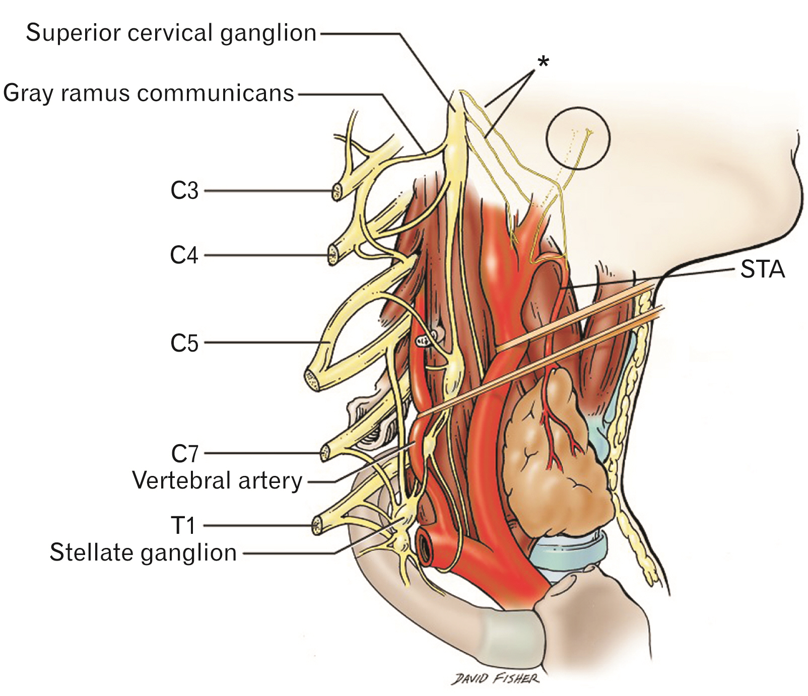
Fig. 2 Schematic drawing of the right side of the neck with structures superficial to the superior cervical ganglion removed for clarity. Note the laryngopharyngeal branches (*) and their ascending (circle) and descending pathways. The ascending branches are thought to terminate primarily in the pharyngeal plexus. A carotid body branch from the laryngopharyngeal nerve is also shown. STA, superficial thyroid artery (By David Fisher).
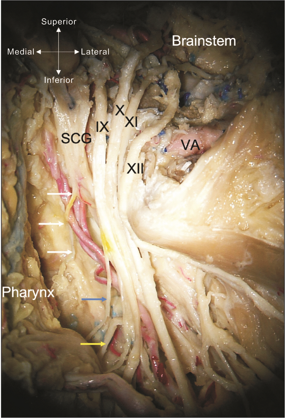
Fig. 3 Cadaveric dissection of the left skull base and opening of the jugular foramen and hypoglossal canal. Note the laryngopharyngeal branch (white arrows) of the superior cervical ganglion (SCG). The IX-XI and XII cranial nerves are seen. Distally, the pharyngeal branch of the IX cranial nerve is shown at the blue arrow and the pharyngeal branch of the X cranial nerve at the yellow arrow. VA, vertebral artery (cadaveric dissection from Tulane University).

Fig. 4 Schematic drawing of the posterior pharynx and pharyngeal plexus. Note the left laryngopharyngeal branch (white arrow) of the superior cervical ganglion (*). The plexus is seen being contributed to by the pharyngeal branches of the glossopharyngeal (IX) and vagus (X) nerves as well as the laryngopharyngeal nerve (By David Fisher).
Reference
-
References
1. Tibbetts PE. 2015; Neuroanatomical terminology: a lexicon of classical origins and historical foundations by Larry W. Swanson. Q Rev Biol. 90:223–4. DOI: 10.1086/681483.2. Willis T. 1664. Cerebri anatome: cui accessit nervorum descriptio et usus]. Typis Tho. Roycroft, Impensis Jo. Martyn & Ja. Allestry;London:3. Vieussens R. 1684. Neurographia universalis. Apud Joannem Certe Lugduni.4. Huber JJ. 1744. Epistola anatomica de nervo intercostali de nervis octavi et noni paris deqve accessorio; nonnvlla tradens ad virvm ilustrem D.D. Wolrath wigand potentissimi svecorvm regis consiliarivm et archiatrvm ioh. Iacobi huberi D. Vandenhoeck;Göttingen:5. Neubauer JE. 1772. Descriptio anatomica nervorum cardiacorum. In Officina Fleischeriana;DOI: 10.1163/1574-9347_dnp_e315900.6. Lobstein JF. 1823. De nervi sympathetici humani fabrica, usu et morbis, commentatio anatomico-physiologico-pathologica. Levrault.7. Moriyama H, Shimada K, Goto N. 1995; Morphometric analysis of neurons in ganglia: geniculate, submandibular, cervical spinal and superior cervical. Okajimas Folia Anat Jpn. 72:185–90. DOI: 10.2535/ofaj1936.72.4_185. PMID: 8570139.
Article8. Kameda Y, Saitoh T, Nemoto N, Katoh T, Iseki S. 2012; Hes1 is required for the development of the superior cervical ganglion of sympathetic trunk and the carotid body. Dev Dyn. 241:1289–300. DOI: 10.1002/dvdy.23819. PMID: 22689348.
Article9. Maningat AL, Munakomi S. 2022. Neuroanatomy, superior cervical ganglion. StatPearls. StatPearls Publishing;DOI: 10.53347/rid-98669.10. Yokota H, Mukai H, Hattori S, Yamada K, Anzai Y, Uno T. 2018; MR imaging of the superior cervical ganglion and inferior ganglion of the vagus nerve: structures that can mimic pathologic retropharyngeal lymph nodes. AJNR Am J Neuroradiol. 39:170–6. DOI: 10.3174/ajnr.A5434. PMID: 29122764. PMCID: PMC7410715.
Article11. Janfaza P. 2001. Surgical anatomy of the head and neck. Lippincott Williams & Wilkins;DOI: 10.1001/archotol.1938.00650030527016.12. Tubbs RS, Salter G, Wellons JC 3rd, Oakes WJ. 2002; Blood supply of the human cervical sympathetic chain and ganglia. Eur J Morphol. 40:283–8. DOI: 10.1076/ejom.40.5.283.28905. PMID: 15101443.
Article13. Goosmann MM, Dalvin M. 2022. Anatomy, head and neck, deep petrosal nerve. StatPearls. StatPearls Publishing;DOI: 10.53347/rid-56609.14. Waxenbaum JA, Reddy V, Bordoni B. 2022. Anatomy, head and neck, cervical nerves. StatPearls. StatPearls Publishing.15. Fazliogullari Z, Kilic C, Karabulut AK, Yazar F. 2016; A morphometric analysis of the superior cervical ganglion and its surrounding structures. Surg Radiol Anat. 38:299–302. DOI: 10.1007/s00276-015-1551-3. PMID: 26364034.
Article16. Gutierrez S, Iwanaga J, Pekala P, Yilmaz E, Clifton WE, Dumont AS, Tubbs RS. 2021; The pharyngeal plexus: an anatomical review for better understanding postoperative dysphagia. Neurosurg Rev. 44:763–72. DOI: 10.1007/s10143-020-01303-5. PMID: 32318923.
Article17. Nurse CA, Piskuric NA. 2013; Signal processing at mammalian carotid body chemoreceptors. Semin Cell Dev Biol. 24:22–30. DOI: 10.1016/j.semcdb.2012.09.006. PMID: 23022231.
Article18. Razipour SE, Zarrintan S, Mathkour M, Iwanaga J, Dumont AS, Tubbs RS. 2021; Review of the external carotid plexus: anatomy, function, and clinical manifestations. Anat Cell Biol. 54:137–42. DOI: 10.5115/acb.20.308. PMID: 33731490. PMCID: PMC8225485.
Article19. Mu L, Sanders I. 2007; Neuromuscular specializations within human pharyngeal constrictor muscles. Ann Otol Rhinol Laryngol. 116:604–17. DOI: 10.1177/000348940711600809. PMID: 17847729.
Article20. Lima AF, Moreira FC, Menezes A, Dias L. 2018; Cervical ganglioneuroma in pediatric age: a case report. Turk Arch Otorhinolaryngol. 56:237–40. DOI: 10.5152/tao.2018.3690. PMID: 30701121. PMCID: PMC6340319. PMID: 170080a138274bfaa143b7d7c47ce9d6.
Article21. Hara I, Tanuma K, Suzuki K. 1993; Morphology of the ganglion cervicale superius in human fetuses and an adult. Kaibogaku Zasshi. 68:544–63. Japanese.22. Nourinezhad J, Mazaheri Y, Biglari Z. 2015; Detailed anatomy of the cranial cervical ganglion in the dromedary camel (Camelus dromedarius). Anat Rec (Hoboken). 298:1479–91. DOI: 10.1002/ar.23169. PMID: 25950508.23. Sisson S, Grossman JD, Getty R. 1975. Sisson and Grossman's the anatomy of the domestic animals. 5th ed. Saunders.24. Ari HH, Soyguder Z, Cinaroglu S. 2010; Macroanatomy of the cranial cervical ganglion in Angora goats. Vet Med. 55:389–93. DOI: 10.17221/2952-VETMED. PMID: 8fec90b10ede40adaab6ecfaaa7245cb.
Article25. Hedger JH, Webber RH. 1976; Anatomical study of the cervical sympathetic trunk and ganglia in the albino rat (Mus norvegicus albinus). Acta Anat (Basel). 96:206–17. DOI: 10.1159/000144674. PMID: 970104.26. Kabak M. 2007; The gross anatomy of the cranial cervical ganglion in the guinea pig (Cavia porcellus). Vet Res Commun. 31:1–7. DOI: 10.1007/s11259-006-3398-x. PMID: 17186407.
Article27. Nieuwenhuys R, Donkelaar HJ, Nicholson C. 1997. The central nervous system of vertebrates. Springer;DOI: 10.1007/978-3-642-18262-4.28. Gaskell WH. 1886; On the structure, distribution and function of the nerves which innervate the visceral and vascular systems. J Physiol. 7:1–80. DOI: 10.1113/jphysiol.1886.sp000207. PMID: 16991419. PMCID: PMC1484971.
Article29. Iwanaga J, Singh V, Ohtsuka A, Hwang Y, Kim HJ, Moryś J, Ravi KS, Ribatti D, Trainor PA, Sañudo JR, Apaydin N, Şengül G, Albertine KH, Walocha JA, Loukas M, Duparc F, Paulsen F, Del Sol M, Adds P, Hegazy A, Tubbs RS. 2021; Acknowledging the use of human cadaveric tissues in research papers: recommendations from anatomical journal editors. Clin Anat. 34:2–4. DOI: 10.1002/ca.23671. PMID: 32808702.
Article30. Iwanaga J, Singh V, Takeda S, Ogeng'o J, Kim HJ, Moryś J, Ravi KS, Ribatti D, Trainor PA, Sañudo JR, Apaydin N, Sharma A, Smith HF, Walocha JA, Hegazy AMS, Duparc F, Paulsen F, Del Sol M, Adds P, Louryan S, Fazan VPS, Boddeti RK, Tubbs RS. 2022; Standardized statement for the ethical use of human cadaveric tissues in anatomy research papers: recommendations from Anatomical Journal Editors-in-Chief. Clin Anat. 35:526–8. DOI: 10.1002/ca.23849. PMID: 35218594.
- Full Text Links
-
- Actions
-
Cited
- CITED
-
- Close
- Share
-
- Similar articles
-
- Mapping Regional Laryngopharyngeal Mechanoreceptor Response
- Pepsin and Laryngeal and Hypopharyngeal Carcinomas
- The Usefulness of Esophagography as a Screening Test for Laryngopharyngeal Reflux
- Neurophysiology of Laryngopharyngeal Reflux and Brainstem Reflex
- The Management of Laryngopharyngeal Reflux Disease
