Dendritic cells resist to disulfiram-induced cytotoxicity, but reduced interleukin-12/23(p40) production
- Affiliations
-
- 1College of Veterinary Medicine, Jeju National University, Jeju 63243, Korea
- 2Veterinary Medical Research Institute, Jeju National University, Jeju 63243, Korea
- KMID: 2545537
- DOI: http://doi.org/10.4196/kjpp.2023.27.5.471
Abstract
- Disulfiram (DSF), a medication for alcoholism, has recently been used as a repurposing drug owing to its anticancer effects. Despite the crucial role of dendritic cells (DCs) in immune homeostasis and cancer therapy, the effects of DSF on the survival and function of DCs have not yet been studied. Therefore, we treated bone marrow-derived DCs with DSF and lipopolysaccharide (LPS) and performed various analyses. DCs are resistant to DSF and less cytotoxic than bone marrow cells and spleen cells. The viability and metabolic activity of DCs hardly decreased after treatment with DSF in the absence or presence of LPS. DSF did not alter the expression of surface markers (MHC II, CD86, CD40, and CD54), antigen uptake capability, or the antigen-presenting ability of LPS-treated DCs. DSF decreased the production of interleukin (IL)-12/23 (p40), but not IL-6 or tumor necrosis factor-α, in LPS-treated DCs. We considered the granulocyte-macrophage colony-stimulating factor (GM-CSF) as a factor to make DCs resistant to DSF-induced cytotoxicity. The resistance of DCs to DSF decreased when GM-CSF was not given or its signaling was inhibited. Also, GM-CSF upregulated the expression of a transcription factor XBP-1 which is essential for DCs’ survival. This study demonstrated for the first time that DSF did not alter the function of DCs, had low cytotoxicity, and induced differential cytokine production.
Figure
-

Fig. 1 The effect of DSF on the cellular metabolic activity. (A) DCs. (B) BMs. (C) Spleen cells. Cells were cultured in a 96-well plate with 0 to 1 µM of DSF with or without LPS. Concentrations of LPS were 0.1 µg/ml on DCs and BMs, and 1 µg/ml on spleen cells. 1 µg/ml of DOX was used as a positive control on DCs. The MTT assay was performed at 3 days after the treatment. The optical density (O.D.) was measured at 570 nm by using a microplate reader. Results are presented as mean ± SD, and statistical significance was performed by two-way ANOVA followed by Dunnett’s multiple comparisons test. *, **, **** indicate p < 0.05, 0.01, 0.0001 respectively compared to control cells (DSF 0 µM). DSF, Disulfiram; DCs, dendritic cells; BMs, bone marrow cells; LPS, lipopolysaccharide; DOX, doxorubicin; MTT, 3-(4,5-dimethylthiazol-2-yl)-2,5-diphenylltetrazolium bromide.

Fig. 2 DSF has little effect on apoptosis and cell viability. After 24 h of treatment, the DCs were stained with Annexin V-FITC and PI. (A) The quadrants of dot plot indicate live cells (annexin V–/PI–), the cells in early apoptosis (annexin V+/PI–), late apoptosis (annexin V+/PI+), and necrosis (annexin V–/PI+). (B) Statistical analysis was performed based on the results of the four experiments conducted under the same conditions. A trypan blue exclusion assay was performed after 3 days of treatment. (C, D) As a result of four experiments under the same conditions, the net number of live and dead cells was counted. The viability was calculated by the ratio of live cells to total cells. Results are presented as mean ± SD, and statistical significance was performed by two-way ANOVA followed by Tukey’s multiple comparisons test (B, C) or Dunnett’s multiple comparisons test (D). * indicates p < 0.05 compared to control DCs (DSF 0 µM). DSF, Disulfiram; DCs, dendritic cells; LPS, lipopolysaccharide; FITC, fluorescein isothiocyanate; PI, propidium iodide.

Fig. 3 DSF does not affect the maturation of LPS-treated DCs. To evaluate the effect of DSF on the maturation of DCs, antigen-uptake capability and expression of maturation-related surface markers of DCs were analyzed. After 3 days of treatments, the DCs were incubated with FITC-dextran for 1 h or stained with the marker-specific antibodies for 30 min as described in the “Methods” section. Anti-CD11c antibody was used for gating DCs. (A) Numbers of histograms represent the geometric mean of fluorescence intensity. (B, C) For statistical analysis of three independent experiments, the mean fluorescence intensity detected in the control DCs was set to 100%. Results are presented as mean ± SD, and statistical significance was performed by two-way ANOVA followed by Šídák’s multiple comparisons test. * indicates p < 0.05 compared to control DCs (DSF 0 µM). DSF, Disulfiram; DCs, dendritic cells; LPS, lipopolysaccharide; FITC, fluorescein isothiocyanate.
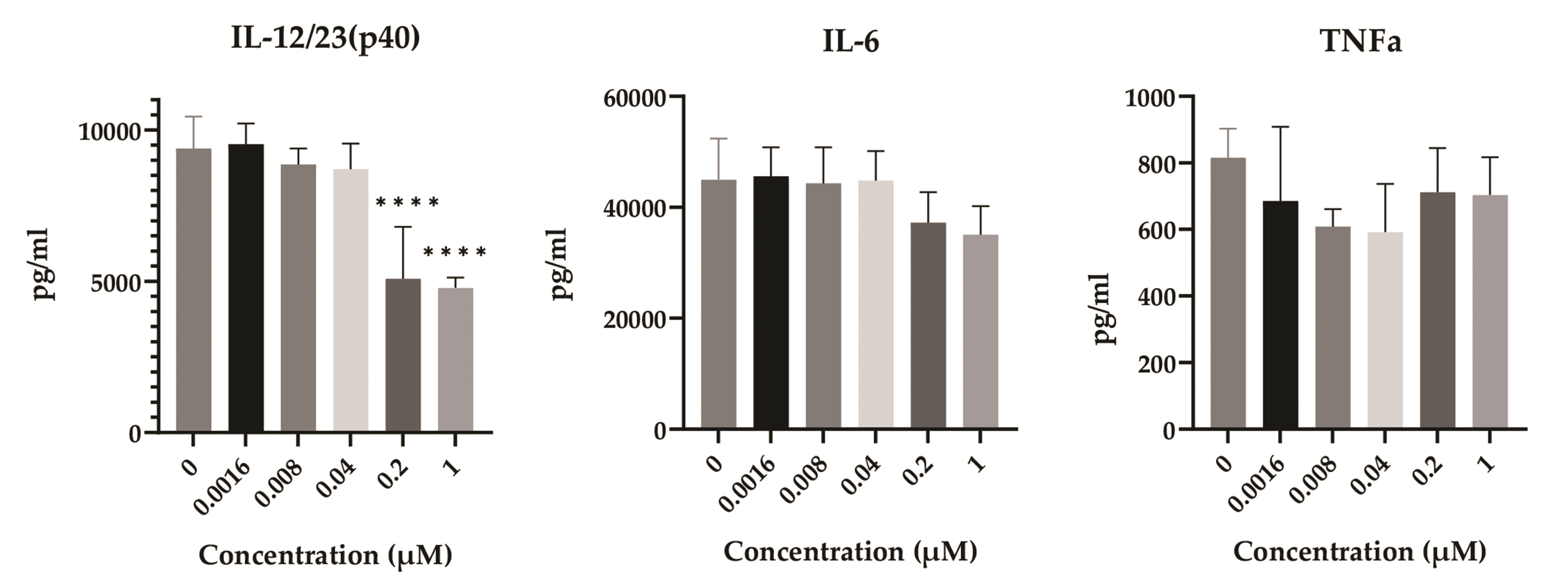
Fig. 4 The effect of DSF on the production of immune-related cytokines. After 2 days of treatments, the supernatant of treated DCs was collected and used for ELISA. The optical density was measured at 450 nm by using a microplate reader. Results are presented as mean ± SD, and statistical significance was performed by ordinary one-way ANOVA followed by Dunnett’s multiple comparisons test. **** indicates p < 0.0001 compared to control DCs (DSF 0 µM). DSF, Disulfiram; DCs, dendritic cells; IL, interleukin; TNF, tumor necrosis factor.

Fig. 5 Effect of DSF on the antigen-presenting capability of DCs. After 3 days of DSF and LPS treatment, the DCs were incubated with 50 µg/ml mitomycin C for 30 min and co-cultured with allogeneic spleen cells. After 5 days, CCK-8 assay was performed and the optical density (O.D.) was measured at 450 nm by using a microplate reader. Results are presented as mean ± SD, and statistical significance was performed by two-way ANOVA followed by Dunnett’s multiple comparisons test. DSF, Disulfiram; DCs, dendritic cells; LPS, lipopolysaccharide.

Fig. 6 GM-CSF is responsible for the resistance of DCs to DSF. (A) Schematic overview of the experiment. (B) DCs were pretreated with or without baricitinib for 4 h. 10 ng/ml of GM-CSF was added (or not) and DCs were incubated for 24 h. 0–1 µM of DSF were treated and incubated for 48 h. The metabolic activity was measured by MTT assay. (C) DCs were cultured in a culture medium without GM-CSF + IL-4 for 48 h. The expression of XBP-1 in DCs was measured using qPCR with or without GM-CSF treatment for 24 h. Results are presented as mean ± SD. Statistical significance was performed by two-way ANOVA with Šídák’s multiple comparisons test and Dunnett’s multiple comparisons test (B), or unpaired t-test (C). **, **** indicate p < 0.01, 0.0001 respectively, compared to control DCs (DSF 0 µM). #, ##, #### indicate p < 0.1, 0.01, 0.0001 compared between the groups at the same concentration of DSF. GM-CSF, granulocyte-macrophage colony-stimulating factor; DSF, Disulfiram; DCs, dendritic cells; MTT, 3-(4,5-dimethylthiazol-2-yl)-2,5-diphenylltetrazolium bromide; qPCR, quantitative real-time PCR; IL, interleukin.
Reference
-
1. Johansson B. 1992; A review of the pharmacokinetics and pharmacodynamics of disulfiram and its metabolites. Acta Psychiatr Scand Suppl. 369:15–26. DOI: 10.1111/j.1600-0447.1992.tb03310.x. PMID: 1471547.
Article2. Lu C, Li X, Ren Y, Zhang X. 2021; Disulfiram: a novel repurposed drug for cancer therapy. Cancer Chemother Pharmacol. 87:159–172. DOI: 10.1007/s00280-020-04216-8. PMID: 33426580.
Article3. McMahon A, Chen W, Li F. 2020; Old wine in new bottles: advanced drug delivery systems for disulfiram-based cancer therapy. J Control Release. 319:352–359. DOI: 10.1016/j.jconrel.2020.01.001. PMID: 31911155.
Article4. Skrott Z, Majera D, Gursky J, Buchtova T, Hajduch M, Mistrik M, Bartek J. 2019; Disulfiram's anti-cancer activity reflects targeting NPL4, not inhibition of aldehyde dehydrogenase. Oncogene. 38:6711–6722. DOI: 10.1038/s41388-019-0915-2. PMID: 31391554.
Article5. Terashima Y, Toda E, Itakura M, Otsuji M, Yoshinaga S, Okumura K, Shand FHW, Komohara Y, Takeda M, Kokubo K, Chen MC, Yokoi S, Rokutan H, Kofuku Y, Ohnishi K, Ohira M, Iizasa T, Nakano H, Okabe T, Kojima H, et al. 2020; Targeting FROUNT with disulfiram suppresses macrophage accumulation and its tumor-promoting properties. Nat Commun. 11:609. DOI: 10.1038/s41467-020-14338-5. PMID: 32001710. PMCID: PMC6992764. PMID: a4d75cebbb8f47a985d666129063bb6d.
Article6. Yip NC, Fombon IS, Liu P, Brown S, Kannappan V, Armesilla AL, Xu B, Cassidy J, Darling JL, Wang W. 2011; Disulfiram modulated ROS-MAPK and NFκB pathways and targeted breast cancer cells with cancer stem cell-like properties. Br J Cancer. 104:1564–1574. DOI: 10.1038/bjc.2011.126. PMID: 21487404. PMCID: PMC3101904.
Article7. Zha J, Chen F, Dong H, Shi P, Yao Y, Zhang Y, Li R, Wang S, Li P, Wang W, Xu B. 2014; Disulfiram targeting lymphoid malignant cell lines via ROS-JNK activation as well as Nrf2 and NF-kB pathway inhibition. J Transl Med. 12:163. DOI: 10.1186/1479-5876-12-163. PMID: 24915933. PMCID: PMC4075939.
Article8. Kannappan V, Ali M, Small B, Rajendran G, Elzhenni S, Taj H, Wang W, Dou QP. 2021; Recent advances in repurposing disulfiram and disulfiram derivatives as copper-dependent anticancer agents. Front Mol Biosci. 8:741316. DOI: 10.3389/fmolb.2021.741316. PMID: 34604310. PMCID: PMC8484884. PMID: fcaf5f20e5dc494488e3a449e9c91401.
Article9. Steinman RM. 2012; Decisions about dendritic cells: past, present, and future. Annu Rev Immunol. 30:1–22. DOI: 10.1146/annurev-immunol-100311-102839. PMID: 22136168.
Article10. Qian C, Cao X. 2018; Dendritic cells in the regulation of immunity and inflammation. Semin Immunol. 35:3–11. DOI: 10.1016/j.smim.2017.12.002. PMID: 29242034.
Article11. Audiger C, Rahman MJ, Yun TJ, Tarbell KV, Lesage S. 2017; The importance of dendritic cells in maintaining immune tolerance. J Immunol. 198:2223–2231. DOI: 10.4049/jimmunol.1601629. PMID: 28264998. PMCID: PMC5343761.
Article12. Kim CW, Kim KD, Lee HK. 2021; The role of dendritic cells in tumor microenvironments and their uses as therapeutic targets. BMB Rep. 54:31–43. DOI: 10.5483/BMBRep.2021.54.1.224. PMID: 33298246. PMCID: PMC7851442.
Article13. Wculek SK, Cueto FJ, Mujal AM, Melero I, Krummel MF, Sancho D. 2020; Dendritic cells in cancer immunology and immunotherapy. Nat Rev Immunol. 20:7–24. DOI: 10.1038/s41577-019-0210-z. PMID: 31467405.
Article14. Fasehee H, Zarrinrad G, Tavangar SM, Ghaffari SH, Faghihi S. 2016; The inhibitory effect of disulfiram encapsulated PLGA NPs on tumor growth: different administration routes. Mater Sci Eng C Mater Biol Appl. 63:587–595. DOI: 10.1016/j.msec.2016.03.023. PMID: 27040254.
Article15. Masten BJ, Yates JL, Pollard Koga AM, Lipscomb MF. 1997; Characterization of accessory molecules in murine lung dendritic cell function: roles for CD80, CD86, CD54, and CD40L. Am J Respir Cell Mol Biol. 16:335–342. DOI: 10.1165/ajrcmb.16.3.9070619. PMID: 9070619.
Article16. Blanco P, Palucka AK, Pascual V, Banchereau J. 2008; Dendritic cells and cytokines in human inflammatory and autoimmune diseases. Cytokine Growth Factor Rev. 19:41–52. DOI: 10.1016/j.cytogfr.2007.10.004. PMID: 18258476. PMCID: PMC2413068.
Article17. Neurath MF. 2007; IL-23: a master regulator in Crohn disease. Nat Med. 13:26–28. DOI: 10.1038/nm0107-26. PMID: 17206128.
Article18. Iwakura Y, Ishigame H. 2006; The IL-23/IL-17 axis in inflammation. J Clin Invest. 116:1218–1222. DOI: 10.1172/JCI28508. PMID: 16670765. PMCID: PMC1451213.
Article19. Opferman JT. 2008; Apoptosis in the development of the immune system. Cell Death Differ. 15:234–242. DOI: 10.1038/sj.cdd.4402182. PMID: 17571082.
Article20. Baird AM, Gerstein RM, Berg LJ. 1999; The role of cytokine receptor signaling in lymphocyte development. Curr Opin Immunol. 11:157–166. DOI: 10.1016/S0952-7915(99)80027-2. PMID: 10322150.
Article21. Zhang N, Hartig H, Dzhagalov I, Draper D, He YW. 2005; The role of apoptosis in the development and function of T lymphocytes. Cell Res. 15:749–769. DOI: 10.1038/sj.cr.7290345. PMID: 16246265.
Article22. Hildeman D, Jorgensen T, Kappler J, Marrack P. 2007; Apoptosis and the homeostatic control of immune responses. Curr Opin Immunol. 19:516–521. DOI: 10.1016/j.coi.2007.05.005. PMID: 17644328. PMCID: PMC4127626.
Article23. Sallusto F, Lanzavecchia A. 1994; Efficient presentation of soluble antigen by cultured human dendritic cells is maintained by granulocyte/macrophage colony-stimulating factor plus interleukin 4 and downregulated by tumor necrosis factor alpha. J Exp Med. 179:1109–1118. DOI: 10.1084/jem.179.4.1109. PMID: 8145033. PMCID: PMC2191432.
Article24. Chen M, Wang J. 2010; Programmed cell death of dendritic cells in immune regulation. Immunol Rev. 236:11–27. DOI: 10.1111/j.1600-065X.2010.00916.x. PMID: 20636805. PMCID: PMC3282617.
Article25. Fujita Y, Matsuoka N, Temmoku J, Furuya-Yashiro M, Asano T, Sato S, Matsumoto H, Watanabe H, Kozuru H, Yatsuhashi H, Kawakami A, Migita K. 2020; JAK inhibitors impair GM-CSF-mediated signaling in innate immune cells. BMC Immunol. 21:35. DOI: 10.1186/s12865-020-00365-w. PMID: 32539713. PMCID: PMC7296727. PMID: 2122a39e70894270b93fd834c7961c85.
Article26. Iwakoshi NN, Pypaert M, Glimcher LH. 2007; The transcription factor XBP-1 is essential for the development and survival of dendritic cells. J Exp Med. 204:2267–2275. DOI: 10.1084/jem.20070525. PMID: 17875675. PMCID: PMC2118458.
Article
- Full Text Links
-
- Actions
-
Cited
- CITED
-
- Close
- Share
-
- Similar articles
-
- IL-12 p40-Expressing Immune Cells Revealed by Cytokine Reporter Mouse System
- Mycobacterium tuberculosis ESAT6 Drives the Activation and Maturation of Bone Marrow-Derived Dendritic Cells via TLR4-Mediated Signaling
- Induction of IL-12 Experession in Bone Marrow-derived Mouse Dendritic Cells
- The Role of Interleukin-12 in Dendritic Cells
- BCG-Induced Dendritic Cell Responses and Suppression of Interleukin-5 Production from T Cells in Atopic Asthmatics
