Solitary Fibrous Tumors of the Mesocolon: A Report of Two Cases and Review of Literature
- Affiliations
-
- 1Departments of Surgical Gastroenterology, Nizam’s Institute of Medical Sciences, Hyderabad, India
- 2Departments of Pathology, Nizam’s Institute of Medical Sciences, Hyderabad, India
- KMID: 2545334
- DOI: http://doi.org/10.4166/kjg.2023.053
Abstract
- Solitary fibrous tumors (SFTs) are an uncommon group of neoplasms. The visceral pleura is the most common site of origin of these tumors. The colonic mesentery is an unusual site of origin of SFTs. A pre-operative diagnosis of SFT is challenging as there are no pathognomonic clinical or radiological signs. Most patients reported thus far were diagnosed post-operatively with the aid of immunohistochemical markers. Complete surgical excision is the treatment of choice for SFTs. Recurrences are uncommon. However, they can occasionally show aggressive behavior. In this report, we describe two cases of rare colonic mesentery SFTs.
Keyword
Figure
-

Fig. 1 (A) Contrast-enhanced computed tomography showing a heterogeneously enhancing mass with lobulated margins and central necrotic components. (B) Specimen showing distal pancreatectomy and splenectomy along with the resected segment of the colon.
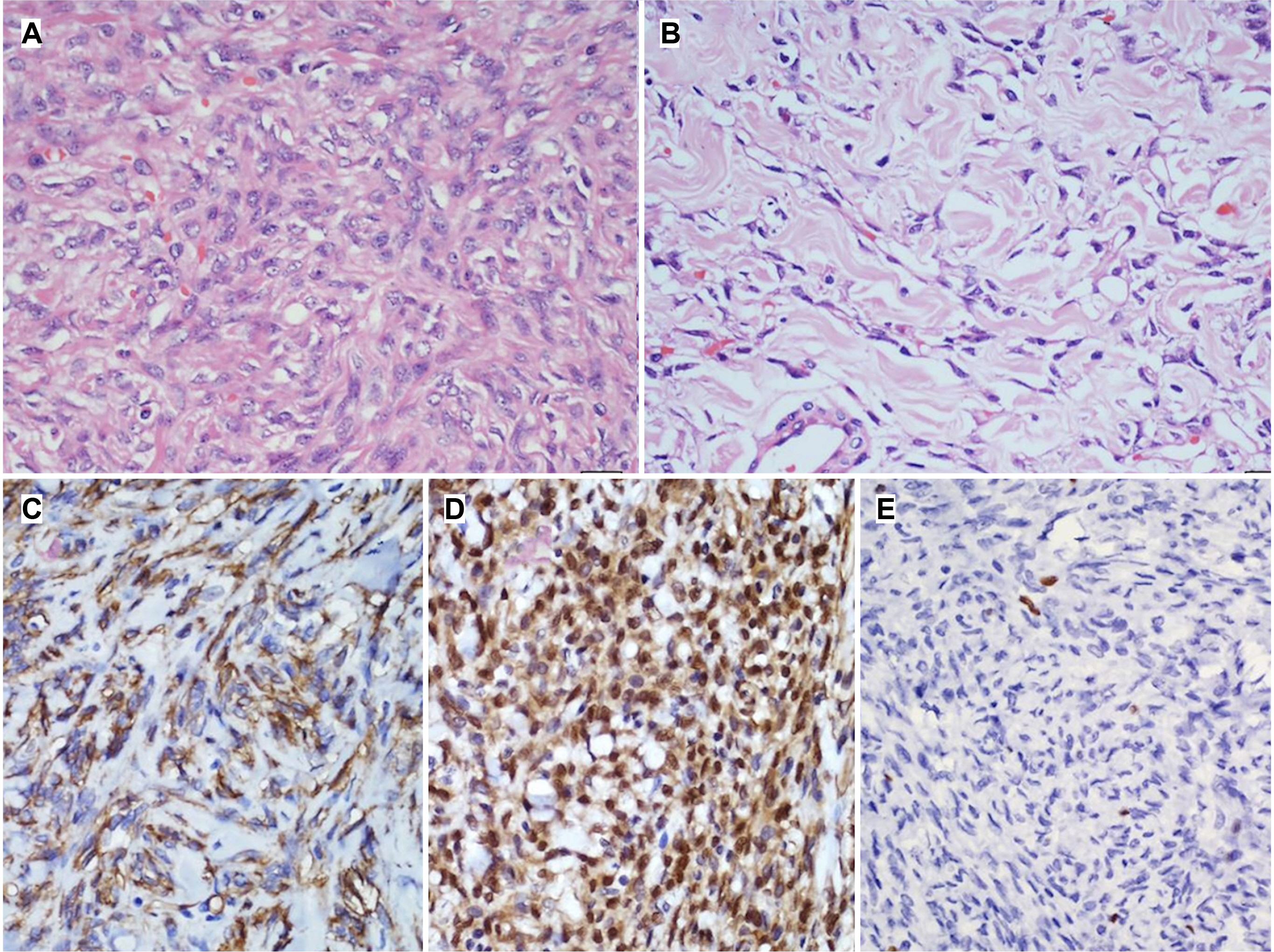
Fig. 2 (A) H&E sections reveal a higher magnification of the cellular area of the tumor showing closely packed cells with little intervening collagenous stroma. Tumor cells have oval to slightly elongated bland nuclei with fine pale chromatin and inconspicuous nucleoli. The cytoplasm is scant to moderate, pale eosinophilic with indistinct borders (×400). (B) Higher magnification of the hypocellular area of the tumor showing prominent hyalinised (keloid-like) collagenous stroma with relatively few tumor cells (×400). Immunohistochemical findings of the tumor. (C) CD34 and (D) STAT6 shows diffuse cytoplasm and nucleus (with some cytoplasmic staining) in the tumor cells respectively (HRP-Polymer; ×400). (E) Immunohistochemical staining with Ki-67 labels occasional tumor cell nuclei indicating low proliferative activity within the tumor (HRP-Polymer; ×400).

Fig. 3 (A) Contrast-enhanced computed tomography showing a heterogeneously enhancing mass occupying the pelvis. (B) Post-operative specimen showing the resected recto-sigmoid colon with the adjacent tumor.

Fig. 4 Hematoxylin and esosin staining. (A) Cells arranged in fascicles separated by collagen (magnification ×100). (B) Cellular lesion with a hemangiopericytomatous pattern; immuno-histochemical staining (magnificantion ×400). (C) Nuclear positivity for STAT6 (HRP-Polymer; ×400). (D) Tumor cell positivity for CD34 (HRP-Polymer; ×400). (E) Low Ki67 index (HRP-Polymer; ×400).
Reference
-
1. Liu JN, Liu Z, Ji PY, Zhang H, Guo SL. 2020; Solitary fibrous tumor of the mesentery: a case report and review of the literature. J Int Med Res. 48:300060520950111. DOI: 10.1177/0300060520950111. PMID: 33050750. PMCID: PMC7710395.
Article2. Mekel G, Balshan E, Traupman F. 2019; Solitary fibrous tumour of the sigmoid colon mesentery. BMJ Case Rep. 12:e228774. DOI: 10.1136/bcr-2018-228774. PMID: 31068346. PMCID: PMC6506114.
Article3. Val-Bernal JF, Mayorga M, Fernández F, Parra A, Crespo J, García-Polavieja M. 2014; Solitary fibrous tumor arising from the mesentery of adult patients. Report of two cases and review of the literature. Rom J Morphol Embryol. 55:203–207.4. Kay S, Warthen HJ. 1953; Hemangiopericytoma of the rectum. Cancer. 6:167–169. DOI: 10.1002/1097-0142(195301)6:1<167::AID-CNCR2820060119>3.0.CO;2-0.
Article5. Genter B, Mir R, Strauss R, et al. 1982; Hemangiopericytoma of the colon: report of a case and review of literature. Dis Colon Rectum. 25:149–156. DOI: 10.1007/BF02553264. PMID: 7067552.6. Nakagawa T, Shinoda Y, Masuko Y, et al. 1997; Hemangiopericytoma of the sigmoid mesentery: report of a case with immunohistochemical findings. Surg Today. 27:64–67. DOI: 10.1007/BF01366942. PMID: 9035303.
Article7. West NJ, Daniels IR, Allum WH. 2004; Haemangiopericytoma of the sigmoid mesentery. Tech Coloproctol. 8:179–181. DOI: 10.1007/s10151-004-0084-2. PMID: 15654526.
Article8. Balaji R, Ramachandran K, Somanathan T. 2009; A rare case of solitary fibrous tumour of the sigmoid mesocolon: imaging features and review of literature. Cancer Imaging. 9:67–69. DOI: 10.3816/CBC.2009.n.034. PMID: 19661047.9. Soda H, Kainuma O, Yamamoto H, et al. 2010; Giant intrapelvic solitary fibrous tumor arising from mesorectum. Clin J Gastroenterol. 3:136–139. DOI: 10.1007/s12328-010-0146-0. PMID: 26190119.
Article10. Medina-Franco H, Cabrera-Mendoza F, Almaguer-Rosales S, Guillén-Pérez F, Chablé-Montero F. 2011; [Mesenteric solitary fibrous tumor: a case report and literature review]. Rev Gastroenterol Mex. 76:186–189. Spanish.11. Nishikawa T, Takei Y, Teshima S, et al. 2013; A hypervascular malignant solitary fibrous tumor in the mesentery of the rectum with good response to preoperative radiation therapy. Int Cancer Conf J. 2:30–35. DOI: 10.1007/s13691-012-0059-5.
Article12. Scarlett A, Stillwell R, Deutscher D, Sengupta SK. 2015; 49. Solitary fibrous tumour of transverse mesocolon: An unusual location. Pathology. 47(Suppl 1):S115. DOI: 10.1097/01.PAT.0000461658.78598.52.
Article13. Ge W, Yu DC, Chen G, Ding YT. 2016; Clinical analysis of 47 cases of solitary fibrous tumor. Oncol Lett. 12:2475–2480. DOI: 10.3892/ol.2016.4967. PMID: 27698815. PMCID: PMC5038456.
Article14. Keser BN, Kırman ÜN, Aktemur G, Alimoĝlu O. 2019; A rare solitary fibrous tumour of the ascending mesocolon: a case report. Ann R Coll Surg Engl. 101:e108–e110. DOI: 10.1308/rcsann.2019.0031. PMID: 30854871. PMCID: PMC6432965.
Article15. Chaouch MA, Jerraya H, Dougaz MW, Haloui N, Bouasker I, Nouira R. 2020; A case report of a right mesocolon solitary fibrosis tumour. J Gastrointest Cancer. 51:351–353. DOI: 10.1007/s12029-019-00294-x. PMID: 31407250.
Article16. Hoseini M, Negahi A, Vosough F, et al. 2020; Solitary fibrous tumor in pelvis extended to transverse mesocolon and peritoneum. Res J Pharm Technol. 13:1943–1950. DOI: 10.5958/0974-360X.2020.00350.9.
Article17. Sousa M, Proença L, Baldaia H. 2020; Intussusception caused by a colonic solitary fibrous tumor. GE Port J Gastroenterol. 27:59–61. DOI: 10.1159/000499137. PMID: 31970245. PMCID: PMC6959109.
Article
- Full Text Links
-
- Actions
-
Cited
- CITED
-
- Close
- Share
-
- Similar articles
-
- A Case of Solitary Fibrous Tumor in the Cheek
- Dynamic Contrast-Enhanced CT Findings of a Extrapleural Solitary Fibrous Tumor in the Spleen: A Case Report and Literature Review
- A Case of Solitary Fibrous Tumor of Lacrimal Gland
- A Case of Solitary Fibrous Tumor That Developed on the Scalp
- Malignant Hepatic Solitary Fibrous Tumor
