Buccal nerve schwannoma mimicking a salivary gland tumor: a rare case report
- Affiliations
-
- 1Department of Oral and Maxillofacial Surgery, Gangnam Severance Hospital, Yonsei University College of Dentistry, Seoul, Korea
- 2Department of Oral Pathology, Oral Cancer Research Institute, Yonsei University College of Dentistry, Seoul, Korea
- KMID: 2544317
- DOI: http://doi.org/10.5125/jkaoms.2023.49.3.148
Abstract
- Schwannomas are benign tumors originating from myelinating cells constituting nerve sheaths but rarely contain cellular elements of the nerve. The authors encountered a 47-year-old female patient with a schwannoma on the anterior mandibular ramus arising from the buccal nerve, measuring 3 cm×4 cm. Surgical resection was performed with preservation of the buccal nerve via microsurgical dissection. After one month, the sensory function of the buccal nerve was recovered without complications.
Keyword
Figure
-
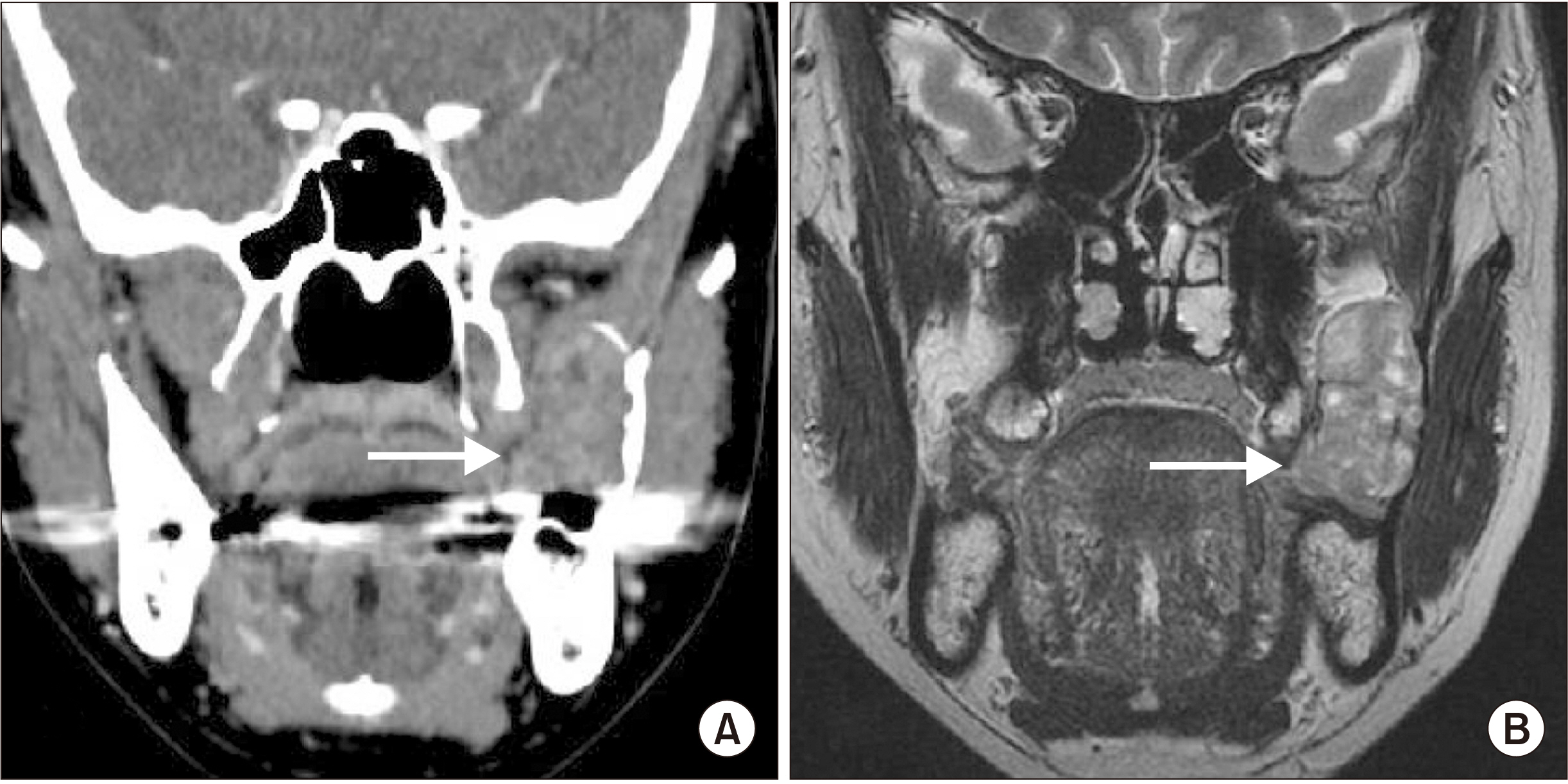
Fig. 1 Clinical images of schwannoma from the long buccal nerve. A. Swelling was noted on the anterior border of the ramus ascending branch. B. The long buccal nerve was detected from the middle portion of the tumor. C. The tumor was resected while preserving the long buccal nerve by dissecting it from the tumor.
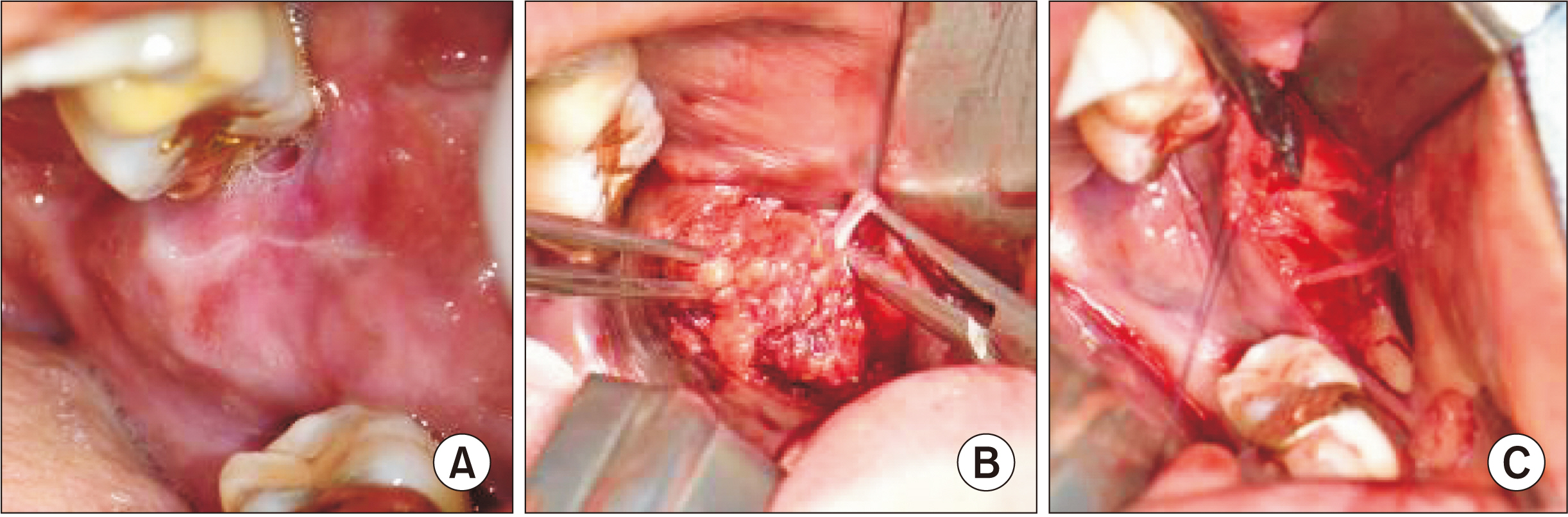
Fig. 2 Preoperative images. A. Computed tomography revealed a homogeneous mass, slightly less dense than the adjacent muscles, and expansion of the ramus with some irregular resorption borders (arrow). B. T2-weighted magnetic resonance imaging showed a multifocal hypointense portion, but the long buccal nerve was not visualized clearly (arrow).
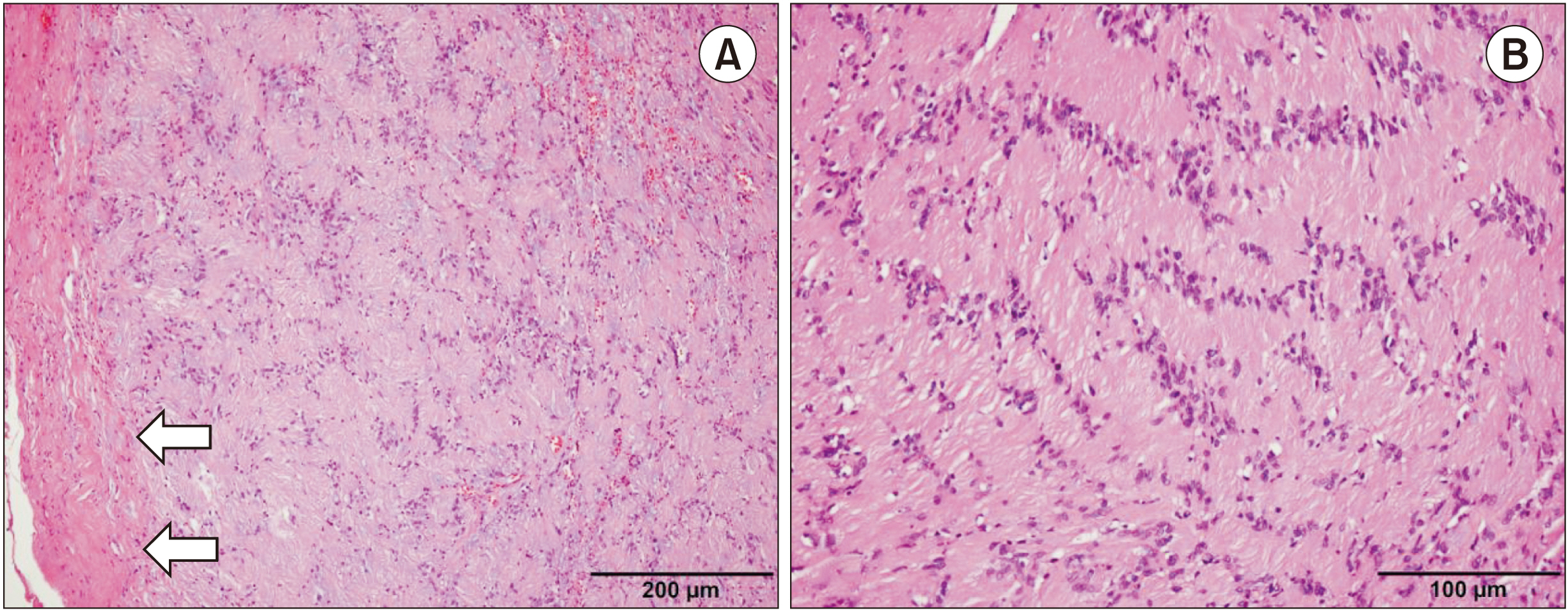
Fig. 3 Histopathology of the surgical specimen (H&E staining). A. The mass was composed of spindle cells with proliferated biphasic growth patterns and was surrounded by a well-formed fibrous capsule (arrows) (×100). Scale bar=200 µm. B. Compact cellular area, called an Antoni A area, showed characteristic nuclear palisading separated by fibrillary cell processes (×100). Scale bar=100 µm.
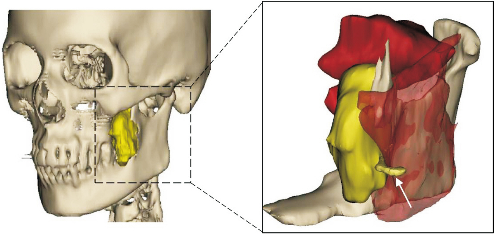
Fig. 4 Reconstructed three-dimensional images from preoperative computed tomography (left) and magnetic resonance imaging (MRI) (right). In the MRI view, the location of the long buccal nerve was suggested based on the intraoperative findings (arrow).
Reference
-
References
1. Fehlings MG, Nater A, Zamorano JJ, Tetreault LA, Varga PP, Gokaslan ZL, et al. 2016; Risk factors for recurrence of surgically treated conventional spinal schwannomas: analysis of 169 patients from a multicenter international database. Spine (Phila Pa 1976). 41:390–8. https://doi.org/10.1097/brs.0000000000001232. DOI: 10.1097/BRS.0000000000001232. PMID: 26555828. PMCID: PMC4769652.
Article2. Crist J, Hodge JR, Frick M, Leung FP, Hsu E, Gi MT, et al. 2017; Magnetic resonance imaging appearance of schwannomas from head to toe: a pictorial review. J Clin Imaging Sci. 7:38. https://doi.org/10.4103/jcis.jcis_40_17. DOI: 10.4103/jcis.JCIS_40_17. PMID: 29114437. PMCID: PMC5651654.
Article3. Kurup S, Thankappan K, Krishnan N, Nair PP. 2012; Intraoral schwannoma--a report of two cases. BMJ Case Rep. 2012:bcr1220115389. https://doi.org/10.1136/bcr.12.2011.5389. DOI: 10.1136/bcr.12.2011.5389. PMID: 22778466. PMCID: PMC3417034.
Article4. Shim SK, Myoung H. 2016; Neurilemmoma in the floor of the mouth: a case report. J Korean Assoc Oral Maxillofac Surg. 42:60–4. https://doi.org/10.5125/jkaoms.2016.42.1.60. DOI: 10.5125/jkaoms.2016.42.1.60. PMID: 26904498. PMCID: PMC4761576.
Article5. Yumuşakhuylu AC, Sari M, Topuz MF, Bağlam T, Binnetoğlu A. 2014; Trigeminal schwannoma extending into the parapharyngeal space. J Craniofac Surg. 25:e328–30. https://doi.org/10.1097/scs.0000000000000593. DOI: 10.1097/SCS.0000000000000593. PMID: 24978683.
Article6. Perkins D, Stiharu TI, Swift JQ, Dao TV, Mainville GN. 2018; Intraosseous schwannoma of the jaws: an updated review of the literature and report of 2 new cases affecting the mandible. J Oral Maxillofac Surg. 76:1226–47. DOI: 10.1016/j.joms.2017.12.017. PMID: 29360457.
Article7. Sun Z, Sun L, Li T, Ma X, Zhang Z. 2011; Intraosseous trigeminal schwannoma of mandible with intracranial extension. J Laryngol Otol. 125:418–22. https://doi.org/10.1017/s0022215110002707. DOI: 10.1017/S0022215110002707. PMID: 21269550.
Article8. Sanna M, Bacciu A, Falcioni M, Taibah A. 2006; Surgical management of jugular foramen schwannomas with hearing and facial nerve function preservation: a series of 23 cases and review of the literature. Laryngoscope. 116:2191–204. https://doi.org/10.1097/01.mlg.0000246193.84319.e5. DOI: 10.1097/01.mlg.0000246193.84319.e5. PMID: 17146395.
Article9. Wippold FJ 2nd, Lubner M, Perrin RJ, Lämmle M, Perry A. 2007; Neuropathology for the neuroradiologist: Antoni A and Antoni B tissue patterns. AJNR Am J Neuroradiol. 28:1633–8. https://doi.org/10.3174/ajnr.a0682. DOI: 10.3174/ajnr.A0682. PMID: 17893219. PMCID: PMC8134199.
Article10. Raper DMS, Sweiss F, Almira-Suarez MI, Helm G, Sheehan JP. 2013; Malignant peripheral nerve sheath tumor at the cerebellopontine angle treated with Gamma Knife radiosurgery: case report and review of the literature. J Radiosurg SBRT. 2:147–53. PMID: 29296354. PMCID: PMC5658887.
- Full Text Links
-
- Actions
-
Cited
- CITED
-
- Close
- Share
-
- Similar articles
-
- Schwannoma Originating From the Lingual Nerve Radiologically Mimicking a Submandibular Gland Tumor
- 6 Cases of Salivary Gland Tumors Arising at Buccal and Masseteric Area
- A Case of Ancient Schwannoma of the Submandibular Gland
- A case report of the salivary duct cyst and review of literatures
- Ancient schwannoma in the parotid gland: A case report and review of the literature
