Chiari Malformation with Surgically Induced Open Neural Tube Defect in Late Chick Embryos : Characterization by Magnetic Resonance Imaging and Histopathological Analysis
- Affiliations
-
- 1Department of Neurosurgery, Seoul National University Hospital, Seoul National University College of Medicine, Seoul, Korea
- 2Division of Pediatric Neurosurgery, Seoul National University Children’s Hospital, Seoul, Korea
- 3Department of Neurosurgery, Jeju National University Hospital, Jeju National University School of Medicine, Jeju, Korea
- KMID: 2543530
- DOI: http://doi.org/10.3340/jkns.2022.0144
Abstract
Objective
: Chiari II malformation (CM II) is still the main cause of severe morbidity and mortality in children with open neural tube defects (ONTDs). The goal of this study was to validate a CM II model in late-stage chick embryos with surgically induced ONTDs.
Methods
: To make the chick embryo model of ONTD, their neural tubes were opened for a length of 5–6 somites at the thoracic level in Hamburger and Hamilton stage 18 chick embryos (n=150). They were reincubated in ovo. up to a total age of 17–21 days. A total of 19 embryos survived and were assigned to either the postoperative day (POD) 14–15 group (n=6) or the POD 17–18 group (n=13). Magnetic resonance imaging (MRI) and histopathologic findings of embryo heads with spinal ONTDs were compared with age-matched normal chick embryos.
Results
: The chick embryos with ONTDs demonstrated definite and constant structural changes, such as downward displacement of the cerebellum to just above the foramen magnum and narrow and small cerebrospinal fluid spaces in the crowded small posterior fossa. These morphologic features were more prominent in the POD 17–18 group than in the POD 14–15 group.
Conclusion
: This is the first description of CM II with spinal ONTD in a late-stage chick embryo model with MRI and histopathological analysis. The morphological changes of the posterior fossa in this study mimic those of CM II associated with spinal ONTD in humans. This model will facilitate investigation of the pathogenesis of CM II.
Keyword
Figure
-
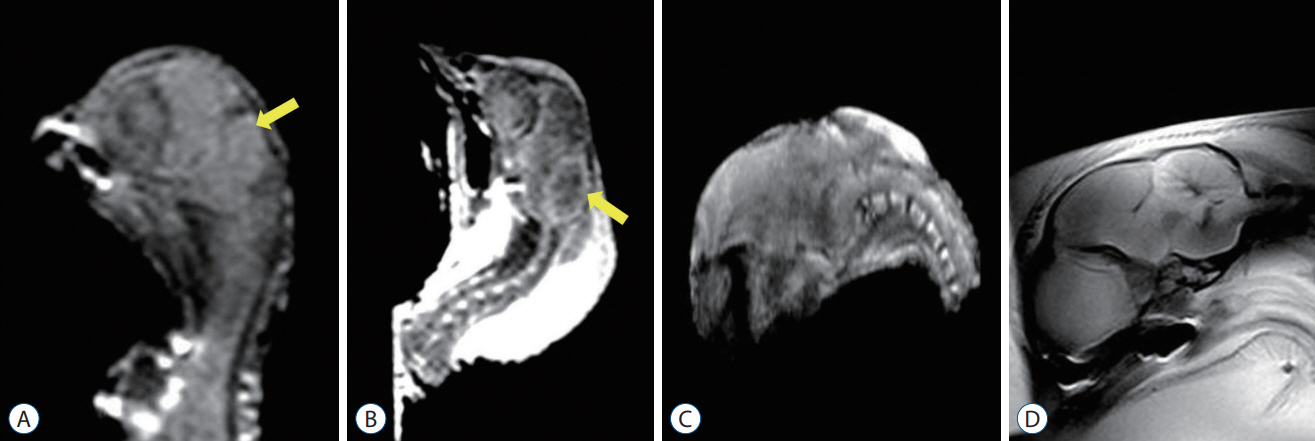
Fig. 1. Mid-sagittal magnetic resonance imaging images of the head of chick embryos. Control group : (A) incubation day (ID) 17–18 and (B) ID 20–21. Surgical group : (C) postoperative day (POD) 14–15 and (D) POD 17–18. T1 weighted image (WI) (A) and T2 WI (B) : sagittal images show the cerebrospinal fluid (CSF) signal (marked with yellow arrows) surrounding the hindbrain and between the cerebellum and brainstem. Significant cerebellar growth occurred in the ID 20–21 group compared to the ID 17–18 group. Embryos in the ID 17–18 group showed physiologic levels of CSF in the posterior fossa and a normal configuration of the cerebellum. The configuration of the cerebellum also becomes elongated rostrally and caudally to some extent. T1 WI (C) and T1 WI (D) : mid-sagittal images demonstrate that the CSF signal surrounding the hindbrain and between the cerebellum and brainstem is absent or barely visible due to overcrowding in the smaller bony posterior fossa. The cerebellum is more elongated rostrally and caudally and is located more caudally. The morphologic changes are more prominent in the POD 17–18 group than in the POD 14–15 group. There is no evidence of hydrocephalus.

Fig. 2. Light microscopic findings of a median section of the posterior fossa of a chick embryo. Control group : (A) incubation day (ID) 17–18 and (B) ID 20–21 (Hematoxylin and Eosin [H&E] stain, ×12.5). Surgical group : (C) postoperative day (POD) 14–15 group and (D) POD 17–18 group (H&E stain, ×12.5). The subarachnoid spaces between the cerebellar cortex and the inner surface of the skull and between the cerebellum and the brainstem (marked with red arrow and yellow arrow, respectively) are narrower in the surgical group than in the control group. The cerebellum is elongated rostrally and caudally and located downward in the overcrowded smaller posterior fossa. There is no evidence of hindbrain herniation or hydrocephalus. The cerebellum increases in size, and a folia pattern appears. There is significant development of the cerebellar hemispheres, along with an increase in volume manifested by the more typical mushroom-shaped configuration in both the surgical and control groups with increased incubation time. Of note, the cerebellum did not completely herniate into the cervical spinal canal.
Cited by 1 articles
-
Editor’s Pick in July 2023
Bum-Tae Kim
J Korean Neurosurg Soc. 2023;66(4):341-343. doi: 10.3340/jkns.2023.0117.
Reference
-
References
1. Bouchard S, Davey MG, Rintoul NE, Walsh DS, Rorke LB, Adzick NS. Correction of hindbrain herniation and anatomy of the vermis after in utero repair of myelomeningocele in sheep. J Pediatr Surg. 38:451–458. discussion 451-458. 2003.
Article2. Briner W, Lieske R. Arnold-Chiari-like malformation associated with a valproate model of spina bifida in the rat. Teratology. 52:306–311. 1995.
Article3. Fontecha CG, Peiro JL, Sevilla JJ, Aguirre M, Soldado F, Fresno L, et al. Fetoscopic coverage of experimental myelomeningocele in sheep using a patch with surgical sealant. Eur J Obstet Gynecol Reprod Biol. 156:171–176. 2011.
Article4. Glenn OA. MR imaging of the fetal brain. Pediatr Radiol. 40:68–81. 2010.
Article5. Griffiths PD, Wilkinson ID, Variend S, Jones A, Paley MN, Whitby E. Differential growth rates of the cerebellum and posterior fossa assessed by post mortem magnetic resonance imaging of the fetus: implications for the pathogenesis of the chiari 2 deformity. Acta Radiol. 45:236–242. 2004.
Article6. Hamburger V, Hamilton HL. A series of normal stages in the development of the chick embryo. J Morphol. 88:49–92. 1951.
Article7. Inagaki T, Schoenwolf GC, Walker ML. Experimental model: change in the posterior fossa with surgically induced spina bifida aperta in mouse. Pediatr Neurosurg. 26:185–189. 1997.
Article8. McLone DG, Knepper PA. The cause of Chiari II malformation: a unified theory. Pediatr Neurosci. 15:1–12. 1989.
Article9. Paek BW, Farmer DL, Wilkinson CC, Albanese CT, Peacock W, Harrison MR, et al. Hindbrain herniation develops in surgically created myelomeningocele but is absent after repair in fetal lambs. Am J Obstet Gynecol. 183:1119–1123. 2000.
Article10. Park TS, Hoffman HJ, Hendrick EB, Humphreys RP. Experience with surgical decompression of the Arnold-Chiari malformation in young infants with myelomeningocele. Neurosurgery. 13:147–152. 1983.
Article11. Sim KB, Cho BK, Chi JG, Wang KC. Morphological study of surgically induced open neural tube defect in old (14 and 21 days) chick embryos. Neurosci Lett. 192:61–64. 1995.
Article12. Sim KB, Hong SK, Cho BK, Choi DY, Wang KC. Experimentally induced Chiari-like malformation with myeloschisis in chick embryos. J Korean Med Sci. 11:509–516. 1996.
Article13. Sim KB, Lee CS, Shin TK. A study for normal development of the posterior cranial fossa in the chick embryos (gestation 14-20 days) with MR images and histopathology. Environmental Mutagens and Carcinogens. 25:25–31. 2005.14. Weber Guimarães Barreto M, Ferro MM, Guimarães Bittencourt D, Violin Pereira LA, Barini R, Sbragia L. Arnold-Chiari in a fetal rat model of dysraphism. Fetal Diagn Ther. 20:437–441. 2005.
Article15. Williams H. A unifying hypothesis for hydrocephalus, Chiari malformation, syringomyelia, anencephaly and spina bifida. Cerebrospinal Fluid Res. 5:7. 2008.
Article16. Wright C, Sibley CP, Baker PN. The role of fetal magnetic resonance imaging. Arch Dis Child Fetal Neonatal Ed. 95:F137–141. 2010.
Article
- Full Text Links
-
- Actions
-
Cited
- CITED
-
- Close
- Share
-
- Similar articles
-
- Re-closure Capacity of Surgically Induced Open Neural Tube Defect in Chick Embryos
- Experimentally induced Chiari-like malformation with myeloschisis in chick embryos
- Re-closure by the Skin Graft of the Surgically Induced Spinal Open Neural Tube Defect in Chick Embryos
- Neural Tube Defect of Chick Embryos by Needle Puncture and Albumen Removal
- The Effect of Ca++ on Neurulation of Early Chick Embryos
