Surgical Treatment of Talar Chondroblastoma via Partial Posterior Medial Malleolar Osteotomy: A Case Report
- Affiliations
-
- 1Departments of Orthopedic Surgery, Ilsan Paik Hospital, Inje Univerisity College of Medicine, Goyang, Korea
- 2Departments of Orthopedic Pathology, Ilsan Paik Hospital, Inje Univerisity College of Medicine, Goyang, Korea
- KMID: 2543005
- DOI: http://doi.org/10.14193/jkfas.2023.27.2.75
Abstract
- During bone tumor resection, many cases require medial malleolar osteotomy to achieve adequate access to the operative field. Various osteotomy methods have been developed to address this issue, including oblique, transverse, reverse V-shape, and step-cut osteotomies. However, medial malleolar osteotomy has several drawbacks, such as the excessive disruption of the joint surface, unstable screw fixation when fixing the medial malleolus, and iatrogenic medial ankle joint arthritis due to articular displacement during the reduction of the osteotomy site. In addition, there is a possibility of injury to the posterior tibial artery, tibial nerve, or posterior tibialis tendon if the osteotomy range is too aggressive. Therefore, the authors propose a new osteotomy method, which has shown promising clinical results, namely, partial posterior medial malleolar osteotomy. This method minimizes articular involvement and provides adequate access to the operative field during talar body bone tumor resection.
Keyword
Figure
-
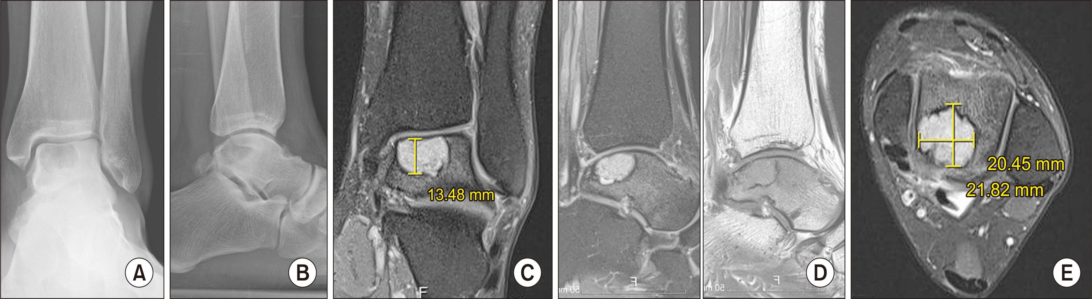
Figure 1 On standing anteroposterior (A) and lateral (B) radiographs, a round, radiolucent intraosseous lesion was detected. Patient’s magnetic resonance imaging (C∼) showed a subchondral tumoral lesion which was low signal intensity in T1-weighted and high signal intensity in T2-weighted images without a communication to joint space.

Figure 2 A posterior half of medial malleolus was cut (A) and articular cartilage was exposed with an intact deltoid ligament as a hinge. A tumoral lesion was curetted under a fluoroscopic guidance (B). Two 3.0-mm cannulated screw to secure the osteotomy site were fixed after bone grafting (C, D). post.: posterior, ant.: anterior.
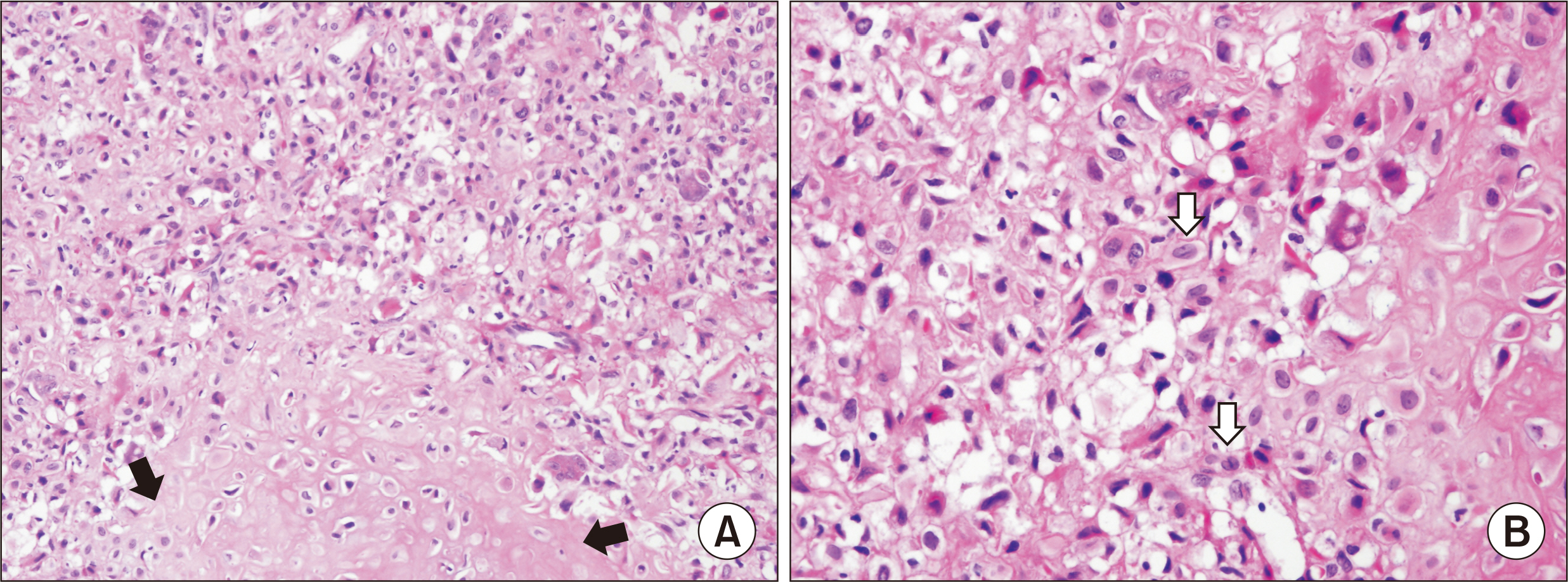
Figure 3 (A) Tumor cells consist of mononuclear chondroblasts, some giant cells and chondroid matrix (black arrows) (H&E, ×200). (B) Neoplastic chondroblasts show coffee-bean nuclear shape (white arrows) (H&E, ×400).

Figure 4 At postoperative 12 months, patient’s radiographs show the bony union of both bone graft and osteotomy site without a recurrence of chondroblastoma (A: anteroposterior; B: lateral).
Reference
-
1. Ningegowda RV, Subramanian K, Suresh I. 2013; Chondroblastoma of the talus. J Foot Ankle Surg. 52:673–7. doi: 10.1053/j.jfas.2013.02.020. DOI: 10.1053/j.jfas.2013.02.020. PMID: 23540757.
Article2. Park JS, Suh JS, Choi JY. 2019; Chondroblastoma of the talus mimicking an aneurysmal bone cyst: a case report. J Korean Foot Ankle Soc. 23:31–4. doi: 10.14193/jkfas.2019.23.1.31. DOI: 10.14193/jkfas.2019.23.1.31.
Article3. Hu Y, Yue C, Li X, Li Z, Zhou D, Xu H, et al. 2021; A novel medial malleolar osteotomy technique for the treatment of osteochondral lesions of the talus. Orthop J Sports Med. 9:2325967121989988. doi: 10.1177/2325967121989988. DOI: 10.1177/2325967121989988. PMID: 34250160. PMCID: PMC8237210.
Article4. Mendicino RW, Lee MS, Grossman JP, Shromoff PJ. 1998; Oblique medial malleolar osteotomy for the management of talar dome lesions. J Foot Ankle Surg. 37:516–23. doi: 10.1016/s1067-2516(98)80029-x. DOI: 10.1016/S1067-2516(98)80029-X. PMID: 9879047.
Article5. Shah R, Geevarughese NM, Shah S. 2021; An improved technique for medial malleolar osteotomy. Tech Foot Ankle Surg. 20:171–4. doi: 10.1097/BTF.0000000000000290. DOI: 10.1097/BTF.0000000000000290.
Article6. Zhang Y, Liang JQ, Wen XD, Liu PL, Lu J, Zhao HM. 2022; Triplane osteotomy combined with talar non-weight-bearing area autologous osteochondral transplantation for osteochondral lesions of the talus. BMC Musculoskelet Disord. 23:79. doi: 10.1186/s12891-022-05043-z. DOI: 10.1186/s12891-022-05043-z. PMID: 35065640. PMCID: PMC8783502.
Article7. Jamshidi K, Kargar Shooroki K, Sharifi Dalooei SMA, Mirzaei A. 2023; Intraosseous ganglion cyst of the talus treated with curettage and bone grafting through a medial malleolus osteotomy. Foot Ankle Int. 44:118–24. doi: 10.1177/10711007221141671. DOI: 10.1177/10711007221141671. PMID: 36571389.
Article8. Tripathy S, Varghese P, Panigrahi S, Puthiyapura LK. 2021; Medial malleolar osteotomy for intralesional curettage and bone grafting of primary aneurysmal bone cyst of the talus. BMJ Case Rep. 14:e242452. doi: 10.1136/bcr-2021-242452. DOI: 10.1136/bcr-2021-242452. PMID: 33962932. PMCID: PMC8108680.
Article9. Magnusson EA, Telfer S, Jackson M, Githens MF. 2021; Does a medial malleolar osteotomy or posteromedial approach provide greater surgical visualization for the treatment of talar body fractures? J Bone Joint Surg Am. 103:2324–30. doi: 10.2106/JBJS.21.00299. DOI: 10.2106/JBJS.21.00299. PMID: 34644268.
Article10. van Bergen CJ, Tuijthof GJ, Sierevelt IN, van Dijk CN. 2011; Direction of the oblique medial malleolar osteotomy for exposure of the talus. Arch Orthop Trauma Surg. 131:893–901. doi: 10.1007/s00402-010-1227-8. DOI: 10.1007/s00402-010-1227-8. PMID: 21165631. PMCID: PMC3117279.
Article
- Full Text Links
-
- Actions
-
Cited
- CITED
-
- Close
- Share
-
- Similar articles
-
- Reverse Chevron Transmalleolar Osteotomy for Exposure of the Medial Talar Dome Lesions: Operative Technique
- Outcome of Type 3 Talar Neck Fractures by Means of Medial Malleolar Osteotomy and Large Distractor
- Results of Operative Treatment for Large Osteochondral Lesion of Medial Talar Dome
- HERBERT SCREW FIXATION FOR NON-COMMINUTED CLOSED MEDIAL MALLEOLAR FRACTURE
- Surgical Treatment of Using Acutrak Screw for Ankle Medial Malleolar Fracture
