An ancient schwannoma misdiagnosed as a dermoid cyst on ultrasound examination: a case report
- Affiliations
-
- 1Department of Orthopaedic Surgery, CHA Gumi Medical Center, CHA University School of Medicine, Gumi, Korea
- 2Department of Orthopaedic Surgery, CHA Bundang Medical Center, CHA University School of Medicine, Seongnam, Korea
- KMID: 2542564
- DOI: http://doi.org/10.12790/ahm.21.0145
Abstract
- Ancient schwannomas are benign, long-standing schwannomas of the neural sheaths. This schwannoma subtype is characterized by cystic and hemorrhagic changes. These degenerative changes are thought to result from the tumor’s long-term progression and have the potential to be misdiagnosed as other soft tissue tumors or sarcomas. A 37-year-old female patient presented to our department with discomfort caused by a mass on the anterior side of her forearm. Approximately 2 weeks prior to her visit, an excisional biopsy was attempted under local anesthesia, but the patient experienced severe tingling sensations up to the wrist during surgery, preventing successful removal of the mass. Ultrasonography and magnetic resonance imaging (MRI) were performed for further differential diagnosis. An excisional biopsy was performed under general anesthesia, and histologic analysis revealed typical degenerative features of ancient schwannoma. A thorough examination, including MRI, is necessary to identify these tumors, prevent potential misdiagnosis, and ensure appropriate treatment.
Figure
-
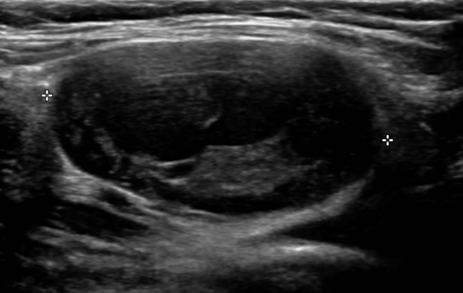
Fig. 1. Ultrasound image from the clinic the patient previously visited. The mass has a round, oval shape, a wall with a relatively clear border, and internal heterogeneity.

Fig. 2. Ultrasound image from our hospital. The examination shows a large well-circumscribed mass (3.8×3.5 cm) with a fluid-solid or fluid-fluid level filled with heterogeneous echoic material.

Fig. 3. Magnetic resonance imaging shows an enhanced, well-demarcated tumor in the left forearm. (A) A well-encapsulated isointense lesion on a T1-weighted image. The cystic and necrotic areas have low signal intensity in the T1 sequences. (B) Heterogeneous enhancement in the central mass with the entering-exiting nerve sign and split-fat sign on a T2 fat-suppression image. The cystic and necrotic areas have high signal intensity in the T2 sequences. In the cystic mass, a fluid-solid level is seen within the central tumorous lesion.
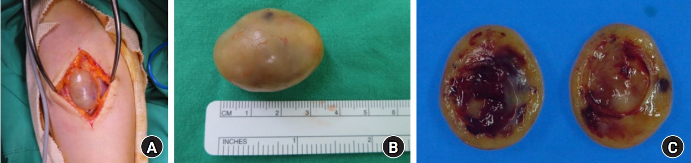
Fig. 4. (A) A well-encapsulated mass anterior to the median nerve was observed. (B) The actual size of the resected tumor was 2.7×2.0×3.5 cm, and it was a firm and yellow oval with a clear border. (C) Macroscopic view of the enucleated tumor. The cut surface revealed myxomatous changes accompanied by cystic degeneration and hematomas.
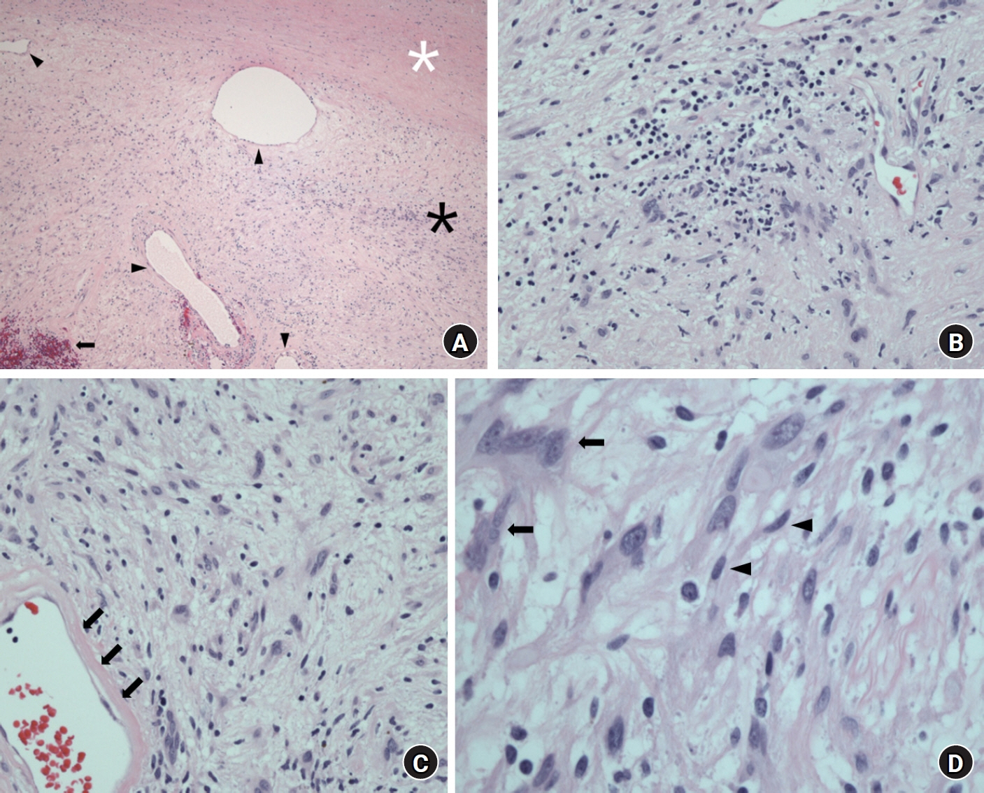
Fig. 5. Microscopic findings. (A) In the low-magnification micrograph, alternating Antoni A (black asterisk) and Antoni B (white asterisk) patterns are seen. Several blood vessels (black arrowheads) and hemorrhages (black arrow) are observed as well (H&E stain, ×40). (B) Multiple inflammatory cells representing degenerative changes are observed (H&E stain, ×100). (C) The thickened blood vessel wall is hyalinized (black arrows) (H&E stain, ×200). (D) Degenerative atypical cells (black arrows) with typical neurogenic tumor cells (black arrowheads) are observed (H&E stain, ×400).
Reference
-
References
1. Weiss SW GJ, Enzinger FM. Enzinger and Weiss’s soft tissue tumours. 4th ed. St. Louis: Mosby;2001. p. 1111–208.2. MacCollin M, Chiocca EA, Evans DG, et al. Diagnostic criteria for schwannomatosis. Neurology. 2005; 64:1838–45.
Article3. Knight DM, Birch R, Pringle J. Benign solitary schwannomas: a review of 234 cases. J Bone Joint Surg Br. 2007; 89:382–7.4. Hide IG, Baudouin CJ, Murray SA, Malcolm AJ. Giant ancient schwannoma of the pelvis. Skeletal Radiol. 2000; 29:538–42.
Article5. Schultz E, Sapan MR, McHeffey-Atkinson B, Naidich JB, Arlen M. Case report 872. “Ancient” schwannoma (degenerated neurilemoma). Skeletal Radiol. 1994; 23:593–5.6. Yasumatsu R, Nakashima T, Miyazaki R, Segawa Y, Komune S. Diagnosis and management of extracranial head and neck schwannomas: a review of 27 cases. Int J Otolaryngol. 2013; 2013:973045.
Article7. Koga H, Matsumoto S, Manabe J, Tanizawa T, Kawaguchi N. Definition of the target sign and its use for the diagnosis of schwannomas. Clin Orthop Relat Res. 2007; 464:224–9.
Article8. Vilanova JR, Burgos-Bretones JJ, Alvarez JA, Rivera-Pomar JM. Benign schwannomas: a histopathological and morphometric study. J Pathol. 1982; 137:281–6.
Article9. Ackerman LV, Taylor FH. Neurogenous tumors within the thorax: a clinicopathological evaluation of forty-eight cases. Cancer. 1951; 4:669–91.
Article10. Rockwell GM, Thoma A, Salama S. Schwannoma of the hand and wrist. Plast Reconstr Surg. 2003; 111:1227–32.
Article
