Evaluation of fracture strength and translucency of 3D printing resin crown for carious primary anterior tooth
- Affiliations
-
- 1Department of Pediatric Dentistry, College of Dentistry, Dankook University, Cheonan, Korea
- KMID: 2541090
- DOI: http://doi.org/10.11149/jkaoh.2023.47.1.40
Abstract
Objectives
The purpose of this study was to compare the fracture strength and traslucency of 3D printing resin crowns according to different thicknesses.
Methods
Resin crowns were designed with CAD software and a 3D scanner, using scanned data of the #61 tooth model. Resin Crowns with different thicknesses were printed using a 3D printer, and subsequently divided into four groups according to thickness (0.3, 0.5, 0.7, and 1.0 mm). Fracture strength was compared among groups with a resin strip crown of 1.0 mm thickness. Compressive force was applied using a universal testing machine at 30° along the lingual surface at 1 mm/min cross head speed. For translucency evaluation, thin square specimens were printed of thicknesses 0.3, 0.5, 0.7, and 1.0 mm, and translucency was measured using a spectrophotometer.
Results
As a result of fracture strength measurement, fracture strength increased as thickness increased, and a significant difference was observed solely between thicknesses of 0.3 and 0.5 mm, and the thicknesses of 0.3 and 0.5 mm (P<0.05). Translucency decreased as thickness increased, and similarly, a significant difference was observed only between thicknesses of 0.3 and 0.5 mm and the thicknesses of 0.7 and 1.0 mm (P<0.05).
Conclusions
A 3D printing resin crown can be used as a clinical option for restoring a primary anterior tooth affected by caries.
Figure
-

Fig. 1 CAD design for primary anterior 3D printing resin crown. (A-C) Designed resin crown view from buccal, below, and lateral. (D) 1.0 mm thickness of resin crowns design protocol.
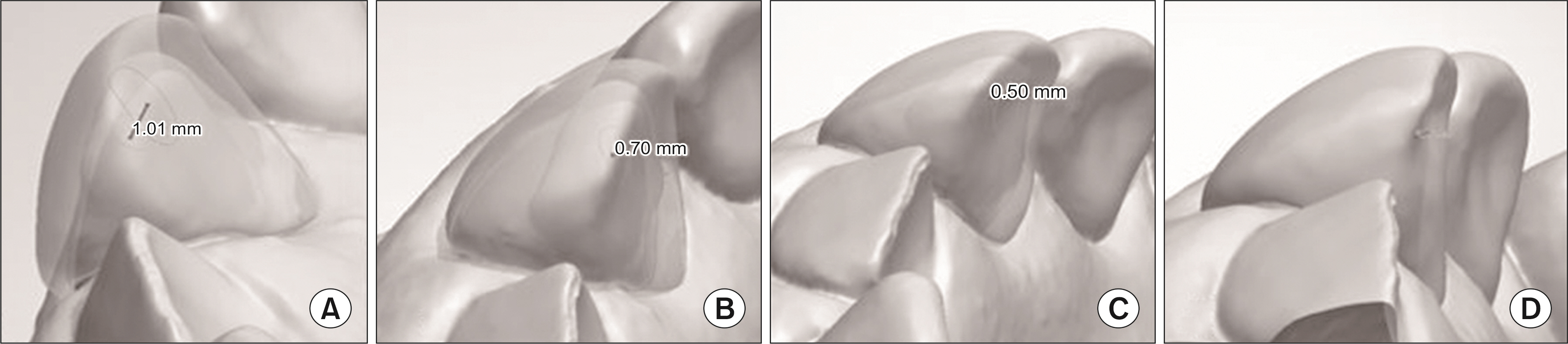
Fig. 2 Designed resin crown for different lingual surface thickness. (A) 1.0 mm, (B) 0.7 mm, (C) 0.5 mm, (D) 0.3 mm.
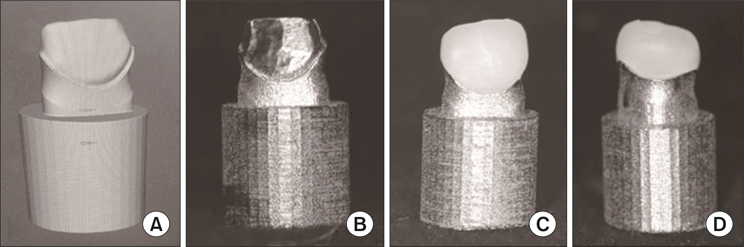
Fig. 3 Design of metal abutment and crown cementation. (A) Design protocol of metal abutment for 1.0 mm thickness 3D printing resin crown, (B) Metal abutment of 1.0 mm thickness 3D printing resin crown, (C) Image of 3D printing resin crown cemented to the abutment (buccal), (D) Image of 3D printing resin crown cemented to the abutment (lingual).
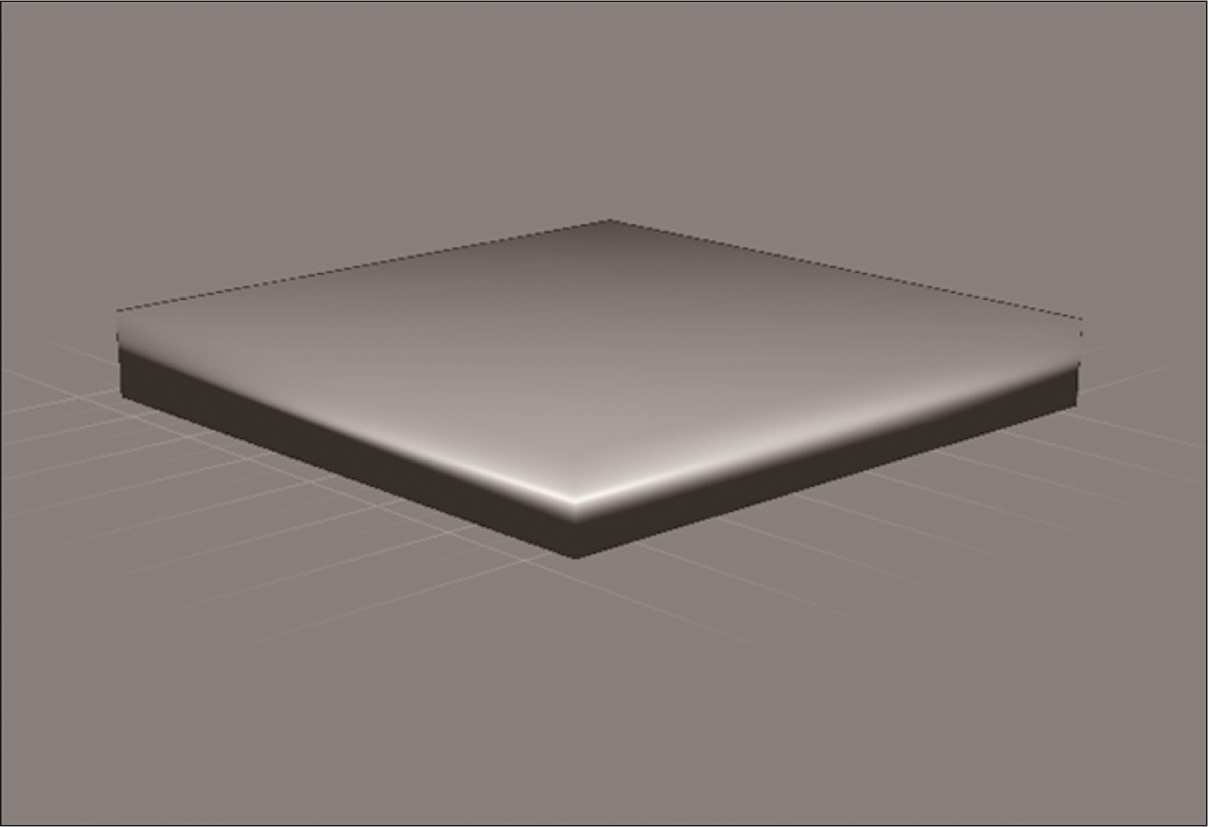
Fig. 4 Design of resin specimens for measuring the translucency parameter.
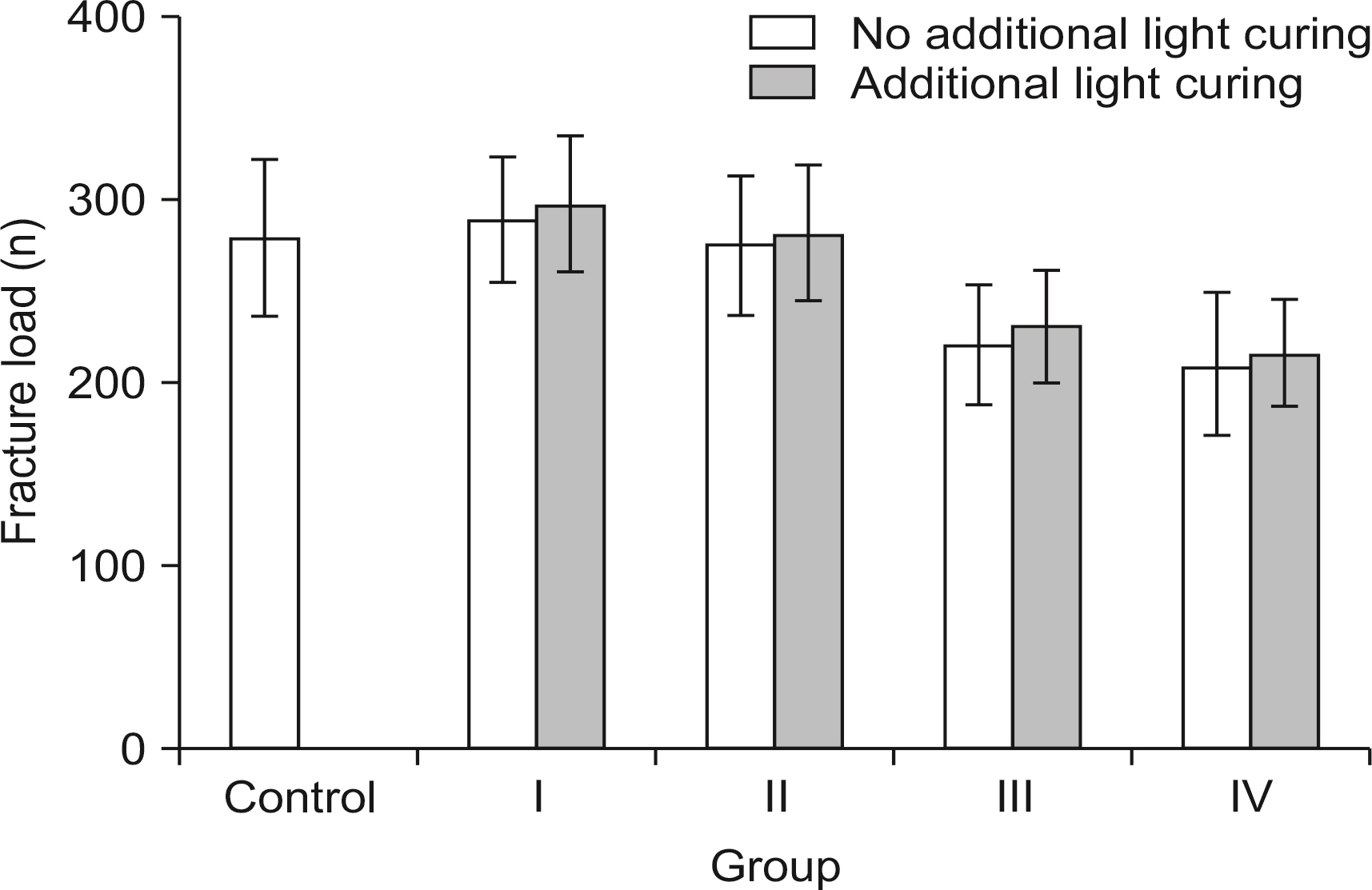
Fig. 5 Comparison of fracture strength between different thickness of 3D printing resin crown and resin strip crown.

Fig. 6 Comparison of Translucency parameter between different thickness of 3D printing resin specimens.
Reference
-
References
1. Li Y, Wang W. 2002; Predicting caries in permanent teeth from caries in primary teeth: an eight-year cohort study. J Dent Res. 81:561–566. DOI: 10.1177/154405910208100812. PMID: 12147748.
Article2. Ram D, Fuks AB. 2006; Clinical performance of resin-bonded composite strip crowns in primary incisors: a restrospective study. Int J Paediatr Dent. 16:49–54. DOI: 10.1111/j.1365-263X.2006.00680.x. PMID: 16364093.
Article3. Margolis FS. 2005; Esthetic restoration of discolored primary incisors. CDS Review. 98:28–31.4. Selwitz RH, Ismail AI, Pitts NB. 2007; Dental caries. Lancet. 369:51–59. DOI: 10.1016/S0140-6736(07)60031-2. PMID: 17208642.
Article5. O'Sullivan DM, Tinanoff N. 1993; Maxillary anterior caries associated with increased caries risk in other primary teeth. J Dent Res. 72:1577–1580. DOI: 10.1177/00220345930720120801. PMID: 8254125.6. Choi WS, Lee SH, Jih MK, Sung MA, Lee NY. 2022; Color comparison of maxillary primary anterior teeth and various composite resins using a spectrophotometer. J Korean Acad Pediatr Dent. 49:1–13. DOI: 10.5933/JKAPD.2022.49.1.1.
Article7. Waggoner WF. 2002; Restoring primary anterior teeth. Pediatr Dent. 24:511–516. DOI: 10.5353/th_b4417339. PMID: 12412967.8. Kim SY, Lim YJ, Lee SH, Lee NY, Jih MK. 2019; Comparison of crown shape and amount of tooth reduction for primary anterior prefabricated crowns. J Korean Acad Pediatr Dent. 46:64–75. DOI: 10.5933/JKAPD.2019.46.1.64.9. Alaki SM, Abdulhadi BS, AbdElBaki MA, Alamoudi NM. 2020; Comparing zirconia to anterior strip crowns in primary anterior teeth in children: a randomized clinical trial. BMC Oral Health. 20:1–11. DOI: 10.1186/s12903-020-01305-1. PMID: 33167954. PMCID: PMC7654025. PMID: f7e3ad045827425e9d55ac974742b837.
Article10. Souza MIAV, Cavalheiro JP, Bussaneli DG, Jeremias F, Zuanon ACC. 2018; Aesthetic rehabilitation with strip crowns in pediatric dentistry: a case report. Odontologia. 31:66–75. DOI: 10.21615/cesodon.31.2.7.
Article11. Kupietzky A. 2002; Bonded resin composite strip crowns for primary incisors: clinical tips for a successful outcome. Pediatr Dent. 24:145–148. PMID: 11991317.12. Clark L, Wells MH, Harris EF, Lou J. 2016; Comparison of amount of primary tooth reduction requred for anterior and posterior zirconia and stainless steel crowns. Pediatr Dent. 38:42–46.13. Corbani K, Hardan L, Skienhe H, Ozcan M, Alharbi N, Salameh Z. 2020; Effect of material thckness on the fracture resistance and failure pattern of 3D-printed composite crowns. Int J Comput Dent. 23:225–233.14. Ong SH, Kim JS, Kim JB, Shin JS, Yoo SH. 2020; Fracture strength and translucency of CAD/CAM zirconia crown for primary anterior tooth. J Korean Acad Pediatr Dent. 47:205–212. DOI: 10.5933/JKAPD.2020.47.2.205.
Article15. Vignesh KC, Eswar K, Muthu MS. 2020; A comparative evaluation of fracture toughness of composite resin vs protemp 4 for use in strip crowns: an in vitro study. Int J Clin Pediatr Dent. 13:57–60. DOI: 10.5005/jp-journals-10005-1711. PMID: 32581481. PMCID: PMC7299896.
Article16. McDonald RE, Avery DR, Dean JA. 2022. Dentistry for the child and adolescent. 11th ed. Elsevier;DOI: 10.1016/0300-5712(84)90075-7.17. Bayindir F, Koseoglu M. 2020; The effect of restoration thickness and resin cement shade on the color and translucency of a high-translucent monolithic zirconia. J Prosthet Dent. 123:149–154. DOI: 10.1016/j.prosdent.2018.11.002. PMID: 31027961.
Article18. Pozo P, Fuks AB. 2014; Zirconia crowns-an esthetic and resistant restorative alternative for ECC affected primary teeth. J Clin Pediatr Dent. 38:193–195. DOI: 10.17796/jcpd.38.3.0255q84jt2851311. PMID: 25095311.19. Gillings B, Buonocore M. 1961; An investigation of enamel thickness in human lower incisor teeth. J D Res. 40:105–118. DOI: 10.1177/00220345610400010201. PMID: 13705376.
Article20. Arangannal P, Chandra B, Hariharan VS, Vishnurekha , Jeevarathan , Vijayaprabha . 2012; Enamel thickness in primary teeth. J Clin Pediatr Dent. 37:177–182. DOI: 10.17796/jcpd.37.2.d6837416076l3334. PMID: 23534326.21. Shobber MZA, Alkhadra TA. 2017; Fracture resistance of different primary anterior esthetic crowns. Saud Dent J. 29:179–184. DOI: 10.1016/j.sdentj.2017.07.006. PMID: 29033529. PMCID: PMC5634802.
Article22. Fernandes NA, Vally ZI, Sykes LM. 2015; The longevity of restorations-a literature review. S Afr Dent J. 70:410–413.23. Mountain G, Wood D, Toumba J. 2011; Bite force measurement in children with primary dentition. Int J Paediatr Dent. 21:112–118. DOI: 10.1111/j.1365-263X.2010.01098.x. PMID: 20731734.
Article24. Gaviao MBD, Raymundo VG, Rentes AM. 2007; Masticatory performance and bite force in children with primary dentition. Braz Oral Res. 21:146–152. DOI: 10.1590/S1806-83242007000200009. PMID: 17589650. PMID: 88269813f1814ae8b52e670edd9c9514.
Article25. Waggoner WF. 1995; Fracture strength of four veneered primary stainless steel crowns. Pediatr Dent. 17:36–40.26. Kim NY, Kim H, Kim IH, Lee JH, Lee KE, Lee HS, et al. 2022; Novel 3D printed resin crowns for primary molars: in vitro study of fracture resistance, biaxial flexural strength, and dynamic mechnical analysis. Children. 9:1–13. DOI: 10.3390/children9101445. PMID: 36291379. PMCID: PMC9600781. PMID: 3280eb415900453c94ed505ece3828bc.27. Tiwary VK, Ravi NJ, Arunkumar P, Shivakumar S, Deshpande AS, Malik VR. 2020; Investigations on friction stir joining of 3D printed parts to overcome bed size limitation and enhance joint quality for unmanned aircraft systems. J Mechanical Engineering Sci. 20:1–15. DOI: 10.1177/0954406220930049.
Article28. Alsandi Q, Ikeda M, Arisaka Y, Nikaido T, Tsuchida Y, Sadr A, et al. 2021; Evaluation of mechanical and physical properties of light and heat polymerized UDMA for DLP 3D printer. Sensors. 21:1–10. DOI: 10.3390/s21103331. PMID: 34064860. PMCID: PMC8151691. PMID: 7ff3c90e783d4af89a2881e0ca616a8c.29. Al-Juaila E, Osman E, Segaan L, Shrebaty M, Farghaly EA. 2018; Comparison of translucency for different thickness of recent types of esthetic zirconia ceramics versus conventional ceramics: in vitro study. Future Dent J. 4:297–301. DOI: 10.1016/j.fdj.2018.05.003.30. Lee YK. 2016; Criteria for clinical translucency evaluation of direct esthetic restorative materials. Restor Dent Endod. 41:159–166. DOI: 10.5395/rde.2016.41.3.159. PMID: 27508156. PMCID: PMC4977345. PMID: 4c1ad6d4a8684de38e965293eff62ba9.
Article
- Full Text Links
-
- Actions
-
Cited
- CITED
-
- Close
- Share
-
- Similar articles
-
- Fracture Strength and Translucency of CAD/CAM Zirconia Crown for Primary Anterior Tooth
- In Vitro Study on the Bond Strength Between 3D-Printed Resin and Resin Cement for Pediatric Crown Restoration
- Comparison of fracture strength, surface hardness, and color stain of conventionally fabricated, 3D printed, and CAD-CAM milled interim prosthodontic materials after thermocycling
- Effect of shell thickness on fracture strength of single implant provisional crowns fabricated by indirect-direct technique using 3D printing
- Effect of surface treatment on shear bond strength between artificial resin teeth and 3D printing denture base resin
