Blood Res.
2023 Mar;58(1):51-60. 10.5045/br.2023.2023007.
Diagnostic approach and use of CTPA in patients with suspected pulmonary embolism in an emergency department in Saudi Arabia
- Affiliations
-
- 1Department of Medicine, College of Medicine, Shaqra University, Shaqra, Saudi Arabia.
- 2Department of Medicine, King Abdulaziz Medical City National Guard Health Affairs Riyadh Saudi Arabia, College of Medicine, King Saud bin Abdulaziz University for Health Science Riyadh Saudi Arabia, Riyadh, Saudi Arabia.
- 3Department of Oncology, King Abdulaziz Medical City, Ministry of National Guard Health Affairs Riyadh, King Abdullah International Medical Research Center, Ministry of National Guard Health Affairs, Riyadh, Saudi Arabia.
- 4Department of Oncology, King Abdulaziz Medical City, Ministry of National Guard Health Affairs Riyadh, Riyadh, Saudi Arabia.
- 5Department of Medicine, Ministry of National Guard Health Affairs, Riyadh, Saudi Arabia.
- 6Department of Medicine, College of Medicine, Imam Mohammad Ibn Saud Islamic University, Riyadh, Saudi Arabia.
- KMID: 2541065
- DOI: http://doi.org/10.5045/br.2023.2023007
Abstract
- Background
In patients with suspected pulmonary embolism (PE), the literature suggests the overuse of computerized tomography pulmonary angiography (CTPA) and underuse of clinical decision rules before imaging request. This study determined the potential for avoidable CTPA using the modified Wells score (mWS) and D-dimer assay in patients with suspected PE.
Methods
This hospital-based retrospective study analyzed the clinical data of 661 consecutive patients with suspected PE who underwent CTPA in the emergency department of a tertiary hospital for the use of a clinical prediction rule (mWS) and D-dimer assay. The score was calculated retrospectively from the available data in the files of patients who did not have a documented clinical prediction rule. Overuse (avoidable) CTPA was defined as D-dimer negativity and PE unlikely for this study.
Results
Of 661 patients’ data examined, clinical prediction rules were documented in 15 (2.3%). In total, 422 patients (63.8%) had required information on modified Wells criteria and D-dimer assays and were included for further analysis. PE on CTPA was present in 22 (5.21%) of PE unlikely (mWS ≤4) and 1 (0.24%) of D-dimer negative patients. Thirty patients (7.11%) met the avoidable CTPA (DD negative+PE unlikely) criteria, and it was significantly associated with dyspnea. The value of sensitivity of avoidable CTPA was 100%, whereas the positive predictive value was 90.3%.
Conclusion
Underutilization of clinical prediction rules before prescribing CTPA is common in emergency departments. Therefore, a mandatory policy should be implemented regarding the evaluation of avoidable CTPA imaging to reduce CTPA overuse.
Keyword
Figure
-
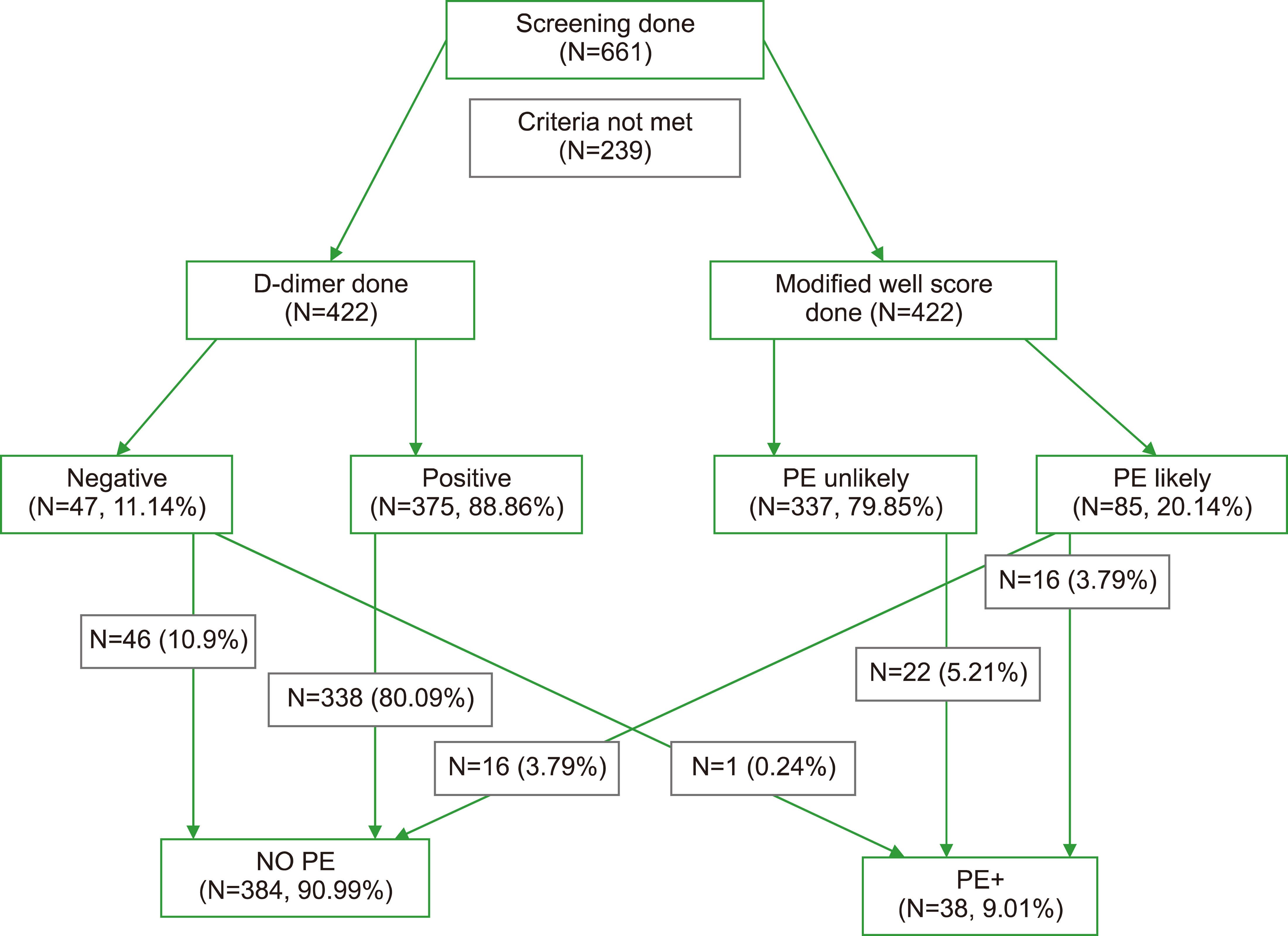
Fig. 1 STROB flow chart.
Reference
-
1. Morris T, Fedullo P. 2016; Pulmonary thromboembolism. In: Courtney Broaddus V, Mason RJ, Ernst JD, et al, eds. Murray and Nadel's textbook of respiratory medicine. 6th ed. Philadelphia, PA:. Elsevier Saunders,. 1001. DOI: 10.1016/B978-1-4557-3383-5.00057-9. PMID: 27324830.2. Silverstein MD, Heit JA, Mohr DN, Petterson TM, O'Fallon WM, Melton LJ 3rd. 1998; Trends in the incidence of deep vein thrombosis and pulmonary embolism: a 25-year population-based study. Arch Intern Med. 158:585–93. DOI: 10.1001/archinte.158.6.585. PMID: 9521222.
Article3. AlEidan FAS, AlManea RK, AlMoneef AT, et al. 2022; Incidence and predictors of recurrence and mortality following first venous thromboembolism among the Saudi population: single-center cohort study. Int J Gen Med. 15:7559–68. DOI: 10.2147/IJGM.S359893. PMID: 36199587. PMCID: PMC9527814.4. El Margoushy N, Al-Suwat R, Al-Otaibi W, Mougrabi M. 2017; Incidence of pulmonary embolism in CCU at King Faisal Hospital, Taif, Saudi Arabia. Egypt J Hosp Med. 68:865–77. DOI: 10.12816/0038185.5. Alharbi SH. 2020; Incidence of pulmonary thromboembolism and its associated comorbidities in Ha'il Region, Northern Saudi Arabia. Med Sci. 24:4352–8.6. Algahtani FH, Bayoumi N, Abdelgadir A, et al. 2013; Clinical characteristics and risk factors of pulmonary embolism: data from a Saudi tertiary-care center. J Thromb Haemost. 11:388–90. DOI: 10.1111/jth.12083. PMID: 23205904.7. Aziz S, Mfarah S, Rehman A, et al. 2020; Epidemiology and pattern of thromboembolism in Aseer Central Hospital, a multispecialty hospital in Aseer region, Abha in Southern Saudi Arabia. J Cardiol Cardiovas Res. 1:1–12. DOI: 10.37191/mapsci-jccr-1(2)-017.8. Stein PD, Fowler SE, Goodman LR, et al. 2006; Multidetector computed tomography for acute pulmonary embolism. N Engl J Med. 354:2317. DOI: 10.1056/NEJMoa052367. PMID: 16738268.9. Mamlouk MD, vanSonnenberg E, Gosalia R, et al. 2010; Pulmonary embolism at CT angiography: implications for appropriateness, cost, and radiation exposure in 2003 patients. Radiology. 256:625. DOI: 10.1148/radiol.10091624. PMID: 20551182.10. Perelas A, Dimou A, Saenz A, et al. 2015; CT pulmonary angiography utilization in the emergency department: diagnostic yield and adherence to current guidelines. Am J Med Qual. 30:571–7. DOI: 10.1177/1062860614543302. PMID: 25037560.11. Crichlow A, Cuker A, Mills AM. 2012; Overuse of computed tomography pulmonary angiography in the evaluation of patients with suspected pulmonary embolism in the emergency department. Acad Emerg Med. 19:1219–26. DOI: 10.1111/acem.12012. PMID: 23167851. PMCID: PMC3506180.12. Molaee S, Ghanaati H, Safavi E, Foroumandi M, Peiman S. 2015; Computed tomography pulmonary angiography for evaluation of patients with suspected pulmonary embolism: use or overuse. Iran J Radiol. 12:e22383. DOI: 10.5812/iranjradiol.12(2)2015.22383. PMID: 26557282.
Article13. Burge AJ, Freeman KD, Klapper PJ, Haramati LB. 2008; Increased diagnosis of pulmonary embolism without a corresponding decline in mortality during the CT era. Clin Radiol. 63:381–6. DOI: 10.1016/j.crad.2007.10.004. PMID: 18325357.14. Haap MM, Gatidis S, Horger M, Riessen R, Lehnert H, Haas CS. 2012; Computed tomography angiography in patients with suspected pulmonary embolism-too often considered? Am J Emerg Med. 30:325–30. DOI: 10.1016/j.ajem.2010.12.013. PMID: 21277141.
Article15. Venkatesh AK, Kline JA, Courtney DM, et al. 2012; Evaluation of pulmonary embolism in the emergency department and consistency with a national quality measure: quantifying the room for improvement. Arch Intern Med. 172:1028–32. DOI: 10.1001/archinternmed.2012.1804. PMID: 22664742. PMCID: PMC3775003.16. Al Dandan O, Hassan A, Alnasr A, et al. 2020; The use of clinical decision rules for pulmonary embolism in the emergency department: a retrospective study. Int J Emerg Med. 13:23. DOI: 10.1186/s12245-020-00281-1. PMID: 32393324. PMCID: PMC7216540. PMID: 4562014cd64845f5b013692d278d2e63.17. Takahashi EA, Yoon HC. 2013; Four-year cumulative radiation exposure in patients undergoing computed tomography angiography for suspected pulmonary embolism. Radiol Res Pract. 2013:482403. DOI: 10.1155/2013/482403. PMID: 23984065. PMCID: PMC3745975. PMID: b7b3369870194449abe46d3062496ac0.18. Mitchell AM, Jones AE, Tumlin JA, Kline JA. 2012; Prospective study of the incidence of contrast-induced nephropathy among patients evaluated for pulmonary embolism by contrast-enhanced computed tomography. Acad Emerg Med. 19:618–25. DOI: 10.1111/j.1553-2712.2012.01374.x. PMID: 22687176. PMCID: PMC5366244.
Article19. Lecumberri R, Alfonso A, Jiménez D, et al. 2013; Dynamics of case-fatalilty rates of recurrent thromboembolism and major bleeding in patients treated for venous thromboembolism. Thromb Haemost. 110:834–43. DOI: 10.1160/TH13-02-0132. PMID: 23846721.20. Wells PS, Anderson DR, Rodger M, et al. 2000; Derivation of a simple clinical model to categorize patients' probability of pulmonary embolism: increasing the model's utility with the SimpliRED D-dimer. Thromb Haemost. 83:416–20. DOI: 10.1055/s-0037-1613830. PMID: 10744147.
Article21. Wells PS, Anderson DR, Rodger M, et al. 2001; Excluding pulmonary embolism at the bedside without diagnostic imaging: management of patients with suspected pulmonary embolism presenting to the emergency department by using a simple clinical model and d-dimer. Ann Intern Med. 135:98–107. DOI: 10.7326/0003-4819-135-2-200107170-00010. PMID: 11453709.22. Kline JA, Courtney DM, Kabrhel C, et al. 2008; Prospective multicenter evaluation of the pulmonary embolism rule-out criteria. J Thromb Haemost. 6:772–80. DOI: 10.1111/j.1538-7836.2008.02944.x. PMID: 18318689.23. Konstantinides SV, Meyer G, Becattini C, et al. 2020; 2019 ESC guidelines for the diagnosis and management of acute pulmonary embolism developed in collaboration with the European Respiratory Society (ERS). Eur Heart J. 41:543–603. DOI: 10.1093/eurheartj/ehz405. PMID: 31504429.24. Righini M, Perrier A, De Moerloose P, Bounameaux H. 2008; D-dimer for venous thromboembolism diagnosis: 20 years later. J Thromb Haemost. 6:1059–71. DOI: 10.1111/j.1538-7836.2008.02981.x. PMID: 18419743.25. Pasha SM, Klok FA, Snoep JD, et al. 2010; Safety of excluding acute pulmonary embolism based on an unlikely clinical probability by the Wells rule and normal D-dimer concentration: a meta-analysis. Thromb Res. 125:e123–7. DOI: 10.1016/j.thromres.2009.11.009. PMID: 19942258.26. United States Government Accountability Office. 2008. Medicare part B imaging services: rapid spending growth and shift to physician offices indicate need for CMS to consider additional management practices. Government Accountability Office;Washington, D.C.: https://www.gao.gov/products/gao-08-452. Accessed December 20, 2022.27. Mitchell AM, Jones AE, Tumlin JA, Kline JA. 2011; Immediate complications of intravenous contrast for computed tomography imaging in the outpatient setting are rare. Acad Emerg Med. 18:1005–9. DOI: 10.1111/j.1553-2712.2011.01152.x. PMID: 21854485.28. Owaidah T, AlGhasham N, AlGhamdi S, et al. 2014; Evaluation of the usefulness of a D dimer test in combination with clinical pretest probability score in the prediction and exclusion of venous thromboembolism by medical residents. Thromb J. 12:28. DOI: 10.1186/s12959-014-0028-7. PMID: 25530719. PMCID: PMC4272774.
Article29. Abolfotouh MA, Almadani K, Al Rowaily MA. 2020; Diagnostic accuracy of D-dimer testing and the revised Geneva score in the prediction of pulmonary embolism. Int J Gen Med. 13:1537–43. DOI: 10.2147/IJGM.S289289. PMID: 33363402. PMCID: PMC7751841.30. Anwar A, Wong HS, Muza R. 2017; Audit on the use of CTPA in pulmonary embolism (PE) diagnosis. Gen Int Med Clin Innov. 2:1–5. DOI: 10.15761/GIMCI.1000144.31. Perera M, Aggarwal L, Scott IA, Cocks N. 2017; Underuse of risk assessment and overuse of computed tomography pulmonary angiography in patients with suspected pulmonary thrombo-embolism. Intern Med J. 47:1154–60. DOI: 10.1111/imj.13524. PMID: 28635149.
Article32. Alharbi SH, Almegren M, Alshammari HA, et al. 2020; Appropriateness of pulmonary CT angiography testing request in patients suspected with pulmonary embolism in Hai'l region, Northern Saudi Arabia. Med Sci. 24:4190–5.33. Kline JA, Garrett JS, Sarmiento EJ, Strachan CC, Courtney DM. 2020; Over-testing for suspected pulmonary embolism in American emergency departments: the continuing epidemic. Circ Cardiovasc Qual Outcomes. 13:e005753. DOI: 10.1161/CIRCOUTCOMES.119.005753. PMID: 31957477.34. Singh B, Mommer SK, Erwin PJ, Mascarenhas SS, Parsaik AK. 2013; Pulmonary embolism rule-out criteria (PERC) in pulmonary embolism--revisited: a systematic review and meta-analysis. Emerg Med J. 30:701–6. DOI: 10.1136/emermed-2012-201730. PMID: 23038695.35. Germini F, Zarabi S, Eventov M, Turcotte M, Li M, de Wit K. 2019; Prevalence of pulmonary embolism in patients accessing the emergency department, reported by country of study: a systematic review and meta-analysis. Res Pract Thromb Haemost (ISTH 2020 Congress Abstracts). 4(Suppl 1):PB2343. DOI: 10.1016/j.thromres.2019.09.013.36. Wiener RS, Schwartz LM, Woloshin S. 2011; Time trends in pulmonary embolism in the United States: evidence of overdiagnosis. Arch Intern Med. 171:831–7. DOI: 10.1001/archinternmed.2011.178. PMID: 21555660. PMCID: PMC3140219.
Article37. Hutchinson BD, Navin P, Marom EM, Truong MT, Bruzzi JF. 2015; Overdiagnosis of pulmonary embolism by pulmonary CT angiography. AJR Am J Roentgenol. 205:271–7. DOI: 10.2214/AJR.14.13938. PMID: 26204274.38. Alhassan S, Sayf AA, Arsene C, Krayem H. 2016; Suboptimal implementation of diagnostic algorithms and overuse of computed tomography-pulmonary angiography in patients with suspected pulmonary embolism. Ann Thorac Med. 11:254–60. DOI: 10.4103/1817-1737.191875. PMID: 27803751. PMCID: PMC5070434. PMID: 9b7d8ad63f7442789d1b3ce064b370be.39. Obradović D, Joveš B, Pena Karan S, Stefanović S, Ivanov I, Vukoja M. 2016; Correlation between the Wells score and the Quanadli index in patients with pulmonary embolism. Clin Respir J. 10:784–90. DOI: 10.1111/crj.12291. PMID: 25763885.40. Kubak MP, Lauritzen PM, Borthne A, Ruud EA, Ashraf H. 2016; Elevated d-dimer cut-off values for computed tomography pulmonary angiography-d-dimer correlates with location of embolism. Ann Transl Med. 4:212. DOI: 10.21037/atm.2016.05.55. PMID: 27386486. PMCID: PMC4916352.41. Alonso Martinez JL, Anniccherico Sánchez FJ, Urbieta Echezarreta MA, García IV, Álvaro JR. 2016; Central versus peripheral pulmonary embolism: analysis of the impact on the physiological parameters and long-term survival. N Am J Med Sci. 8:134–42. DOI: 10.4103/1947-2714.179128. PMID: 27114970. PMCID: PMC4821092.42. Laporte S, Mismetti P, Décousus H, et al. 2008; Clinical predictors for fatal pulmonary embolism in 15,520 patients with venous thromboembolism: findings from the Registro Informatizado de la Enfermedad TromboEmbolica venosa (RIETE) Registry. Circulation. 117:1711–6. DOI: 10.1161/CIRCULATIONAHA.107.726232. PMID: 18347212.43. Torres-Macho J, Mancebo-Plaza AB, Crespo-Giménez A, et al. 2013; Clinical features of patients inappropriately undiagnosed of pulmonary embolism. Am J Emerg Med. 31:1646–50. DOI: 10.1016/j.ajem.2013.08.037. PMID: 24060320.44. Carrier M, Righini M, Le Gal G. 2012; Symptomatic subsegmental pulmonary embolism: what is the next step? J Thromb Haemost. 10:1486–90. DOI: 10.1111/j.1538-7836.2012.04804.x. PMID: 22672341.
Article45. Carrier M, Righini M, Wells PS, et al. 2010; Subsegmental pulmonary embolism diagnosed by computed tomography: incidence and clinical implications. A systematic review and meta-analysis of the management outcome studies. J Thromb Haemost. 8:1716–22. DOI: 10.1111/j.1538-7836.2010.03938.x. PMID: 20546118.
Article46. Goy J, Lee J, Levine O, Chaudhry S, Crowther M. 2015; Sub-segmental pulmonary embolism in three academic teaching hospitals: a review of management and outcomes. J Thromb Haemost. 13:214–8. DOI: 10.1111/jth.12803. PMID: 25442511.
Article47. Konstantinides SV, Torbicki A, Agnelli G, et al. 2014; 2014 ESC guidelines on the diagnosis and management of acute pulmonary embolism. Eur Heart J. 35:3033–69. 3069a–k. DOI: 10.1093/eurheartj/ehu283. PMID: 25173341.48. Miniati M, Cenci C, Monti S, Poli D. 2012; Clinical presentation of acute pulmonary embolism: survey of 800 cases. PLoS One. 7:e30891. DOI: 10.1371/journal.pone.0030891. PMID: 22383978. PMCID: PMC3288010. PMID: c10de22305584a958a25052c69a780bf.
Article49. Raj K, Chandna S, Doukas SG, et al. 2021; Combined use of wells scores and D-dimer levels for the diagnosis of deep vein thrombosis and pulmonary embolism in COVID-19: a retrospective cohort study. Cureus. 13:e17687. DOI: 10.7759/cureus.17687. PMID: 34650862. PMCID: PMC8487632.
Article50. Rohacek M, Buatsi J, Szucs-Farkas Z, et al. 2012; Ordering CT pulmonary angiography to exclude pulmonary embolism: defense versus evidence in the emergency room. Intensive Care Med. 38:1345–51. DOI: 10.1007/s00134-012-2595-z. PMID: 22584801.51. Donohoo JH, Mayo-Smith WW, Pezzullo JA, Egglin TK. 2008; Utilization patterns and diagnostic yield of 3421 consecutive multidetector row computed tomography pulmonary angiograms in a busy emergency department. J Comput Assist Tomogr. 32:421–5. DOI: 10.1097/RCT.0b013e31812e6af3. PMID: 18520550.
Article52. Shujaat A, Shapiro JM, Eden E. 2013; Utilization of CT pulmonary angiography in suspected pulmonary embolism in a major urban emergency department. Pulm Med. 2013:915213. DOI: 10.1155/2013/915213. PMID: 24078873. PMCID: PMC3783975. PMID: 73c99a678556460db491011c4b106c12.
Article53. Green DB, Raptis CA, Alvaro Huete Garin I, Bhalla S. 2015; Negative computed tomography for acute pulmonary embolism: important differential diagnosis considerations for acute dyspnea. Radiol Clin North Am. 53:789–99. ixDOI: 10.1016/j.rcl.2015.02.014. PMID: 26046511.54. Ozakin E, Kaya FB, Acar N, Cevik AA. 2014; An analysis of patients that underwent computed tomography pulmonary angiography with the prediagnosis of pulmonary embolism in the emergency department. ScientificWorldJournal. 2014:470358. DOI: 10.1155/2014/470358. PMID: 24955406. PMCID: PMC4052481. PMID: e2da53490157409fb400168e37e7089d.
Article55. Zarabi S, Chan TM, Mercuri M, et al. 2021; Physician choices in pulmonary embolism testing. CMAJ. 193:E38–46. DOI: 10.1503/cmaj.201639. PMID: 33431544. PMCID: PMC7773048.
Article
- Full Text Links
-
- Actions
-
Cited
- CITED
-
- Close
- Share
-
- Similar articles
-
- Scintigraphic Evidence for Overdiagnosis of Small PE on CT Pulmonary Angiography
- The Diagnostic Accuracy and Impact of Computer-Aided Diagnosis of Pulmonary Emboli Using CT Pulmonary Angiography
- Early Use of Thrombolytics for Massive Pulmonary Embolism by a Comparison of Cases
- Multislice CT Virtual Intravascular Endoscopy for Assessing Pulmonary Embolisms: a Pictorial Review
- Venous air emboli during esophagoscopy confirmed by computed tomographic pulmonary angiography -a case report-
