A Novel Human Brainstem Map Based on True-Color Sectioned Images
- Affiliations
-
- 1Department of Anatomy, Dongguk University School of Medicine, Gyeongju, Korea
- KMID: 2540426
- DOI: http://doi.org/10.3346/jkms.2023.38.e76
Abstract
- Background
Existing atlases for the human brainstem were generated from magnetic resonance images or traditional histologically stained slides, but both are insufficient for the identification of detailed brainstem structures at uniform intervals.
Methods
A total of 319 sectioned images of the brainstem were selected from whole-body axial sectioned images, then coronal and sagittal sectioned images were reconstructed from the horizontal images. The fine and detailed structures were annotated in PowerPoint slides, then the volume model was produced and some white matter fibers were traced using MRIcroGL.
Results
In this study, a novel brainstem atlas based on sectioned images was generated that shows the true color and shape, as well as the accurate location of the nuclei and tracts; it reveals the striking contrast between gray and white matter, as well as fine structures. In total, 212 structures, including nuclei and tracts, were annotated in axial, coronal, and sagittal plane views of sectioned images (48-bit true color; 0.2 mm intervals, 0.06 mm × 0.06 mm pixel size). To verify the accuracy of the annotations, a volume model of the brainstem was constructed for independent observations of the three planes.
Conclusion
In this paper, we describe several interesting structures included in the atlas. By depicting the fine structures of the human brainstem in detail, this atlas allows comprehensive understanding of the complicated topographies of the brainstem. As such, it will be of value for neuroanatomy education and research, in addition to enriching the literature on the human brain.
Figure
-
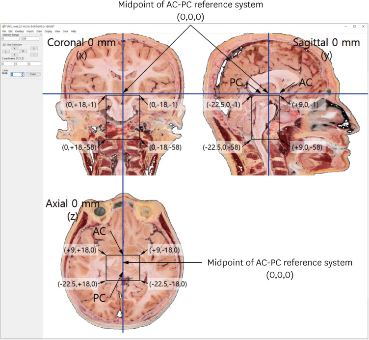
Fig. 1 Axial, coronal, and sagittal images and the 0 mm point defined by the AC-PC reference system. In whole head sectioned images, brainstem sectioned images in black rectangle box in each plane were used for this study.AC-PC = anterior commissure-posterior commissure.
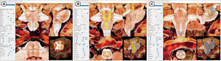
Fig. 2 3D volume models of brainstem with volumes-of-interest view of MRIcroGL. (A) Axial, coronal, and sagittal views. (B) Medial lemniscus (yellow) models. (C) Pyramidal tract (gray) models.
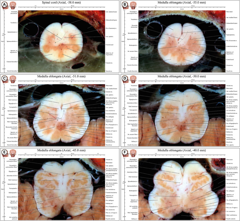
Fig. 3 Axial images of the spinal cord (−58 mm) to the medulla oblongata (−37 mm) in the novel human brainstem atlas. (A) All structures mentioned in the result are noted in underline. Three colonies of gray matter (cuneate nucleus, gracile nucleus, and spinal nucleus of trigeminal nerve) are observed in posterior horn of spinal cord, whereas there is gray matter of a colony in anterior horn of spinal cord. (B) Pyramidal decussation is identified by a curve of anterior spinal artery. In three colonies of gray matter in posterior area, spinal nucleus of trigeminal nerve is still larger than other two nuclei. (C) Three colonies of gray matter in posterior area become similar in size. There are internal arcuate fibers for decussating of medial lemniscus. (D) Inferior olivary complex compose to principal olivary nucleus, medial accessory olivary nucleus, and posterior accessory nucleus. Cuneate and gracile nuclei and tracts exist from (E) caudal end of medulla oblongata to (F) rostral medulla oblongata with inferior olivary complex.

Fig. 4 Axial images of the junction of the rostral medulla oblongata and pons (−36.4 mm) to the junction of the pons and midbrain (−13 mm) in the novel human brainstem atlas. All structures mentioned in the result are noted in underline. In anterior area, pyramidal tract of round shape is located in posterior side of pontocerebellar fibers but the tract is not encircled fully in the fibers unlike them in medulla oblongata. Posterior cochlear nucleus appear in lateral area in (A) and medial vestibular nucleus can be seen continuously from rostral medulla oblongata in (E). Medial lemniscus is moved from medial area to between anterior area and posterior area in (A-F). Sites of nucleus and fibers of facial nerve and nucleus of abducens nerve are lateral area and posterior area, respectively. (B) In posterior area, fibers of facial nerve wrap around behind the nucleus of abducens nerve and then moved forward in. (C) In lateral area, principal sensory nucleus, motor nucleus, and fibers of trigeminal nerve are situated and lateral lemniscus and its nucleus are situated in their lateral side in. (D) In posterior area, locus coeruleus of dark red color is located with lateral side of periaqueductal gray matter. (E) In lateral area, spinotectal tract, spinothalamic tract, and medial lemniscus are connected obliquely. (F) In central area, decussations of trochlear nerve in posterior area and superior cerebellar peduncle are observed.
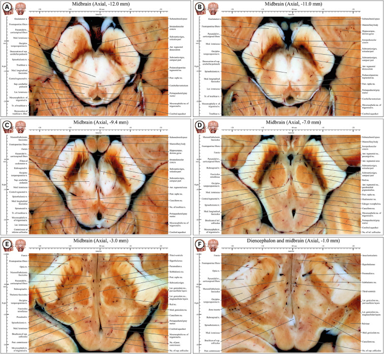
Fig. 5 Axial images of the caudal midbrain (−12 mm) to the rostral midbrain (−1 mm) in the novel human brainstem atlas. All structures mentioned in the result are noted in underline. (A) Substantia nigra is appearing and anterior tegmental nucleus, cerebellar peduncle, and trochlear nerve fibers are decussating. (B) Substantia nigra is divided into in compact and reticular parts. Tract of trochlear nerve is connected with trochlear nerve outside midbrain. (C) Fibers of oculomotor nerve move forward to interpeduncular fossa after the fiber penetrates substantia nigra. Nucleus of trochlear nerve is appearing. (D) Oculomotor nucleus, Edinger-Westphal nucleus, and sensory tracts have well defined features. (E) Subthalamic nucleus and medial and lateral geniculate nuclei are appearing whereas substantia nigra is disappearing. Both diencephalon including lateral geniculate nucleus, medial geniculate nucleus, subthalamic nucleus and midbrain including nucleus of superior colliculus can be seen. (F) This junctional plane has more properties of diencephalon than midbrain. Crus cerebri is clearly divided into frontopontine fibers, pyramidal tract (corticospinal fibers), and occipitotemperopontine tract in entire midbrain.
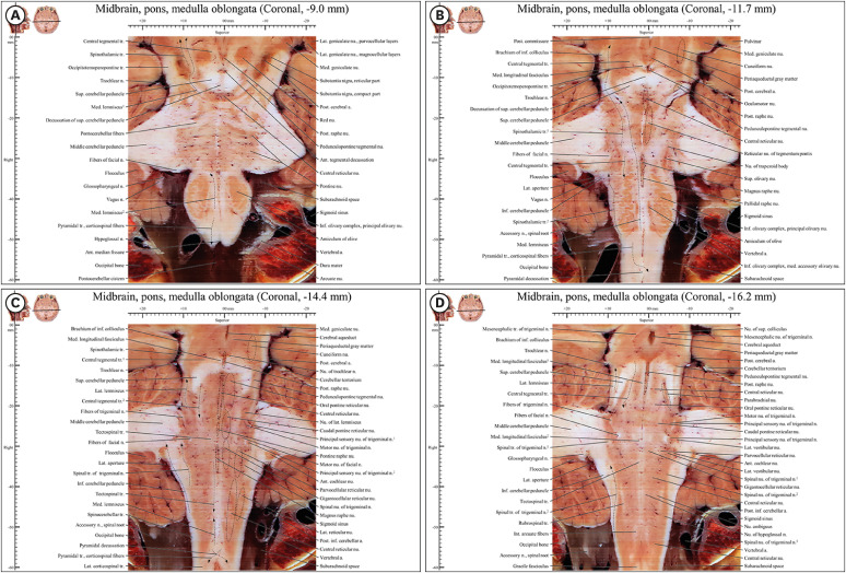
Fig. 6 Coronal images from −9.0 mm to −16 mm in the novel human brainstem atlas. (A) In anterior region (pyramid) of medulla oblongata, there is only pyramidal tract. (B) The medial lemniscus and the spinothalamic tract are located distantly at a distance in medulla oblongata and closely in pons and midbrain because of inferior olivary complex, superior olivary nucleus, and nucleus of trapezoid body. (C) In junction of medulla oblongata and spinal cord, pyramidal decussation can be observed. In middle region, various nuclei of reticular formation can be observed. (D) Posterior raphe nucleus, central reticular nucleus, and parabrachial nucleus are observed.
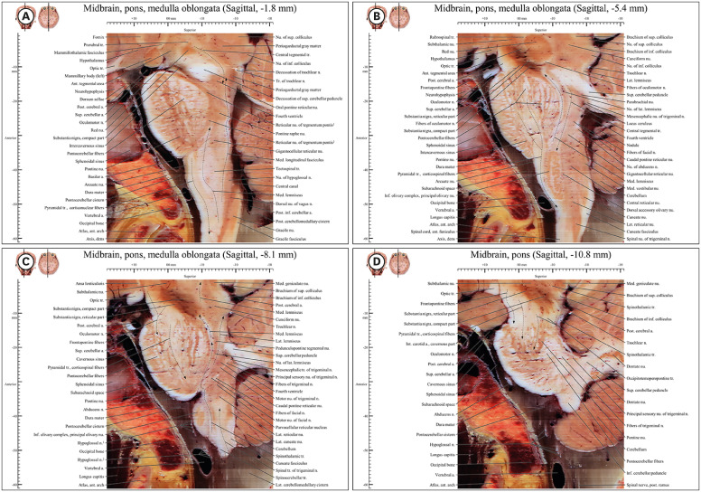
Fig. 7 Sagittal images from −1.8 mm to −10.8 mm in the novel human brainstem atlas. (A) In anterior region (pyramid) of medulla oblongata, there is pyramidal tract in front of medial lemniscus like coronal plane in (Fig. 6A). In posterior region, there is descending fiber of central tegmental tract in (A, B) in front of medial lemniscus. (C) Motor pathway and sensory pathway are located mainly anterior region and posterior region respectively. (D) In pons and midbrain, there are frontopontine fibers, pyramidal tract, and occipitotemoperopontine tract.
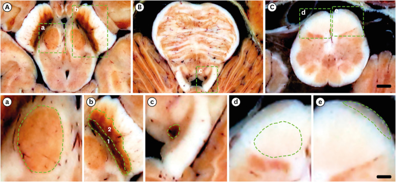
Fig. 8 Specific color of some structures in brainstem. a, b, c, d, e are magnifications of square areas in A, B, C, respectively. (A) It come from the atlas of axial −7.0 mm (Fig. 4D). (B) It come from the atlas of axial −18.0mm (Fig. 3D). (C) It come from the atlas of axial −50.0mm (Fig. 5D). (a) It is the magnifications of red nucleus with red color. (b) It is the magnifications of substantia nigra (compact part) (1) with dark color, and substantia nigra (reticular part) (2) with dark red color. (c) It is the magnifications of locus coeruleus, with dark red color. (d) It is the magnifications of corticospinal fibers with light red color. (e) It is the magnifications of arcuate nucleus with brown color. Scale bar of (A-C): 3 mm; scale bar of (a-e): 1 mm.
Cited by 1 articles
-
Lymph Node Stations of Pancreas Which Are Identified in Real Color Sectioned Images of a Cadaver With Pancreatic Cancer
Chung Yoh Kim, Yongwook Jung, Jin Seo Park
J Korean Med Sci. 2023;38(46):e392. doi: 10.3346/jkms.2023.38.e392.
Reference
-
1. Carpenter MB, Sutin J. Human Neuroanatomy. 8th ed. Baltimore, MD, USA: Williams & Wilkins;1983.2. Noback CR, Strominger NL, Demarest RJ, Ruggiero DA. Brainstem: medulla, pons, and midbrain. Noback CR, Strominger NL, Demarest RJ, Ruggiero DA, editors. The Human Nervous System: Structure and Function. Totowa, NJ, USA: Humana Press;2005. p. 219–242.3. Donaldson SS, Laningham F, Fisher PG. Advances toward an understanding of brainstem gliomas. J Clin Oncol. 2006; 24(8):1266–1272. PMID: 16525181.4. Adil SM, Calabrese E, Charalambous LT, Cook JJ, Rahimpour S, Atik AF, et al. A high-resolution interactive atlas of the human brainstem using magnetic resonance imaging. Neuroimage. 2021; 237:118135. PMID: 33951517.5. Bianciardi M, Toschi N, Eichner C, Polimeni JR, Setsompop K, Brown EN, et al. In vivo functional connectome of human brainstem nuclei of the ascending arousal, autonomic, and motor systems by high spatial resolution 7-Tesla fMRI. Magn Reson Mater Biol Phys Med. 2016; 29(3):451–462.6. Nguyen TH, Vaussy A, Le Gaudu V, Aboab J, Espinoza S, Curajos I, et al. The brainstem in multiple sclerosis: MR identification of tracts and nuclei damage. Insights Imaging. 2021; 12(1):151. PMID: 34674050.7. Rushmore RJ, Wilson-Braun P, Papadimitriou G, Ng I, Rathi Y, Zhang F, et al. 3D exploration of the brainstem in 50-micron resolution MRI. Front Neuroanat. 2020; 14:40. PMID: 33071761.8. Singh K, Cauzzo S, García-Gomar MG, Stauder M, Vanello N, Passino C, et al. Functional connectome of arousal and motor brainstem nuclei in living humans by 7 Tesla resting-state fMRI. Neuroimage. 2022; 249:118865. PMID: 35031472.9. Coulombe V, Saikali S, Goetz L, Takech MA, Philippe É, Parent A, et al. A topographic atlas of the human brainstem in the ponto-mesencephalic junction plane. Front Neuroanat. 2021; 15:627656. PMID: 34483849.10. DeArmond SJ, Fusco MM, Dewey MM. Structure of the Human Brain: A Photographic Atlas. 3rd ed. New York, NY, USA: Oxford University Press, Inc.;1989.11. Leigh RJ. From Nissl stains to modern concepts of brainstem function. Brain. 2014; 138(2):501–503.12. Sclocco R, Beissner F, Bianciardi M, Polimeni JR, Napadow V. Challenges and opportunities for brainstem neuroimaging with ultrahigh field MRI. Neuroimage. 2018; 168:412–426. PMID: 28232189.13. Naidich TP, Duvernoy HM, Delman BN, Sorensen AG, Kollias SS, Haacke EM. Duvernoy's Atlas of the Human Brain Stem and Cerebellum: High-Field MRI, Surface Anatomy, Internal Structure, Vascularization and 3 D Sectional Anatomy. Berlin, Germany: Springer Verlag;2009.14. Talairach J, Tournoux P. Co-Planar Stereotaxic Atlas of the Human Brain: 3-Dimensional Proportional System: An Approach to Cerebral Imaging. 1st ed. Stuttgart, Germany: Thieme Stuttgart;1988.15. Park JS, Chung MS, Park HS, Shin DS, Har DH, Cho ZH, et al. A proposal of new reference system for the standard axial, sagittal, coronal planes of brain based on the serially-sectioned images. J Korean Med Sci. 2010; 25(1):135–141. PMID: 20052359.16. You Y, Kim CY, Kim SK, Chung BS, Har D, Choi J, et al. Advanced-sectioned images obtained by microsectioning of the entire male body. Clin Anat. 2022; 35(1):79–86. PMID: 34591338.17. Park JS, Chung MS, Hwang SB, Lee YS, Har DH, Park HS. Visible Korean human: improved serially sectioned images of the entire body. IEEE Trans Med Imaging. 2005; 24(3):352–360. PMID: 15754985.18. Chung BS, Park JS. Real-color volume models made from real-color sectioned images of Visible Korean. J Korean Med Sci. 2019; 34(10):e86. PMID: 30886552.19. Paxinos G, Huang XF. Atlas of the Human Brainstem. San Diego, CA, USA: Elsevier Inc.;1995.20. Federative Committee on Anatomical Terminology. Terminologia Anatomica: International Anatomical Terminology. 1st ed. New York, NY, USA: Thieme;1998.21. Kiernan JA, Rajakumar R. Barr’s the Human Nervous System: An Anatomical Viewpoint. 10th ed. Philadelphia, PA, USA: Lippincott Williams & Wilkins;2004.22. Park HS, Chung MS, Shin DS, Jung YW, Park JS. Accessible and informative sectioned images, color-coded images, and surface models of the ear. Anat Rec (Hoboken). 2013; 296(8):1180–1186. PMID: 23713007.23. Park HS, Chung MS, Shin DS, Jung YW, Park JS. Whole courses of the oculomotor, trochlear, and abducens nerves, identified in sectioned images and surface models. Anat Rec (Hoboken). 2015; 298(2):436–443. PMID: 25212480.
- Full Text Links
-
- Actions
-
Cited
- CITED
-
- Close
- Share
-
- Similar articles
-
- Lymph Node Stations of Pancreas Which Are Identified in Real Color Sectioned Images of a Cadaver With Pancreatic Cancer
- Serial Slice Images and Segmented Images of the Brainstem for Recognizing the Stereoscopic Morphology of its Nuclei and Tracts
- Automated Techniques for the Sectioned Images of Visible Korean
- Dawn of the Visible Monkey: Segmentation of the Rhesus Monkey for 2D and 3D Applications
- Registration of Cadaver's Sectioned Images to Patient's Head MRIs
