The effects of rebamipide, sucralfate, and rifaximin against inflammation and apoptosis in radiation-induced murine intestinal injury
- Affiliations
-
- 1Department of Internal Medicine, Kosin University College of Medicine, Busan, Korea
- 2Department of Radiation Oncology, Kosin University College of Medicine, Busan, Korea
- 3Kosin University College of Medicine, Busan, Korea
- KMID: 2538863
- DOI: http://doi.org/10.7180/kmj.22.140
Abstract
- Background
Radiotherapy improves overall survival in patients with abdominopelvic malignancies. However, the toxic effects of radiation restrict the maximum dose that can be given, and there are no well-established preventive or therapeutic strategies. This study was conducted to evaluate whether rebamipide, sucralfate, and rifaximin have a suppressive effect on acute ionizing radiation (IR)-induced inflammation in the intestines of mice.
Methods
Thirty-six ICR mice were divided into a vehicle-treated group with sham irradiation; a vehicle-treated group with irradiation; rebamipide, sucralfate, or rifaximin-treated groups with irradiation; and a rebamipide-treated group with sham irradiation. The expression of proinflammatory, anti-inflammatory, proapoptotic, and antiapoptotic factors was investigated.
Results
The downregulated expression of nicotinamide phosphoribosyltransferase by IR was attenuated by all drugs (p<0.05). All drugs suppressed the IR-induced activation of NF-κB and phosphorylation of MAPKs (p<0.05) and attenuated the production of TNF-α, IL-1β, and IL-6 in response to IR (p<0.05). The administration of all drugs markedly attenuated IR-induced increases in iNOS, COX-2, and PGE2 (p<0.05), as well as [Ca2+] oscillations that were increased by IR. The expression of proapoptotic genes and antiapoptotic genes was suppressed and induced, respectively, by all drugs. IR treatment increased the release of cytochrome C, which was attenuated by all drugs (p<0.05). All drug treatments resulted in a significant decrease in the expression of caspase-3 and caspase-7 (p<0.05), which were both upregulated following IR treatment.
Conclusions
The administration of rebamipide, sucralfate, or rifaximin prior to radiation therapy may prevent or attenuate acute radiation-induced enterocolitis.
Keyword
Figure
-
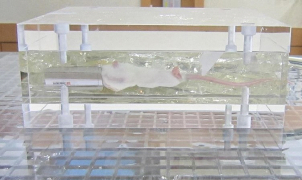
Fig. 1. Mice in the phantom. Three mice at a time were fixed in a supine position in the center of a specially designed, box-shaped acrylic phantom containing two inner jelly-filled bags to ensure a homogeneous density.
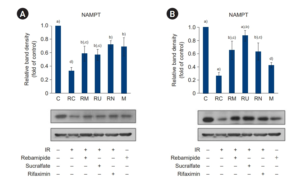
Fig. 2. Effects of rebamipide, sucralfate, and rifaximin on the ionizing radiation (IR)-induced expression of nicotinamide phosphoribosyltransferase (NAMPT) in the large intestine (A) and small intestine (B). The expression of NAMPT was measured by immunoblotting. Bands were quantified using densitometry, and band intensities were compared with controls. Values are presented as mean±standard deviation (n=6). A vehicle-treated control group before sham irradiation (C); a vehicle-treated control group before irradiation (RC); a rebamipide-treated group before irradiation (RM); a sucralfate-treated group before irradiation (RU); a rifaximin-treated group before irradiation (RN); a rebamipide-treated control group before sham irradiation (M). a)-e) Bars with different letters are significantly different at p<0.05 according to the Tukey test.
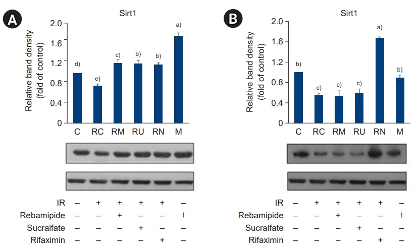
Fig. 3. Effects of rebamipide, sucralfate, and rifaximin on the ionizing radiation (IR)-induced expression of silent information regulator factor 2-related enzyme (Sirt)1 in the large intestine (A) and small intestine (B). The expression of Sirt1 was measured by immunoblotting. Bands were quantified using densitometry, and band intensities were compared with controls. Values are presented as mean± standard deviation (n=6). A vehicle-treated control group before sham irradiation (C); a vehicle-treated control group before irradiation (RC); a rebamipide-treated group before irradiation (RM); a sucralfate-treated group before irradiation (RU); a rifaximin-treated group before irradiation (RN); a rebamipide-treated control group before sham irradiation (M). a)-e)Bars with different letters are significantly different at p<0.05 according to the Tukey test.
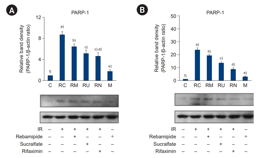
Fig. 4. Effects of rebamipide, sucralfate, and rifaximin on the ionizing radiation (IR)-induced expression of poly-ADP-ribose polymerase (PARP)-1 in the large intestine (A) and small intestine (B). The expression of PARP-1 was measured by immunoblotting. Bands were quantified using densitometry, and band intensities were compared with controls. Values are presented as mean±standard deviation (n=6). A vehicle-treated control group before sham irradiation (C); a vehicle-treated control group before irradiation (RC); a rebamipide-treated group before irradiation (RM); a sucralfate-treated group before irradiation (RU); a rifaximin-treated group before irradiation (RN); a rebamipide-treated control group before sham irradiation (M). a)-f)Bars with different letters are significantly different at p<0.05 according to the Tukey test.
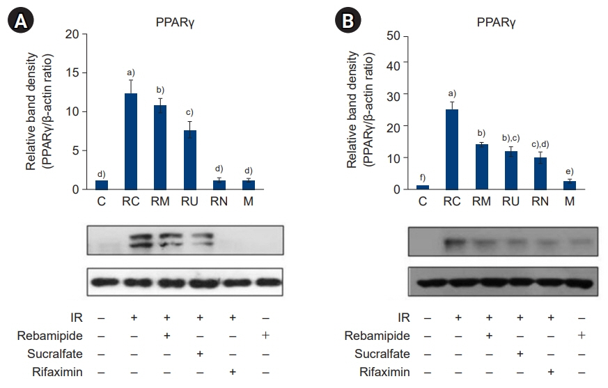
Fig. 5. Effects of rebamipide, sucralfate, and rifaximin on the ionizing radiation (IR)-induced expression of peroxisome proliferator-activated receptor (PPAR)γ in the large intestine (A) and small intestine (B). The expression of PPARγ was measured by immunoblotting. Bands were quantified using densitometry, and band intensities were compared with controls. Values are presented as mean±standard deviation (n=6). A vehicle-treated control group before sham irradiation (C); a vehicle-treated control group before irradiation (RC); a rebamipide-treated group before irradiation (RM); a sucralfate-treated group before irradiation (RU); a rifaximin-treated group before irradiation (RN); a rebamipide-treated control group before sham irradiation (M). a)-f)Bars with different letters are significantly different at p<0.05 according to the Tukey test.
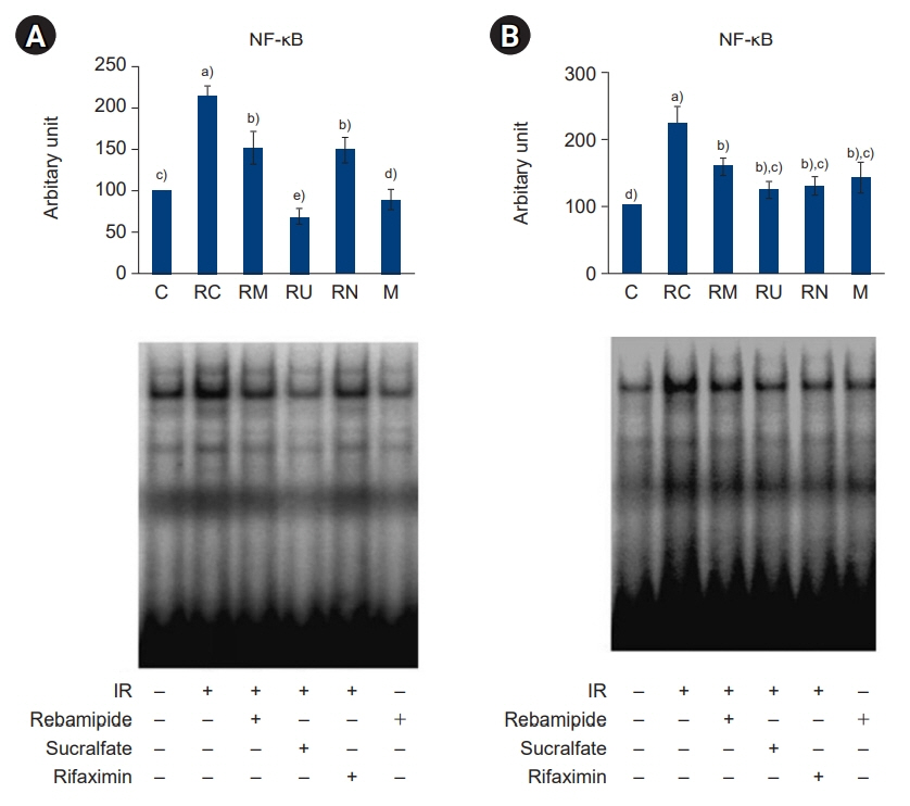
Fig. 6. Effects of rebamipide, sucralfate, and rifaximin on the ionizing radiation (IR)-induced DNA binding activity of NF-κB in the large intestine (A) and small intestine (B) as determined by electrophoretic mobility shift assay analysis. The intensities of the bands were determined by densitometry analysis and are expressed as relative radioactive intensities. Values are presented as mean±standard deviation (n=6). A vehicle-treated control group before sham irradiation (C); a vehicle-treated control group before irradiation (RC); a rebamipide-treated group before irradiation (RM); a sucralfate-treated group before irradiation (RU); a rifaximin-treated group before irradiation (RN); a rebamipide-treated control group before sham irradiation (M). a)-c)Bars with different letters are significantly different at p<0.05 according to the Tukey test.
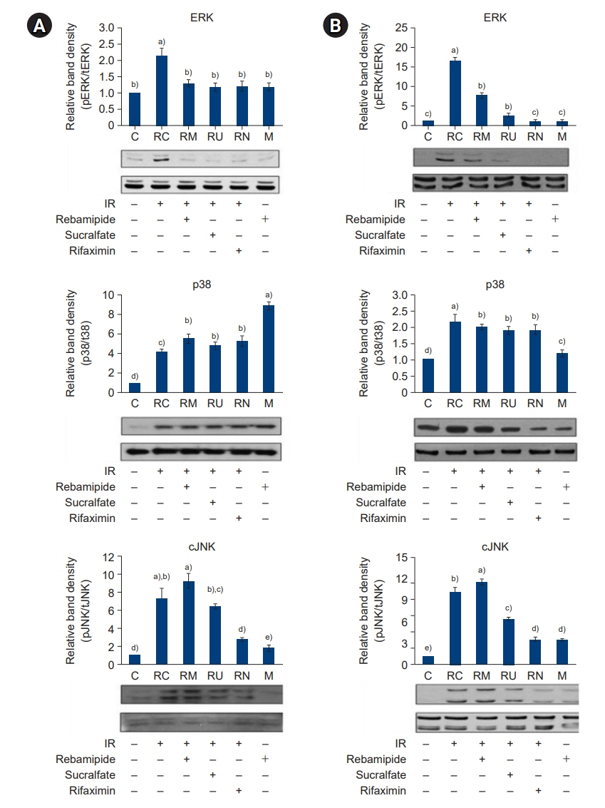
Fig. 7. Effects of rebamipide, sucralfate, and rifaximin on the ionizing radiation (IR)-induced mitogen-activated protein kinase (MARK) activation in the large intestine (A) and small intestine (B). The activation of MAPKs was measured by immunoblotting. Bands were quantified using densitometry, and band intensities were compared with controls. Values are presented as mean±standard deviation (n=6). A vehicle-treated control group before sham irradiation (C); a vehicle-treated control group before irradiation (RC); a rebamipide-treated group before irradiation (RM); a sucralfate-treated group before irradiation (RU); a rifaximin-treated group before irradiation (RN); a rebamipide-treated control group before sham irradiation (M). a)-e)Bars with different letters are significantly different at p<0.05 according to the Tukey test.
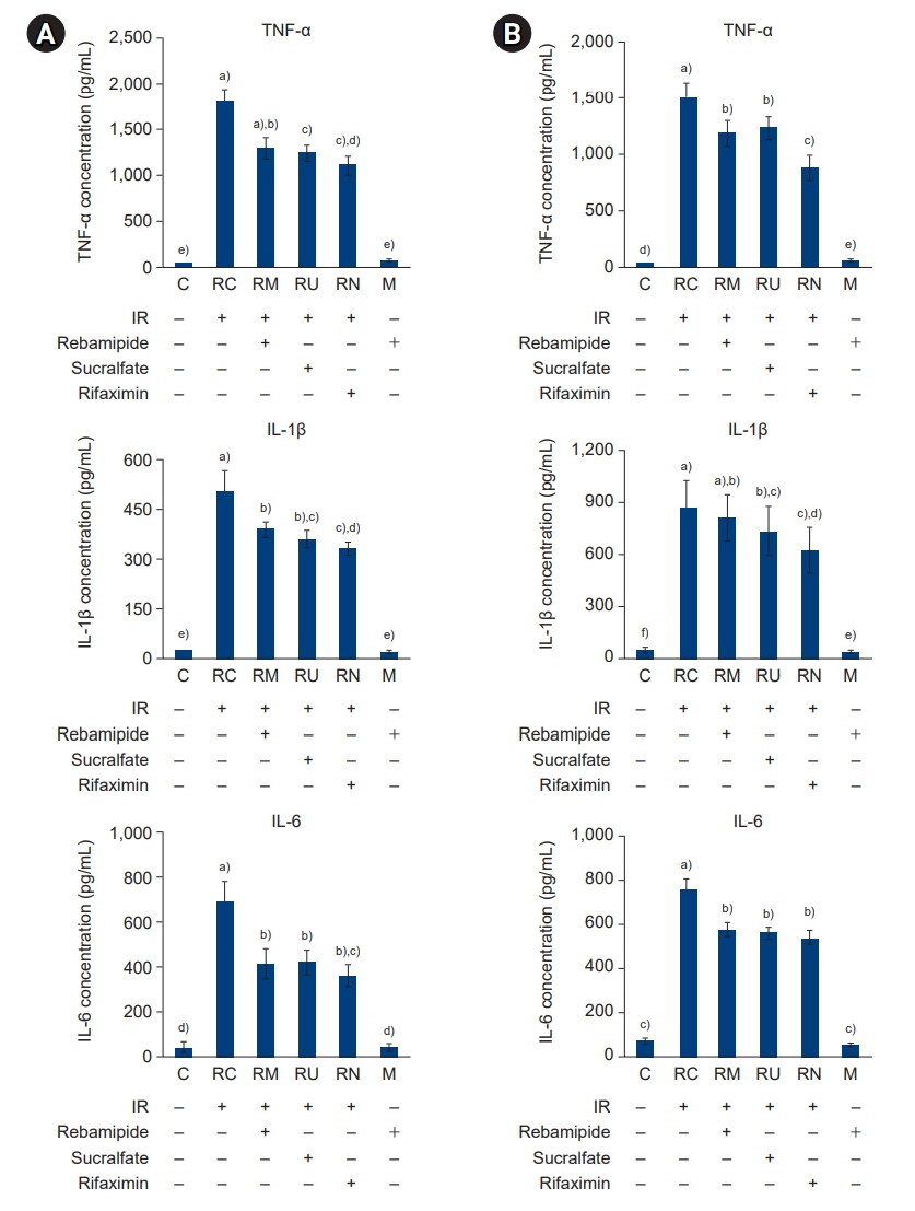
Fig. 8. Effects of rebamipide, sucralfate, and rifaximin on the ionizing radiation (IR)-induced production of TNF-α, IL-1β, and IL-6 in the large intestine (A) and small intestine (B). The production of TNF-α, IL-1β, and IL-6 was measured using enzyme-linked immunosorbent assay kits according to the manufacturer’s instructions. Values are presented as mean±standard deviation (n=6). A vehicle-treated control group before sham irradiation (C); a vehicle-treated control group before irradiation (RC); a rebamipide-treated group before irradiation (RM); a sucralfate-treated group before irradiation (RU); a rifaximin-treated group before irradiation (RN); a rebamipide-treated control group before sham irradiation (M). a)-f)Bars with different letters are significantly different at p<0.05 according to the Tukey test.
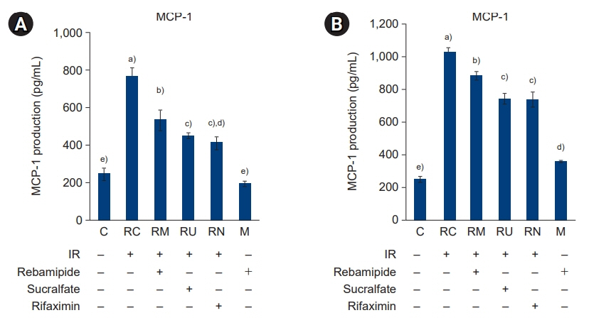
Fig. 9. Effects of rebamipide, sucralfate, and rifaximin on the ionizing radiation (IR)-induced production of major capsid protein (MCP)-1 in the large intestine (A) and small intestine (B). The production of MCP-1 was measured using an enzyme-linked immunosorbent assay kit according to the manufacturer’s instructions. Values are presented as mean±standard deviation (n=6). A vehicle-treated control group before sham irradiation (C); a vehicle-treated control group before irradiation (RC); a rebamipide-treated group before irradiation (RM); a sucralfate-treated group before irradiation (RU); a rifaximin-treated group before irradiation (RN); a rebamipide-treated control group before sham irradiation (M). a)-e)Bars with different letters are significantly different at p<0.05 according to the Tukey test.
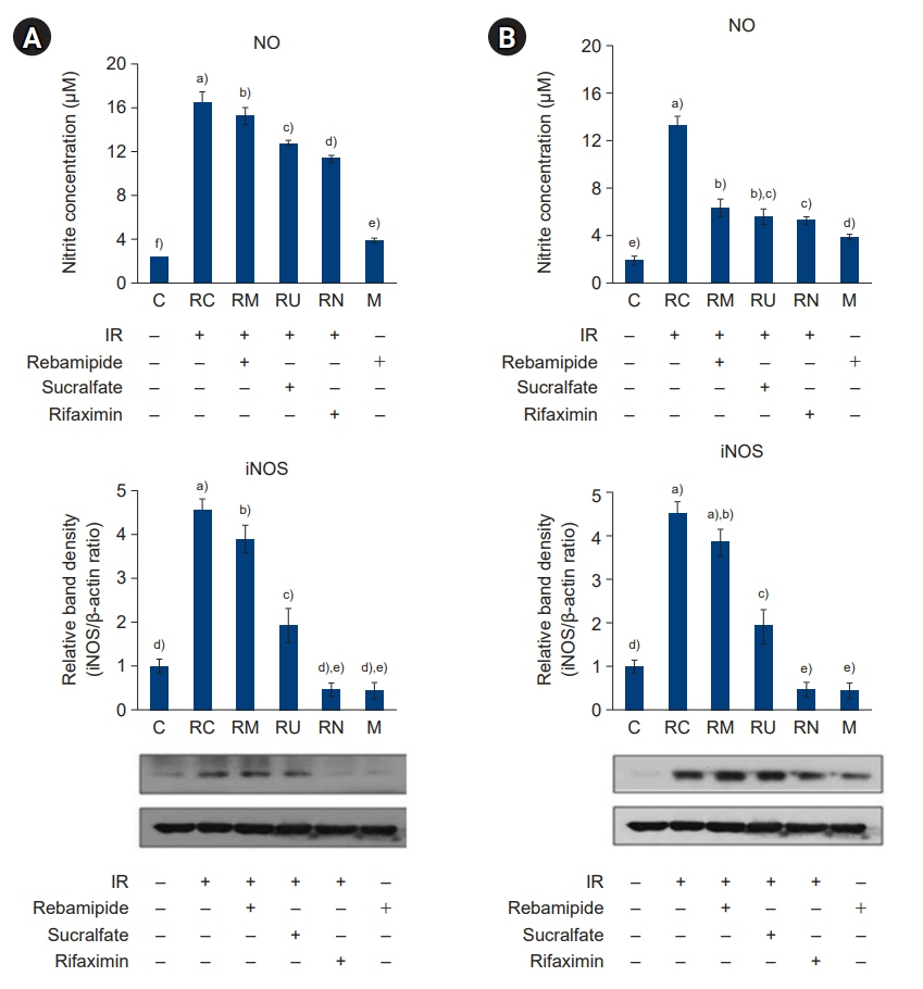
Fig. 10. Effects of rebamipide, sucralfate, and rifaximin on the ionizing radiation (IR)-induced production of nitric oxide (NO) and expression of inducible NO synthase (iNOS) in the large intestine (A) and small intestine (B). The production of NO and expression of iNOS were measured using the Griess reagent system and an immunoblotting assay, respectively. Bands were quantified using densitometry, and band intensities were compared with controls. Values are presented as mean±standard deviation (n=6). A vehicle-treated control group before sham irradiation (C); a vehicle-treated control group before irradiation (RC); a rebamipide-treated group before irradiation (RM); a sucralfate-treated group before irradiation (RU); a rifaximin-treated group before irradiation (RN); a rebamipide-treated control group before sham irradiation (M). a)-f)Bars with different letters are significantly different at p<0.05 according to the Tukey test.
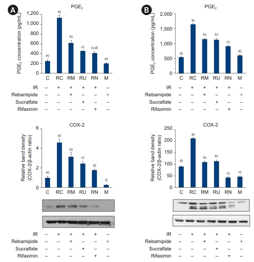
Fig. 11. Effects of rebamipide, sucralfate, and rifaximin on the ionizing radiation (IR)-induced production of PGE2 and expression of COX-2 in the large intestine (A) and small intestine (B). The production of PGE2 and expression of COX-2 were measured using an enzyme-linked immunosorbent assay kit according to the manufacturer’s instructions and an immunoblotting assay, respectively. Bands were quantified using densitometry, and band intensities were compared with controls. Values are presented as mean±standard deviation (n=6). A vehicle-treated control group before sham irradiation (C); a vehicle-treated control group before irradiation (RC); a rebamipide-treated group before irradiation (RM); a sucralfate-treated group before irradiation (RU); a rifaximin-treated group before irradiation (RN); a rebamipide-treated control group before sham irradiation (M). a)-f)Bars with different letters are significantly different at p<0.05 according to the Tukey test.
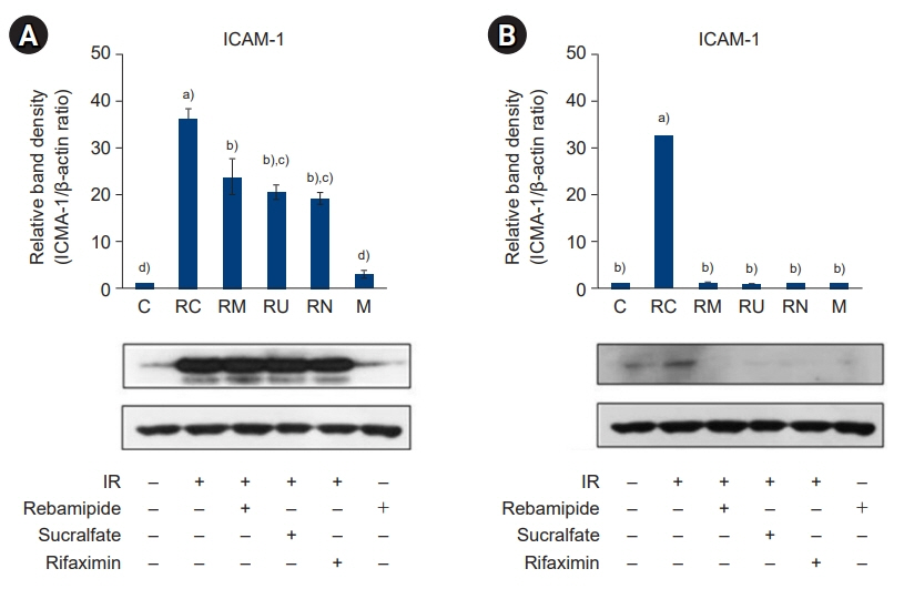
Fig. 12. Effects of rebamipide, sucralfate, and rifaximin on the ionizing radiation (IR)-induced expression of intercellular adhesion molecule 1 (ICAM-1) in the large intestine (A) and small intestine (B). The expression of ICAM-1 was measured by immunoblotting. Bands were quantified using densitometry, and band intensities were compared with controls. Values are presented as mean±standard deviation (n=6). A vehicle-treated control group before sham irradiation (C); a vehicle-treated control group before irradiation (RC); a rebamipide-treated group before irradiation (RM); a sucralfate-treated group before irradiation (RU); a rifaximin-treated group before irradiation (RN); a rebamipide-treated control group before sham irradiation (M). a)-d)Bars with different letters are significantly different at p<0.05 according to the Tukey test.
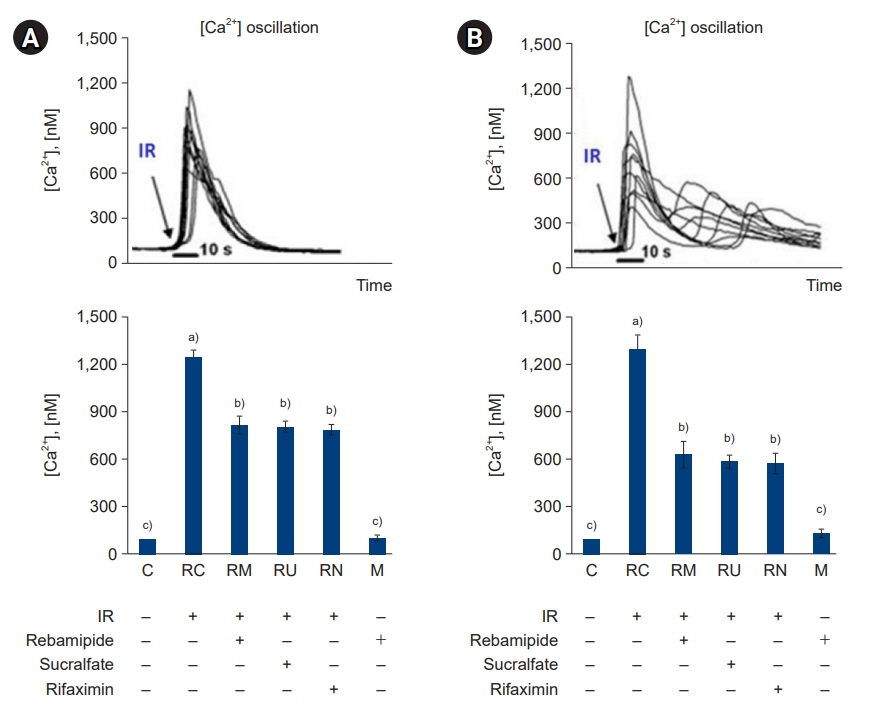
Fig. 13. Effects of rebamipide, sucralfate, and rifaximin on the ionizing radiation (IR)-induced [Ca2+] oscillations in the large intestine (A) and small intestine (B). IR induced an increase in [Ca2+] in the colons and small intestines of mice. Tissue cells were freshly isolated and stained with fluo-3 acetoxymethyl ester, attached to glass slides, and placed under a confocal microscope. The changes in fluorescence were monitored every 1.1 seconds for 100 seconds. The results are representative of 10 randomly chosen cells. Values are presented as mean±standard deviation (n=6). A vehicle-treated control group before sham irradiation (C); a vehicle-treated control group before irradiation (RC); a rebamipide-treated group before irradiation (RM); a sucralfate-treated group before irradiation (RU); a rifaximin-treated group before irradiation (RN); a rebamipide-treated control group before sham irradiation (M). a)-c)Bars with different letters are significantly different at p<0.05 according to the Tukey test.
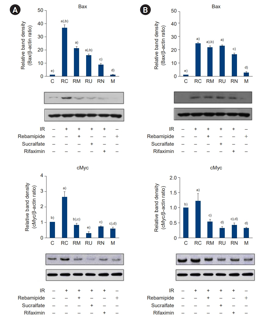
Fig. 14. Effects of rebamipide, sucralfate, and rifaximin on the ionizing radiation (IR)-induced expression of Bax and c-Myc in the large intestine (A) and small intestine (B). The expression of Bax and c-Myc was measured by immunoblotting. Bands were quantified using densitometry, and band intensities were compared with controls. Values are presented as mean±standard deviation (n=6). A vehicle-treated control group before sham irradiation (C); a vehicle-treated control group before irradiation (RC); a rebamipide-treated group before irradiation (RM); a sucralfate-treated group before irradiation (RU); a rifaximin-treated group before irradiation (RN); a rebamipide-treated control group before sham irradiation (M). a)-e)Bars with different letters are significantly different at p<0.05 according to the Tukey test.
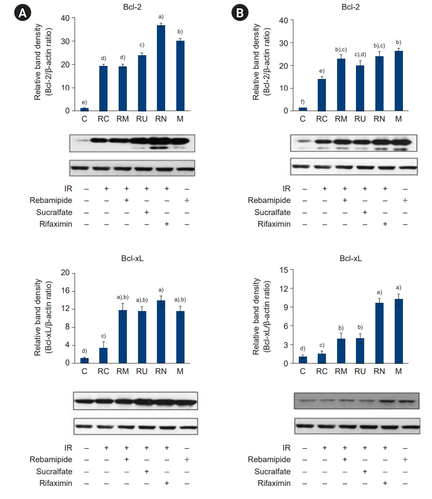
Fig. 15. Effects of rebamipide, sucralfate, and rifaximin on the ionizing radiation (IR)-induced expression of Bcl-2 and Bcl-xL in the large intestine (A) and small intestine (B). The expression of Bcl-2 and Bcl-xL was measured by immunoblotting. Bands were quantified using densitometry, and band intensities were compared with controls. Values are presented as mean±standard deviation (n=6). A vehicle-treated control group before sham irradiation (C); a vehicle-treated control group before irradiation (RC); a rebamipide-treated group before irradiation (RM); a sucralfate-treated group before irradiation (RU); a rifaximin-treated group before irradiation (RN); a rebamipide-treated control group before sham irradiation (M). a)-f)Bars with different letters are significantly different at p<0.05 according to the Tukey test.
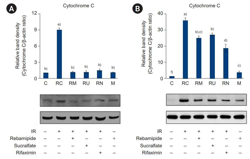
Fig. 16. Effects of rebamipide, sucralfate, and rifaximin on the ionizing radiation (IR)-induced expression of cytochrome C in the large intestine (A) and small intestine (B). The expression of cytochrome C was measured by immunoblotting. Bands were quantified using densitometry, and band intensities were compared with controls. Values are presented as mean±standard deviation (n=6). A vehicle-treated control group before sham irradiation (C); a vehicle-treated control group before irradiation (RC); a rebamipide-treated group before irradiation (RM); a sucralfate-treated group before irradiation (RU); a rifaximin-treated group before irradiation (RN); a rebamipide-treated control group before sham irradiation (M). a)-f)Bars with different letters are significantly different at p<0.05 according to the Tukey test.

Fig. 17. Effects of rebamipide, sucralfate, and rifaximin on the ionizing radiation (IR)-induced expression of caspase-3 and caspase-7 in the large intestine (A) and small intestine (B). The expression of caspase-3 and caspase-7 was measured by immunoblotting. Bands were quantified using densitometry, and the intensity of bands was compared with controls. Values are presented as mean±standard deviation (n=6). A vehicle-treated control group before sham irradiation (C); a vehicle-treated control group before irradiation (RC); a rebamipide-treated group before irradiation (RM); a sucralfate-treated group before irradiation (RU); a rifaximin-treated group before irradiation (RN); a rebamipide-treated control group before sham irradiation (M). a)-f)Bars with different letters are significantly different at p<0.05 according to the Tukey test.
Reference
-
References
1. Koh SB. The ideal strategies of chemotherapy for the treatment of cervical cancer. Kosin Med J. 2018; 33:283–8.
Article2. Potten CS. Extreme sensitivity of some intestinal crypt cells to X and gamma irradiation. Nature. 1977; 269:518–21.
Article3. Mann WJ. Surgical management of radiation enteropathy. Surg Clin North Am. 1991; 71:977–90.
Article4. Zimmermann FB, Feldmann HJ. Radiation proctitis: clinical and pathological manifestations, therapy and prophylaxis of acute and late injurious effects of radiation on the rectal mucosa. Strahlenther Onkol. 1998; 174 Suppl 3:85–9.5. Naito Y, Yoshikawa T. Rebamipide: a gastrointestinal protective drug with pleiotropic activities. Expert Rev Gastroenterol Hepatol. 2010; 4:261–70.
Article6. Kochhar R, Sriram PV, Sharma SC, Goel RC, Patel F. Natural history of late radiation proctosigmoiditis treated with topical sucralfate suspension. Dig Dis Sci. 1999; 44:973–8.7. Livstone EM, Hersh T, Spiro HM, Floch MH. The gastrointestinal microflora of irradiated mice. II. Effect of oral antibiotic administration on the colonic flora and survival of adult mice. Yale J Biol Med. 1970; 42:448–54.8. Ojetti V, Lauritano EC, Barbaro F, Migneco A, Ainora ME, Fontana L, et al. Rifaximin pharmacology and clinical implications. Expert Opin Drug Metab Toxicol. 2009; 5:675–82.
Article9. Kang G, Kim SE. How to write an original article in medicine and medical science. Kosin Med J. 2022; 37:96–101.
Article10. Kim DJ, Kil SY, Son J, Lee HS. How to conduct well-designed clinical research. Kosin Med J. 2022; 37:187–91.
Article11. Dignam JD, Lebovitz RM, Roeder RG. Accurate transcription initiation by RNA polymerase II in a soluble extract from isolated mammalian nuclei. Nucleic Acids Res. 1983; 11:1475–89.
Article12. D’Amours D, Desnoyers S, D’Silva I, Poirier GG. Poly(ADP-ribosyl)ation reactions in the regulation of nuclear functions. Biochem J. 1999; 342(Pt 2):249–68.
Article13. Luk T, Malam Z, Marshall JC. Pre-B cell colony-enhancing factor (PBEF)/visfatin: a novel mediator of innate immunity. J Leukoc Biol. 2008; 83:804–16.
Article14. Li Y, Zhang Y, Dorweiler B, Cui D, Wang T, Woo CW, et al. Extracellular NAMPT promotes macrophage survival via a nonenzymatic interleukin-6/STAT3 signaling mechanism. J Biol Chem. 2008; 283:34833–43.
Article15. Michan S, Sinclair D. Sirtuins in mammals: insights into their biological function. Biochem J. 2007; 404:1–13.
Article16. Yeung F, Hoberg JE, Ramsey CS, Keller MD, Jones DR, Frye RA, et al. Modulation of NF-kappaB-dependent transcription and cell survival by the SIRT1 deacetylase. EMBO J. 2004; 23:2369–80.
Article17. Leibiger IB, Berggren PO. Sirt1: a metabolic master switch that modulates lifespan. Nat Med. 2006; 12:34–6.
Article18. Zong WX, Ditsworth D, Bauer DE, Wang ZQ, Thompson CB. Alkylating DNA damage stimulates a regulated form of necrotic cell death. Genes Dev. 2004; 18:1272–82.
Article19. Wullaert A, Bonnet MC, Pasparakis M. NF-κB in the regulation of epithelial homeostasis and inflammation. Cell Res. 2011; 21:146–58.
Article20. Linard C, Marquette C, Clarencon D, Galonnier M, Mathieu J, Pennequin A, et al. Acute ileal inflammatory cytokine response induced by irradiation is modulated by subdiaphragmatic vagotomy. J Neuroimmunol. 2005; 168:83–95.
Article21. Ostrau C, Hulsenbeck J, Herzog M, Schad A, Torzewski M, Lackner KJ, et al. Lovastatin attenuates ionizing radiation-induced normal tissue damage in vivo. Radiother Oncol. 2009; 92:492–9.
Article22. Hong EH, Lee SJ, Kim JS, Lee KH, Um HD, Kim JH, et al. Ionizing radiation induces cellular senescence of articular chondrocytes via negative regulation of SIRT1 by p38 kinase. J Biol Chem. 2010; 285:1283–95.
Article23. Kumar S, Boehm J, Lee JC. p38 MAP kinases: key signalling molecules as therapeutic targets for inflammatory diseases. Nat Rev Drug Discov. 2003; 2:717–26.
Article24. Van Deventer SJ. Tumour necrosis factor and Crohn's disease. Gut. 1997; 40:443–8.
Article25. Sen A, Paine SK, Chowdhury IH, Mukherjee A, Choudhuri S, Saha A, et al. Impact of interleukin-6 promoter polymorphism and serum interleukin-6 level on the acute inflammation and neovascularization stages of patients with Eales’ disease. Mol Vis. 2011; 17:2552–63.26. Deshmane SL, Kremlev S, Amini S, Sawaya BE. Monocyte chemoattractant protein-1 (MCP-1): an overview. J Interferon Cytokine Res. 2009; 29:313–26.27. Barnes PJ, Liew FY. Nitric oxide and asthmatic inflammation. Immunol Today. 1995; 16:128–30.
Article28. Kolli VK, Abraham P, Rabi S. Methotrexate-induced nitrosative stress may play a critical role in small intestinal damage in the rat. Arch Toxicol. 2008; 82:763–70.
Article29. Courtois F, Seidman EG, Delvin E, Asselin C, Bernotti S, Ledoux M, et al. Membrane peroxidation by lipopolysaccharide and iron-ascorbate adversely affects Caco-2 cell function: beneficial role of butyric acid. Am J Clin Nutr. 2003; 77:744–50.
Article30. Jones DA, Carlton DP, McIntyre TM, Zimmerman GA, Prescott SM. Molecular cloning of human prostaglandin endoperoxide synthase type II and demonstration of expression in response to cytokines. J Biol Chem. 1993; 268:9049–54.
Article31. Brzozowski T, Konturek PC, Konturek SJ, Brzozowska I, Pawlik T. Role of prostaglandins in gastroprotection and gastric adaptation. J Physiol Pharmacol. 2005; 56 Suppl 5:33–55.32. Muller-Decker K, Furstenberger G. The cyclooxygenase-2-mediated prostaglandin signaling is causally related to epithelial carcinogenesis. Mol Carcinog. 2007; 46:705–10.
Article33. Gretzer B, Ehrlich K, Maricic N, Lambrecht N, Respondek M, Peskar BM. Selective cyclo-oxygenase-2 inhibitors and their influence on the protective effect of a mild irritant in the rat stomach. Br J Pharmacol. 1998; 123:927–35.
Article34. Molla M, Gironella M, Miquel R, Tovar V, Engel P, Biete A, et al. Relative roles of ICAM-1 and VCAM-1 in the pathogenesis of experimental radiation-induced intestinal inflammation. Int J Radiat Oncol Biol Phys. 2003; 57:264–73.
Article35. Wang HG, Pathan N, Ethell IM, Krajewski S, Yamaguchi Y, Shibasaki F, et al. Ca2+-induced apoptosis through calcineurin dephosphorylation of BAD. Science. 1999; 284:339–43.
Article36. Wei MC, Zong WX, Cheng EH, Lindsten T, Panoutsakopoulou V, Ross AJ, et al. Proapoptotic BAX and BAK: a requisite gateway to mitochondrial dysfunction and death. Science. 2001; 292:727–30.
Article37. Lakhani SA, Masud A, Kuida K, Porter GA Jr, Booth CJ, Mehal WZ, et al. Caspases 3 and 7: key mediators of mitochondrial events of apoptosis. Science. 2006; 311:847–51.
Article
- Full Text Links
-
- Actions
-
Cited
- CITED
-
- Close
- Share
-
- Similar articles
-
- Effect of Small Dose of Radiation on Induction of Apoptosis in Murine Tumors
- Effect of Sucralfate of the Prophylaxia of Adriamycin Induced Mucositis in the Rat
- Rebamipide Protects TNBS Induced Colonic Damage Through Down-regulation of NF-kappaB Activation and Induction of Heme Oxygenase -1 Expression
- The Effect of Sucralfate on the Reduction of Radiation Esophagitis: Clinical and Laboratory Data
- Rebamipide-induced downregulation of phospholipase D inhibits inflammation and proliferation in gastric cancer cells
